. 1997 Aug 15;11(16):2090-100.
doi: 10.1101/gad.11.16.2090.

Inhibition of CDK activity and PCNA-dependent DNA replication by p21 is blocked by interaction with the HPV-16 E7 oncoprotein

Affiliations

Inhibition of CDK activity and PCNA-dependent DNA replication by p21 is blocked by interaction with the HPV-16 E7 oncoprotein

J O Funk et al. Genes Dev. .

Abstract

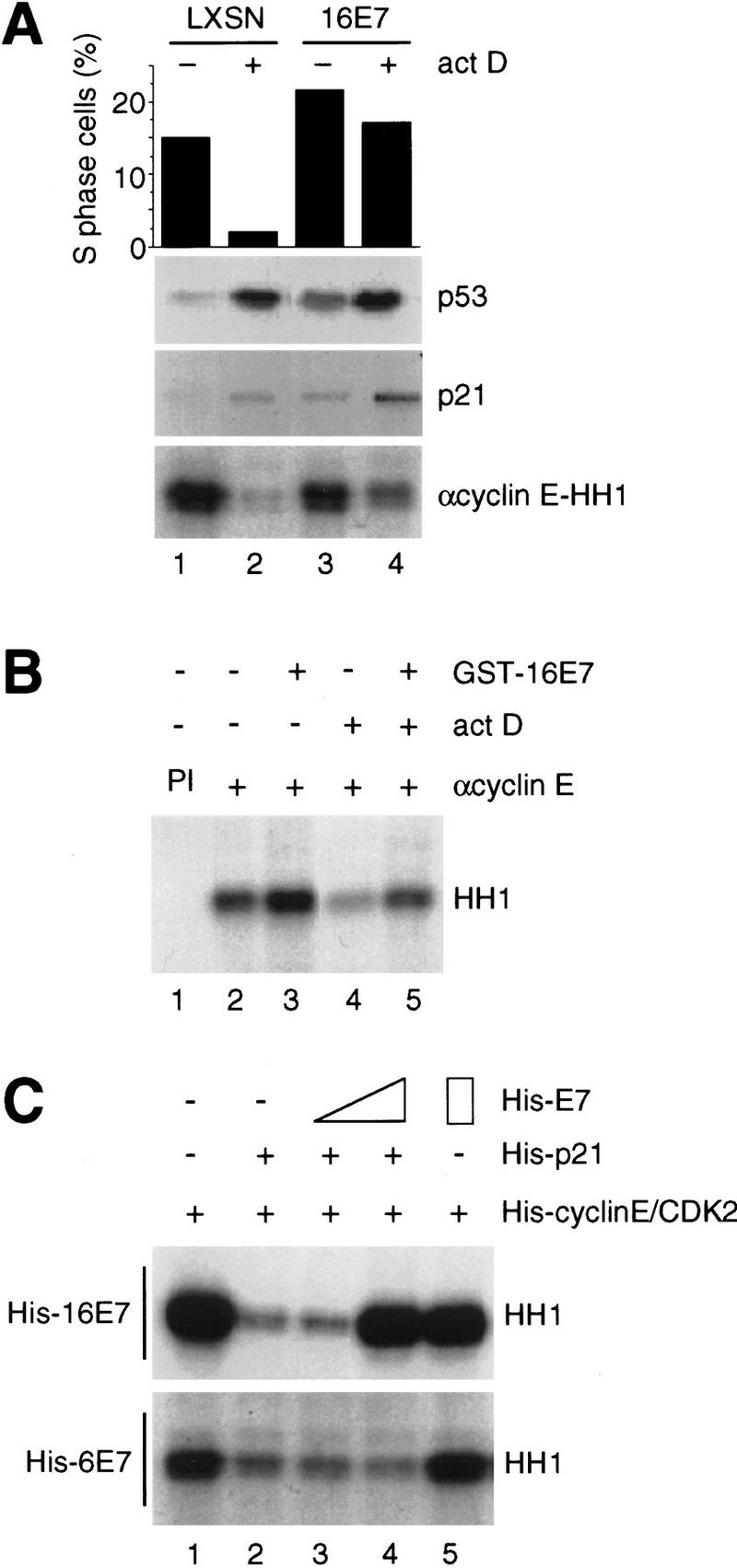
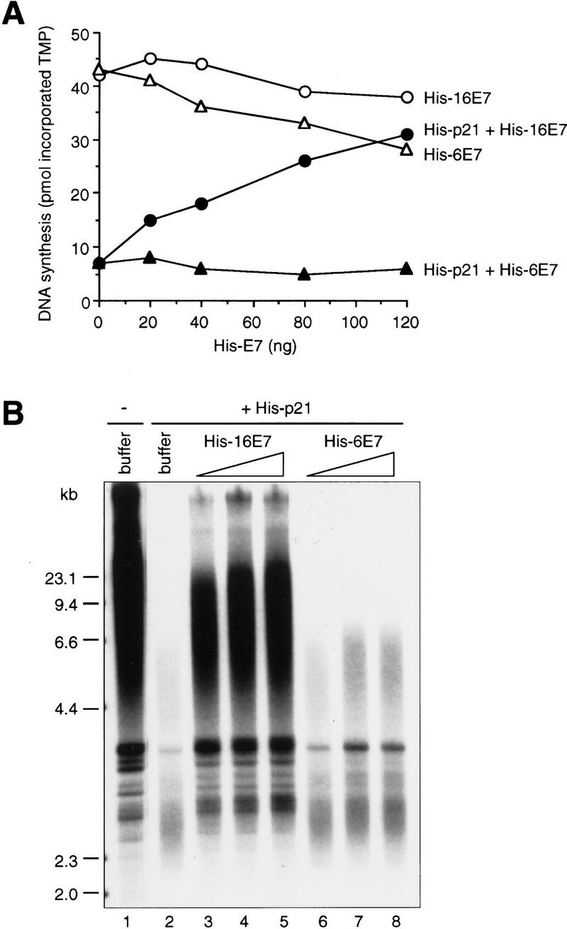
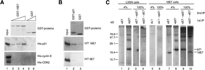
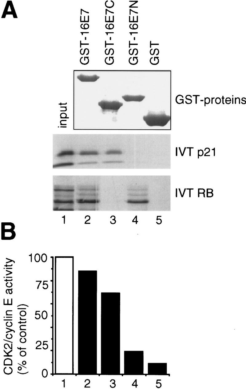
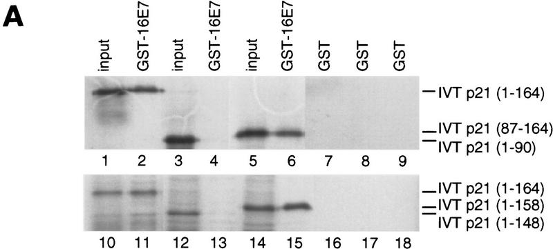
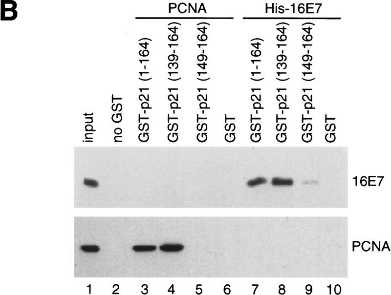
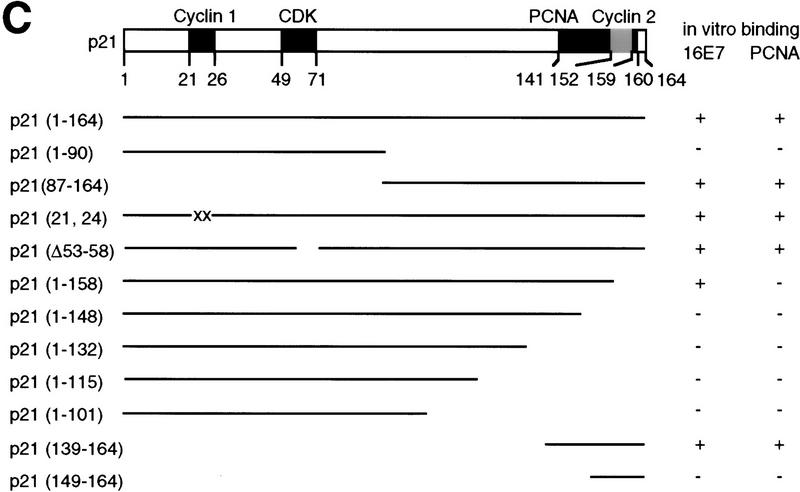
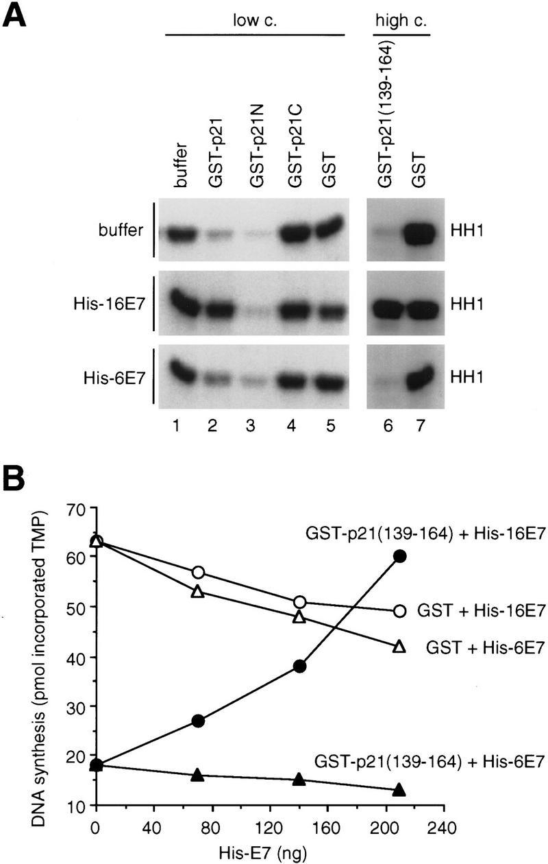
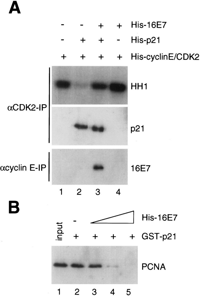
p21 inhibits cyclin-dependent kinase (CDK) activity and proliferating cell nuclear antigen (PCNA)-dependent DNA replication by binding to CDK/cyclin complexes and to PCNA through distinct domains. The human papillomavirus (HPV)-16 E7 oncoprotein (16E7) abrogated a DNA damage-induced cell cycle arrest in vivo, despite high levels of p21. Using cell lysates and purified proteins we show that 16E7 prevented p21 both from inhibiting CDK2/cyclin E activity and PCNA-dependent DNA replication, whereas the nononcogenic HPV-6 E7 had reduced effects. Inactivation of both inhibitory functions of p21 was attained through binding between 16E7 and sequences in the carboxy-terminal end of p21 that overlap with the PCNA-binding site and the second p21 cyclin-binding motif. These data imply that the carboxyl terminus of p21 simultaneously modulates both CDK activity and PCNA-dependent DNA replication and that a single protein, 16E7, can override this modulation to disrupt normal cell cycle control.


Figures

Figure 1
Figure 1
Effect of HPV-16 E7 and HPV-6 E7 on inhibition of CDK activity by p21 in vivo and in vitro. (A) Human keratinocytes were infected with retroviral constructs alone (LXSN) or expressing 16E7 and were treated with actinomycin D (act D) for 24 hr. Cells were harvested, and a fraction was used for cell cycle analysis by flow cytometry to determine the fraction of S-phase cells (top); the remainder was used to prepare extracts for immunoblots with anti-p53 or anti-p21 antibodies (middle) and in vitro kinase assays after anti-cyclin E immunoprecipitations (bottom). (B) Extracts of untreated or actinomycin D-treated (act D) human keratinocytes (200 μg) were coincubated with GST–16E7 (850 ng); then cyclin E was immunoprecipitated [lanes 2–5; (PI) preimmune serum in lane 1] and kinase assays were performed for 20 min at 30°C using histone H1 (HH1) as a substrate. (C) Purified recombinant His–cyclin E/CDK2 complexes, produced in baculovirus-infected cells, were coincubated with His–p21 (10 ng) alone or with His–p21 preincubated with increasing amounts (20 and 200 ng) of His–16E7 (top) or His–6E7 (bottom), and kinase assays were performed.
Figure 2
Figure 2
Effect of HPV-16 E7 and HPV-6 E7 on inhibition of PCNA-dependent DNA replication by p21 in vitro. (A) Increasing amounts of His–16E7 or His–6E7 were preincubated with (50 ng; closed symbols) or without (open symbols) His–p21 for 60 min at room temperature, then mixed with PCNA (10 ng) and DNA polymerase δ (∼15 ng) and further incubated for 30 min on ice. DNA synthesis was started by adding [α32P]TTP and oligo(dT)/poly(dA) at 37°C. The amount of DNA synthesis is expressed as picomoles of TMP incorporated in a 25-μl reaction in 30 min. (B) Increasing amounts (250 ng, 500 ng, 1 μg) of His–16E7 or His–6E7 were preincubated with or without His–p21 (330 ng) for 60 min at room temperature, then mixed with a 293 human cell extract (19.5 μg) and further incubated for 30 min on ice. DNA replication was started by adding purified T antigen, an SV40 ori-containing plasmid pSVO11 (Prelich and Stillman 1988), and nucleotides including [α32P]dATP and incubated at 37°C. Analysis by neutral agarose gel electrophoresis of purified replication products is shown. Lane 1 shows SV40 DNA replication in the absence of p21 or E7.
Figure 3
Figure 3
Analysis of the HPV-16 E7–p21 interaction in vitro and in vivo. (A) In vitro binding reactions with GST–16E7 or GST (100, 200, 400 ng) immobilized on glutathione–Sepharose and His–p21, His–cyclin E, or His–CDK2 (200 ng) were performed, and the resulting protein complexes were visualized by Coomassie staining (top) and Western blotting (bottom) with anti-His antibodies. Lane 1 represents 10% of the input. (B) In vitro binding reactions with GST–p21 or GST (1 μg) immobilized on glutathione–Sepharose and IVT 16E7 or IVT 6E7 were performed, and the resulting protein complexes were visualized by Coomassie staining (top) and fluorography (bottom). Lane 1 represents 10% of the input. (C) Actinomycin D-treated, 16E7-expressing keratinocytes were metabolically labeled with [35S]Protein Labeling Mix, and extracts were prepared. After a first immunoprecipitation (IP) of extracts (900 μg; anti-16E7 or anti-p21 antibody) under low-stringency conditions (data not shown), the associated proteins were eluted under high-stringency conditions (IP shown after the elution; lanes 7,8) and reimmunoprecipitated with a second antibody (anti-p21 and anti-16E7, respectively; lanes 9,10). Vector (LXSN)-infected cells (lanes 1–4) or an irrelevant antibody (anti-16L1; lanes 5,6) were used as negative controls. Four percent of the first IP after the high-stringency elution and 100% of the second IP are shown.
Figure 4
Figure 4
Analysis of binding sites in HPV-16 E7 proteins. (A) In vitro binding reactions with GST–16E7 mutants (1.5 μg) immobilized on glutathione–Sepharose and IVT p21 or IVT RB were performed, and the resulting protein complexes were visualized by Coomassie staining (top) and fluorography (bottom). Lane 1 represents 10% (IVT p21) or 20% (IVT RB) of the input. (B) Purified recombinant His–cyclin E/CDK2 complexes were coincubated with buffer (□) or coincubated with His–p21 (10 ng, ▪) alone or with His–p21 preincubated with GST fusion proteins (300 ng; as indicated in A, lanes 2–5), and kinase assays were performed. The quantitation from PhorphorImager analysis is shown.
Figure 5
Figure 5
Analysis of binding sites in p21 proteins. (A) In vitro binding reactions with GST–16E7 or GST (1 μg) immobilized on glutathione–Sepharose and IVT p21 mutant proteins were performed, and the resulting protein complexes were visualized by fluorography. The mutants used were p21(full-length, 1–164) (lanes 1,2,7,10,11,16), p21(1–90) (lanes 3,4,8), p21(87–164) (lanes 5,6,9), p21(1–148) (lanes 12,13,17), and p21(1–158) (lanes 14,15,18). Ten percent of the input is shown. (B) In vitro binding reactions with GST–p21 mutants (100 ng) immobilized on glutathione–Sepharose and His–16E7 (200 ng) or PCNA (40 ng) were performed, and the resulting protein complexes were visualized by Western blotting with anti-16E7 (top) and anti-PCNA (bottom) antibodies. Lane 1 represents 20% of the input. (C) Schematic representation of p21 and summary of in vitro binding analyses with p21 mutants using purified and IVT proteins. The binding motifs for cyclin (cyclin 1), CDK, and PCNA are shown as solid boxes, the second cyclin binding motif (cyclin 2) overlapping the PCNA binding motif is shown as a shaded box.
Figure 5
Figure 5
Analysis of binding sites in p21 proteins. (A) In vitro binding reactions with GST–16E7 or GST (1 μg) immobilized on glutathione–Sepharose and IVT p21 mutant proteins were performed, and the resulting protein complexes were visualized by fluorography. The mutants used were p21(full-length, 1–164) (lanes 1,2,7,10,11,16), p21(1–90) (lanes 3,4,8), p21(87–164) (lanes 5,6,9), p21(1–148) (lanes 12,13,17), and p21(1–158) (lanes 14,15,18). Ten percent of the input is shown. (B) In vitro binding reactions with GST–p21 mutants (100 ng) immobilized on glutathione–Sepharose and His–16E7 (200 ng) or PCNA (40 ng) were performed, and the resulting protein complexes were visualized by Western blotting with anti-16E7 (top) and anti-PCNA (bottom) antibodies. Lane 1 represents 20% of the input. (C) Schematic representation of p21 and summary of in vitro binding analyses with p21 mutants using purified and IVT proteins. The binding motifs for cyclin (cyclin 1), CDK, and PCNA are shown as solid boxes, the second cyclin binding motif (cyclin 2) overlapping the PCNA binding motif is shown as a shaded box.
Figure 6
Figure 6
Analysis of the function of the carboxy-terminal domain of p21 in the inactivation by HPV-16 E7. (A) Purified recombinant His–cyclin E/CDK2 complexes were coincubated with buffer (lane 1) or GST proteins at low or high concentration preincubated with buffer (top), His–16E7 (middle), or His–6E7 (bottom). Twenty nanograms of GST or GST–p21 (lanes 2–5), or 2.5 μg of GST or GST–p21(139–164) (lanes 6,7) were used, which had been preincubated with 400 ng (lanes 2–5) or 6 μg (lanes 6,7) of His–16E7 or His–6E7, and kinase assays were performed for 20 min at 30°C. (B) Increasing amounts of His–16E7 or His–6E7 were preincubated with GST or GST–p21(139–164) (120 ng). The subsequent DNA synthesis reaction was carried out as described in Fig. 2A.
Figure 7
Figure 7
Analysis of protein complexes in the absence and presence of HPV-16 E7. (A) Purified recombinant His–cyclin E/CDK2 complexes were coincubated with His–16E7 and/or His–p21 as described in Fig. 1C, and after an immunoprecipitation (IP) with anti-CDK2 antibodies kinase assays were performed (top). One-half of the anti-CDK2 immunoprecipitate was analyzed by immunoblotting with anti-p21 antibodies (middle). In parallel, an immunoprecipitate with anti-cyclin E antibodies was analyzed by immunoblotting with anti-16E7 antibodies (bottom). (B) Recombinant PCNA (50 ng) was coincubated with GST–p21 (100 ng), immobilized on glutathione–Sepharose alone or in the presence of increasing amounts (200, 400, 800 ng) of His–16E7, and the PCNA bound to GST–p21 beads was analyzed by immunoblotting with anti-PCNA antibodies. Lane 1 represents 10% of the input.

Similar articles

Cited by

References

    1. Abraham SE, Carter MC, Moran E. Transforming growth factor β1 (TGFβ1) reduces cellular levels of p34cdc2, and this effect is abrogated by adenovirus independently of the E1A-associated pRB binding activity. Mol Cell Biol. 1992;3:655–665. - PMC - PubMed
    1. Adams PD, Sellers WR, Sharma SK, Wu AD, Nalin CM, Kaelin WG. Identification of a cyclin-cdk2 recognition motif present in substrates and p21-like cyclin-dependent kinase inhibitors. Mol Cell Biol. 1996;16:6623–6633. - PMC - PubMed
    1. Balbín M, Hannon GJ, Pendás AM, Ferrando AA, Vizoso F, Fueyo A, López-Otín C. Functional analysis of a p21WAF1,CIP1,SDI1 mutant (Arg94 → Trp) identified in a human breast carcinoma. Evidence that the mutation impairs the ability of p21 to inhibit cyclin-dependent kinases. J Biol Chem. 1996;271:15782–15786. - PubMed
    1. Ball KL, Lain S, Fåhraeus R, Smythe C, Lane DP. Cell-cycle arrest and inhibition of cdk4 activity by small peptides basd on the carboxy-terminal domain of p21WAF1. Curr Biol. 1996;7:71–80. - PubMed
    1. Blackwood EM, Lüscher B, Eisenman RN. Myc and Max associate in vivo. Genes & Dev. 1992;6:71–80. - PubMed

Publication types

MeSH terms

Substances

LinkOut - more resources