. 1997 Sep 8;138(5):1023-40.
doi: 10.1083/jcb.138.5.1023.

Kinesin-related KIP3 of Saccharomyces cerevisiae is required for a distinct step in nuclear migration

Affiliations

Kinesin-related KIP3 of Saccharomyces cerevisiae is required for a distinct step in nuclear migration

T M DeZwaan et al. J Cell Biol. .

Abstract

Spindle orientation and nuclear migration are crucial events in cell growth and differentiation of many eukaryotes. Here we show that KIP3, the sixth and final kinesin-related gene in Saccharomyces cerevisiae, is required for migration of the nucleus to the bud site in preparation for mitosis. The position of the nucleus in the cell and the orientation of the mitotic spindle was examined by microscopy of fixed cells and by time-lapse microscopy of individual live cells. Mutations in KIP3 and in the dynein heavy chain gene defined two distinct phases of nuclear migration: a KIP3-dependent movement of the nucleus toward the incipient bud site and a dynein-dependent translocation of the nucleus through the bud neck during anaphase. Loss of KIP3 function disrupts the unidirectional movement of the nucleus toward the bud and mitotic spindle orientation, causing large oscillations in nuclear position. The oscillatory motions sometimes brought the nucleus in close proximity to the bud neck, possibly accounting for the viability of a kip3 null mutant. The kip3 null mutant exhibits normal translocation of the nucleus through the neck and normal spindle pole separation kinetics during anaphase. Simultaneous loss of KIP3 and kinesin-related KAR3 function, or of KIP3 and dynein function, is lethal but does not block any additional detectable movement. This suggests that the lethality is due to the combination of sequential and possibly overlapping defects. Epitope-tagged Kip3p localizes to astral and central spindle microtubules and is also present throughout the cytoplasm and nucleus.


Figures

Figure 1
Figure 1
Kip3p sequence alignment and coiled-coil formation probability. (A) Alignment of Kip3p residues 86-464 with human kinesin (huKIN) (Navone et al., 1992) residues 1–340 was performed using the Gap program (Genetics Computer Group, Inc., Madison, WI). Vertical lines indicate amino acid identities, and dots indicate gaps introduced to improve alignment. Horizontal lines indicate key residues of the ATP-binding site (GX4GKT) and amino acids suspected to play a role in nucleotide phosphate sensing and microtubule binding. (B) The probability of coiled-coil formation in the predicted Kip3p amino acid sequence was calculated using the algorithm of Lupas et al. (1991) with a window size of 14 amino acids.
Figure 2
Figure 2
Increased benomyl resistance in the kip3Δ mutant. Strains wild-type (DS140), kar3Δ102 (DS276), kip3Δ (DS614), and dyn1Δ (DS730) were incubated at 23°C for 2 d in liquid YPD medium. Serial dilutions were prepared, and ∼104, ∼103, ∼102, and ∼10 cells were spotted in horizontal rows on solid YPD medium containing 1% DMSO and benomyl at the indicated concentration and photographed after 3 d of growth at 30°C.
Figure 3
Figure 3
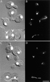
Disruption of KIP3 affects nuclear migration. kip3Δ strain DS614 (A and B) and wild-type strain DS141 (C and D) were grown in YPD medium at 30°C to mid-logarithmic phase and prepared for immunofluorescence microscopy. (A and C) DIC images overlaid with corresponding DAPI-stained images to show position of nuclear DNA. (B and D) Antitubulin staining to visualize microtubules. A and B show kip3Δ cells with nuclei located distal to the neck and with brightly staining astral microtubules extending into the bud (arrows in B). Also present is a mother cell with two nuclei (arrows in A) that are still connected by a mitotic spindle. The small-budded wild-type cells in C and D contain significantly fewer prominent astral microtubules. Bar, 5 μm.
Figure 4
Figure 4
Loss of KIP3 function causes mislocalization of undivided nuclei. (A) Histograms of nuclear migration indices of wild-type (DS141), kip3Δ (DS614), dyn1Δ (DS730), and kar3Δ (DS276) strains grown to mid-logarithmic phase at the indicated temperature. (B) Histograms of kip3(ts) kar3Δ (DS752) and kip3(ts) dynΔ (DS765) strains grown to mid-logarithmic phase at the permissive temperature of 30°C and shifted to the nonpermissive temperature of 37°C for 4 h. The mean nuclear migration index and one standard deviation (mean ± SD) is inset on each histogram. The nuclear migration index of a cell is proportional to the proximity of its nucleus to the bud neck and is calculated as the distance between the neck and the nearest edge of the nucleus visualized by DAPI staining divided by the distance between the neck and the most distal edge of the cell wall (Jacobs et al., 1988). The statistical significance of the difference between the variances of the nuclear migration indices was tested with a variance ratio test. The variance ratio for the kip3Δ and wild-type 30°C cultures was found to be 3.4, well above the 95% significance point of 1.5, indicating that we have significant evidence that the position of nuclei in the kip3Δ mutant is more random than in wild-type. The position of nuclei in the kip3Δ mutant grown at 11 and 37°C was also significantly more random than in wild-type. The variance ratio for the kip3(ts) dyn1Δ double mutant and wild-type cultures was 2.5, above the 95% significance point of 1.5, but there was no significant difference between the kip3(ts) dyn1Δ double mutant and the kip3Δ single mutant. 50–100 cells were measured for each strain and temperature.
Figure 5
Figure 5
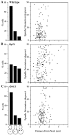
Loss of KIP3 function causes misorientation of preanaphase spindles. The histograms on the left indicate the frequency of cells with bipolar spindles that are oriented from 0–30 degrees of the mother–daughter axis (first bar), from >30–60 degrees (second bar), and from >60–90 degrees (third bar). The scatter plots on the right contain all of the data shown in histogram format and in addition show the relationship between spindle orientation and distance between the neck and the neck-proximal spindle pole. There was little correlation between spindle orientation and nuclear location in all three strains. (A) Wild-type (DS723), (B) kip3Δ (DS786), and (C) dyn1Δ (DS940) strains were grown at 30°C in YPD medium and fixed for microscopy. The location of the spindle poles and spindle orientation was determined by overlaying images of Nuf2p–GFP fusion fluorescence onto DIC images of the corresponding cells. Spindle orientation was measured in all cells in the population that had a preanaphase spindle with both spindle pole bodies in a single focal plane and a pole-to-pole length of 0.8–1.2 μm. More than 120 preanaphase spindles from two independent cultures were measured for each strain.
Figure 6
Figure 6
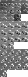
Large oscillations in position of nuclei after spindle disassembly in the kip3Δ mutant. (A, C, and E) A time-lapse series of DIC micrographs of wild-type (A), kip3Δ (C), and dynΔ (E) cells. The elapsed time (min) is indicated, and T = 0 is the time of maximal anaphase spindle elongation. The position of the spindle poles is indicated by open circle symbols, and the position of the bud neck of the new daughter cell is indicated by the asterisk. At early times in the series when the bud had not yet emerged, the prebud site was defined by comparison to later time points. Spindle pole position was defined by overlaying images of Nuf2p– GFP fluorescence onto corresponding DIC images. To obtain DIC images in which the bud was always in focus, images of two focal planes spaced 1 μm apart were projected onto a single plane. (B, D, and F) Two examples of the distance of the spindle pole in the mother cell from the new bud neck, plotted as a function of time. The closed symbols represent pole position in the mother cells shown in A, C, and E. The open symbols represent pole position from an independent set of micrographs (not shown), to convey the range of movements observed. Note that in addition to nuclear migration, small changes in pole position could also be caused by nuclear rotation. Bar, 5 μm.
Figure 6
Figure 6
Large oscillations in position of nuclei after spindle disassembly in the kip3Δ mutant. (A, C, and E) A time-lapse series of DIC micrographs of wild-type (A), kip3Δ (C), and dynΔ (E) cells. The elapsed time (min) is indicated, and T = 0 is the time of maximal anaphase spindle elongation. The position of the spindle poles is indicated by open circle symbols, and the position of the bud neck of the new daughter cell is indicated by the asterisk. At early times in the series when the bud had not yet emerged, the prebud site was defined by comparison to later time points. Spindle pole position was defined by overlaying images of Nuf2p– GFP fluorescence onto corresponding DIC images. To obtain DIC images in which the bud was always in focus, images of two focal planes spaced 1 μm apart were projected onto a single plane. (B, D, and F) Two examples of the distance of the spindle pole in the mother cell from the new bud neck, plotted as a function of time. The closed symbols represent pole position in the mother cells shown in A, C, and E. The open symbols represent pole position from an independent set of micrographs (not shown), to convey the range of movements observed. Note that in addition to nuclear migration, small changes in pole position could also be caused by nuclear rotation. Bar, 5 μm.
Figure 7
Figure 7
Normal kinetics of anaphase spindle elongation in the kip3Δ mutant. (A, D, and G) Pole-to-pole distance as a function of time in an individual cell of the indicated strain. Each cell exhibited the fast and slow phases of anaphase spindle elongation, followed by a reduction of the pole to pole distance upon spindle disassembly. (B, E, and H) Data from the same cells and recordings in A, D, and G, with the first 16 min of anaphase shown on an expanded time axis. To illustrate movement of the spindle poles relative to the neck, distances between the neck and the pole in the mother cell were plotted as positive numbers, and distances between the neck and the pole in the bud were plotted as negative numbers. (C, F, and I) Data recorded from different cells. Measurement of spindle pole position relative to the neck was performed on images of Nuf2p–GFP fluorescence overlaid onto corresponding DIC images. The haploid strains used were wild-type strain DS723 (A–C), kip3Δ strain DS786 (D–F), and dyn1Δ strain DS940 (G–I).
Figure 8
Figure 8
(A) Synthetic lethality of kip3, dyn1, and kar3. Strains kip3, dyn1, kar3, kip3 dyn1 (DS732), kip3 kar3 (DS716), and dyn1 kar3 (DS743) carry a centromere-based plasmid with the markers indicated in parentheses. To test whether the plasmids are essential for viability, the strains were grown on YPD medium for 3 d at 30°C to allow for possible plasmid loss, the strains were plated on minimal medium lacking uracil (selects for plasmid) and on 5-FOA medium (selects against plasmid), and the plates were photographed after 3 d of incubation at 30°C. (B) Temperature-sensitive alleles of kip3. Strains dyn1 (DS749), kip3-20(ts) dyn1 (DS765), kar3 (DS750), and kip3-30(ts) kar3 (DS752) were grown to saturation and spotted on YPD medium at the indicated temperature. Plates were photographed after 7 d at 16°C and after 3 d at 30 or 37°C. (C) Benomyl partially suppresses the temperature sensitivity of kip3(ts) dyn1Δ and kip3(ts) kar3Δ strains. Strains wild-type (DS138), kip3Δ (DS613), dynΔ (DS749), kip3-20(ts) dyn1Δ (DS765), kar3Δ102 (DS750), and kip3-30(ts) kar3Δ (DS752) were incubated at 23°C for 2 d in liquid YPD medium. Approximately 104 cells were spotted on solid YPD medium containing 1% DMSO and benomyl at the indicated concentration, and photographed after 3 d of growth at the indicated temperature. (D) KIP3 and KAR3 differ in their genetic interactions with KIP1 and CIN8. Strains kip1Δ cin8-101 (DS49), wild-type (DS141), kar3Δ (DS276), kip3Δ (DS614), kar3Δ kip1Δ cin8-101 (DS689), and kip3Δ kip1Δ cin8-101 (DS737) were incubated for 2 d at 23°C in YPD medium. Approximately 5 × 10⁴ cells were spotted on YPD medium and incubated for 4 d at the indicated temperature. kar3Δ remediates the temperature-sensitive spindle assembly defect of the cin8-101 kip1Δ double mutant, whereas kip3Δ does not.
Figure 9
Figure 9
Morphology of kip3 dyn1 and kip3 kar3 double mutants. (A) The indicated strains were grown to mid-logarithmic phase at 30°C in YPD medium and fixed for microscopy. (B) The same cultures shifted to 37°C for 4 h. The columns labeled DIC/DAPI are DIC images overlaid with corresponding DAPI-stained images to show position of nuclear DNA, and the columns labeled IF are corresponding images of antitubulin staining to visualize microtubules. Binucleate mother cells are present in the dyn1, kip3(ts) dyn1, and kip3(ts) kar3 mutants but not in the kar3 mutant. The strain designations are the same as in Fig. 8. Bar, 5 μm.
Figure 10
Figure 10
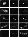
Epitope-tagged Kip3p localizes to astral and spindle microtubules and is present in the cytoplasm and nucleoplasm. The cells are from an asynchronous culture containing epitope-tagged Kip3p expressed from a centromere plasmid. The first column (A, D, G, J, and M) shows staining with 9E10, an anti-myc antibody that recognizes epitope-tagged Kip3p. The second column (B, E, H, K, and N) shows staining with an antitubulin antibody. The third column (C, F, I, L, and O) shows staining with DAPI to localize DNA. (A–C) A G1 cell with colocalization of Kip3p with astral microtubules. (D–F) A preanaphase cell with spindle staining. (G–L) Cells with an elongated anaphase spindle with concentration of Kip3p in the central spindle. (M–O) A cell of an isogenic strain carrying Kip3p lacking the epitope tag, which showed no signal when stained and photographed under conditions identical to those used to localize epitope-tagged Kip3p.
Figure 11
Figure 11
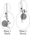
Distinct roles of Kip3p and Dyn1p in the two phases of nuclear migration. Based on the phenotypes of kip3 and dyn1 mutants, Kip3p primarily functions during phase 1 of nuclear migration to (1) orient the nucleus toward the nascent bud and (2) position the nucleus ∼1 μm from the bud neck. During phase 2 of nuclear migration, Dyn1p primarily functions to (3) insert the anaphase-stage nucleus through the neck and (4) mediate forward and reverse oscillations of the nucleus within the neck.

Comment in

Similar articles

Cited by

References

    1. Baker J, Theurkauf WE, Schubiger G. Dynamic changes in microtubule configuration correlate with nuclear migration in the preblastoderm Drosophilaembryo. J Cell Biol. 1993;122:113–121. - PMC - PubMed
    1. Baudin A, Ozier-Kalogeropoulos O, Denouel A, Lacroute F, Cullin C. A simple and efficient method for direct gene deletion in Saccharomyces cerevisiae. . Nucleic Acids Res. 1993;21:3329–3330. - PMC - PubMed
    1. Blacketer MJ, Madaule P, Myers AM. Mutational analysis of morphologic differentiation in Saccharomyces cerevisiae. . Genetics. 1995;140:1259–1275. - PMC - PubMed
    1. Boeke JD, Trueheart J, Natsoulis G, Fink GR. 5-Fluoro-orotic acid as a selective agent in yeast molecular genetics. Methods Enzymol. 1987;154:164–175. - PubMed
    1. Byers B, Goetsch L. Behavior of spindles and spindle plaques in the cell cycle and conjugation in Saccharomyces cerevisiae. . J Bacteriol. 1975;124:511. - PMC - PubMed

Publication types

MeSH terms