. 1997 Aug 25;138(4):861-75.
doi: 10.1083/jcb.138.4.861.

The synaptic protein syntaxin1 is required for cellularization of Drosophila embryos

Affiliations

The synaptic protein syntaxin1 is required for cellularization of Drosophila embryos

R W Burgess et al. J Cell Biol. .

Abstract

Syntaxins are membrane proteins involved in vesicle trafficking and are required for the release of neurotransmitter at nerve terminals. The presence of syntaxins on target membranes has been hypothesized to confer specificity to targeting and fusion via interactions with complementary vesicle-associated proteins, the synaptobrevins or VAMPS. We have mutagenized syntaxin1 in Drosophila and have found that it links the mechanism of synaptic transmission to a distinct cell biological process: the cellularization of early embryos. This specialized form of cell division separates the 6,000 nuclei of the syncytial blastoderm into separate cells through the invagination of the surface membrane of the embryo. During this process, syntaxin1 protein is present on the newly forming lateral cell surfaces and invaginating cleavage furrows. This protein is derived both from maternal deposition of mRNA and protein and from early zygotic transcription. To analyze syntaxin1's role in early development, female germ line mosaics mutant for syntaxin1 expression were generated by mitotic recombination to reduce the maternal contribution. Visualizing the actin cytoskeleton and glycosylated surface proteins reveals that embryos with insufficient syntaxin1 have large acellular patches. The patches do not appear until cellularization begins, and the process fails entirely within these regions. These results provide genetic evidence that membrane trafficking is required for the cellularization of the syncytial blastoderm. We propose that the invagination of the surface membrane proceeds by the fusion of intracellular membrane vesicles with the surface. This reaction uses the same syntaxin1 protein as is required for neurotransmitter secretion at synapses. Thus, a single syntaxin can participate in trafficking steps that are functionally as distinct as synaptic transmission and cell division.


Figures
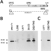
Figure 3
Figure 3
(A) P-element insertions in the syntaxin1 transcription unit. The P36-23 P-element insertion is located ∼1,500 bp upstream of the syntaxin1 open reading frame and is homozygous viable. This insertion was mobilized to generate a series of P-element insertions that disrupt the syntaxin1 transcription unit and are homozygous lethal. The allele syx L266 carries both the original P36-23 insertion and a second insertion in or immediately adjacent to the 5′ untranslated sequence. syx L371 has two P-elements: the original P36-23 and a second, within the syntaxin1 open reading frame, interrupting the codon for amino acid K95. The intervening sequence is deleted as determined by Southern blotting, PCR, and plasmid rescue of the insertions. (B) Syntaxin1 protein in mutant embryos. In late stage embryos homozygous for the parent chromosome P36-23, the protein is present at normal levels, by comparison to control strains (CXD/TM3 or yw). The protein is dramatically reduced in syx L266/syx L266 and syx L371/syx L371 alleles. The syx L266/syx L266 level is higher than that of syx L371/ syx L371; this allele therefore produces a small amount of full-length protein. Full-length syntaxin1 is present in syx L371/syx L371 embryos because of the remaining maternal contribution and can be seen on longer exposures of the gel in B (not shown) or on gels with additional protein layered per lane (C). Homozygous mutant embryos from heterozygous parents were selected based on their failure to hatch after 27–33 h for both the syx L266 and the syx L371 alleles. Age-matched embryos that failed to hatch from CXD/TM3 heterozygous parents were used as a control. Homozygous P36-23 and yw embryos were collected at 24 h. 60 μg of protein was layered per lane in B, corresponding to ∼25 embryos. The protein extracted from 45 embryos was loaded in each lane of C.
Figure 1
Figure 1
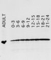
Syntaxin1 levels during embryogenesis. Protein was extracted from timed embryo collections of the ages indicated (hours AED) and from adult heads and separated by SDS-PAGE. Per lane, the protein extracted from 30 embryos or one half of an adult head was loaded. The gels were blotted onto Immobilon membranes and were then probed with the monoclonal antibody 8C3. Syntaxin1 is present in 0–3-h embryos, the stage during which cellularization occurs, and levels increase as the nervous system matures. In both embryos and adults, the protein has a molecular mass of 35 kD, as predicted.
Figure 2
Figure 2
Syntaxin1 localization by antibody staining and confocal microscopy in whole-mount embryos. (A and B) Embryos in early stages of cellularization. As the invaginating cleavage furrows form, syntaxin1 is localized to the newly formed furrows seen in superficial (A) or deep (B) confocal sections. (C) Syntaxin1 distribution as cellularization nears completion. When the cleavage furrows have progressed well beyond the nuclei, syntaxin1 is present along the length of the lateral membrane. Antisyntaxin immunostaining was performed with monoclonal antibody 8C3. The signal was visualized with a Texas red–conjugated secondary (see Materials and Methods). Bar, 10 μm.
Figure 4
Figure 4
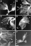
Ovaries from mitotic recombinations in syx/ovo D1 heterozygotes. ovo D1 is a dominant mutation that arrests ovariole development before stage 8. Examples of these rudimentary ovarioles are marked throughout with arrowheads and can be seen in A. Recombination in a P36-23/ovo D1 fly produces P36-23 homozygous clones in which follicles reach maturity and oocytes are produced (B, and marked with arrows throughout) alongside ovo D1-arrested ovaries. Mature ovarioles are also seen in syx L266 clones (C) but were not seen in syxL371 clones (D). syx L371 is therefore either a rudimentary ovary mutant comparable to ovo D1, or syx L371 is cell lethal, leaving only ovo D1 follicles. Mature oocytes are produced from syx L371 ovary clones that express syx on a second chromosome transgene (E). The genotype of these flies is HS-FLP; HS-GAL4:UAS-syx; syx L371-82BFRT/ovo D1-82BFRT. Siblings of these flies that do not carry the transgene show no developed ovarioles (F). All of the ovaries shown were stained with FITC-conjugated phalloidin to allow visualization of the ovarioles. Bar, 200 μm.
Figure 5
Figure 5
Abnormalities in developing embryos are manifest only in those that are maternally and zygotically mutant for syntaxin1. (A) The early lethality of embryos from syx L266/syx L266 maternal germ line clones is dependent on inheriting a syx L371 chromosome from the father. Females bearing homozygous syx L266 germ line clones were crossed to syx L371/TM3, lacZ males. 14–17-h-old embryos were collected, examined to determine if they had successfully progressed through development, and stained with X-gal to determine which embryos received the paternal syx + chromosome (white bars) and which had received the syx L371 chromosome (black bars). (B) Embryos that received the syx + paternal chromosome (lacZ positive, arrow) developed generally normally, with the majority reaching the appropriate stages (16–17) (144 embryos examined). (C) Those embryos with the syx L371 paternal chromosome (lacZ negative) arrested at very early stages, typically at or before germ band extension (169 embryos examined). As shown, the position of the yolk often suggested that some morphogenetic movements had been attempted, but normal germ band extension had not occurred and there had been no subsequent elaboration of internal organs. Anterior is to the left in both pictures. The dark, grainy material in the center of each embryo is the yolk.
Figure 5
Figure 5
Abnormalities in developing embryos are manifest only in those that are maternally and zygotically mutant for syntaxin1. (A) The early lethality of embryos from syx L266/syx L266 maternal germ line clones is dependent on inheriting a syx L371 chromosome from the father. Females bearing homozygous syx L266 germ line clones were crossed to syx L371/TM3, lacZ males. 14–17-h-old embryos were collected, examined to determine if they had successfully progressed through development, and stained with X-gal to determine which embryos received the paternal syx + chromosome (white bars) and which had received the syx L371 chromosome (black bars). (B) Embryos that received the syx + paternal chromosome (lacZ positive, arrow) developed generally normally, with the majority reaching the appropriate stages (16–17) (144 embryos examined). (C) Those embryos with the syx L371 paternal chromosome (lacZ negative) arrested at very early stages, typically at or before germ band extension (169 embryos examined). As shown, the position of the yolk often suggested that some morphogenetic movements had been attempted, but normal germ band extension had not occurred and there had been no subsequent elaboration of internal organs. Anterior is to the left in both pictures. The dark, grainy material in the center of each embryo is the yolk.
Figure 6
Figure 6
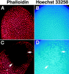
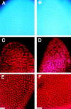
The disruption of cellularization by mutations in syntaxin1 can be seen in the distribution of nuclei and filamentous actin. Females with germ line clones homozygous for either syx L266 (C and D) or the P36-23 control chromosome (A and B) were crossed to syx L371/ TM3, lacZ males and examined with Hoechst 33258 to visualize nuclei (blue) and with rhodamine phalloidin to visualize actin (red). During cellularization, embryos from P36-23 clones (A and B) continue to develop normally; however, half of the embryos from syx L266 clones (C and D) show large acellular patches in which the nuclei were disorganized (D) and the network of filamentous actin was completely absent (C). The disorganization of the nuclei and cytoskeleton corresponded exactly (large arrows). In addition, small defects are also sometimes seen (small arrow, C). Rhodamine phalloidin images were gathered by confocal microscopy.
Figure 7
Figure 7
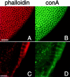
The defects in cellularization also affect surface membrane. Embryos from syx L266 clones were stained with rhodamine-conjugated phalloidin (red) and FITC-conjugated concanavalinA (green) to mark the actin cytoskeleton and the surface membrane, respectively. On the embryo's surface, the two markers label the honeycomb of newly forming cells (A and B). In a deeper section of the same embryo, a large acellular patch was present. Both markers fail to label anything within an acellular patch (C and D), indicating a failure of both the cytoskeleton to form and the surface membrane to invaginate in these areas. Bars: (A and B) 20 μm; (C and D) 10 μm.
Figure 8
Figure 8
Defects due to insufficient syntaxin do not appear before cellularization. The nuclei (A and B) migrate to the surface, and actin caps form above them (C and D) in syx L266/syx L371 embryos derived from homozygous syx L266 germline clones (B and D), just as they do in control embryos derived from P36-23 clones (A and C). The formation of metaphase furrows also progresses normally during mitotic cycles 10–13. The furrows of embryos in mitotic cycle 12 from both P36-23 (E) and syx L266 clones (F) are shown. Furrows and caps are visualized with rhodamine-conjugated phalloidin, and nuclei are stained with Hoechst 33258. In both genotypes, minor irregularities in actin are seen. These regions correspond to nuclei that have fallen away from the cortex (not shown). The frequency of these defects was not significantly different between embryos from P36-23 and syx L266 clones, and was similar to previously reported results for wild type (see text). Bars, 20 μm.
Figure 9
Figure 9
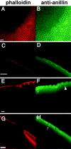
Defects arise at the onset of cellularization and persist throughout in embryos from syx L266 clones. As soon as membrane invagination begins in mitotic cycle 14, defects in the actin cytoskeleton are apparent (A and C). Anillin still shows a hexagonal grid early in cellularization (B), although it does not appear enriched in areas lacking F actin (D). The acellular patch begins near the left edge of C and D. By the midpoint of cellularization, defective regions still show no actin organization (E). Anillin is also disorganized in these regions, and shows a stronger nuclear localization than usual (small arrow, F). Anillin still localizes to the invaginating furrow in those regions undergoing cellularization (large arrowhead F). By the very late stages of cellularization, cytoskeletal organization is still completely lacking in defective regions (G). Anillin is almost completely localized to the nuclei in these regions (arrow, H). Bars: (A and B, C and D, and E and F) 10 μm; (G and H) 20 μm.
Figure 10
Figure 10
Gastrulation can occur in embryos with acellular regions. The posterior end of an embryo stained with rhodamine-conjugated phalloidin is shown. The extent of cellularization on the posterior end is indicated by arrows. In addition, the entire dorsal surface is acellular (wide arrow). However, caudal cells have involuted and migrated dorsally, as would normally occur during gastrulation. Such embryos typically arrest at approximately this stage.

Similar articles

Cited by

References

    1. Aalto MK, Ronne H, Keranen S. Yeast syntaxins Sso1p and Sso2p belong to a family of related membrane proteins that function in vesicular transport. EMBO (Eur Mol Biol Organ) J. 1993;12:4095–4104. - PMC - PubMed
    1. Ashburner, M. 1989. Drosophila, A Laboratory Manual. Cold Spring Harbor Laboratory Press, Cold Spring Harbor, NY. 434 pp.
    1. Bennett MK, Scheller RH. The molecular machinery for secretion is conserved from yeast to neurons. Proc Natl Acad Sci USA. 1993;90:2559–2563. - PMC - PubMed
    1. Bennett MK, Calakos N, Scheller RH. Syntaxin: a synaptic protein implicated in docking of synaptic vesicles at presynaptic active zones. Science (Wash DC) 1992;257:255–259. - PubMed
    1. Bier E, Vaessin H, Shepherd S, Lee K, McCall K, Barbel S, Ackerman L, Carretto R, Uemura T, Grell E, et al. Searching for pattern and mutation in the Drosophila genome with a P-lacZvector. Genes Dev. 1989;3:1273–1287. - PubMed

Publication types

MeSH terms

LinkOut - more resources