. 1997 Aug 11;138(3):671-80.
doi: 10.1083/jcb.138.3.671.

Novel beta-secretase cleavage of beta-amyloid precursor protein in the endoplasmic reticulum/intermediate compartment of NT2N cells

Affiliations

Novel beta-secretase cleavage of beta-amyloid precursor protein in the endoplasmic reticulum/intermediate compartment of NT2N cells

A S Chyung et al. J Cell Biol. .

Abstract

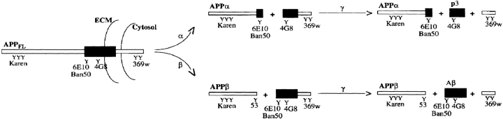
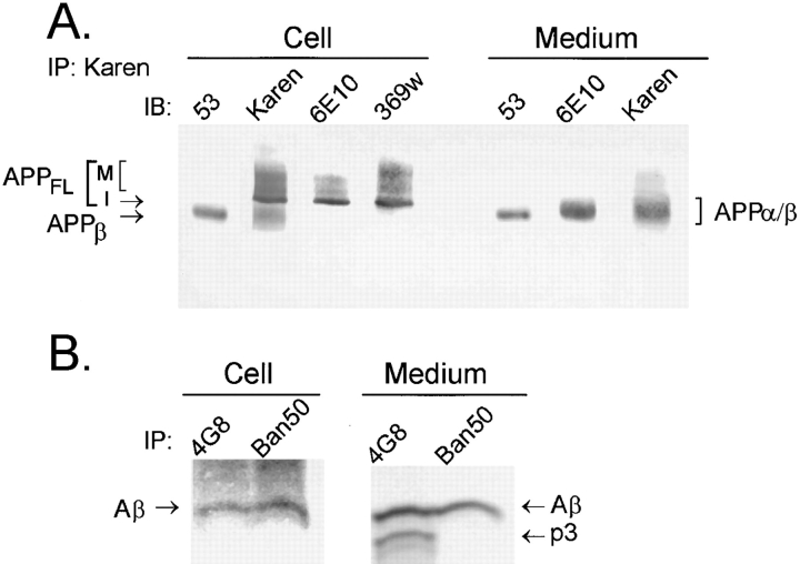
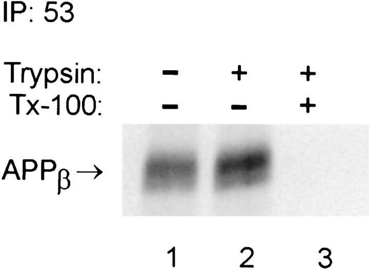
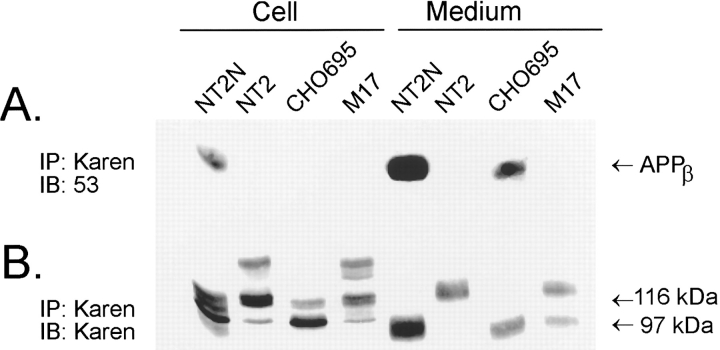
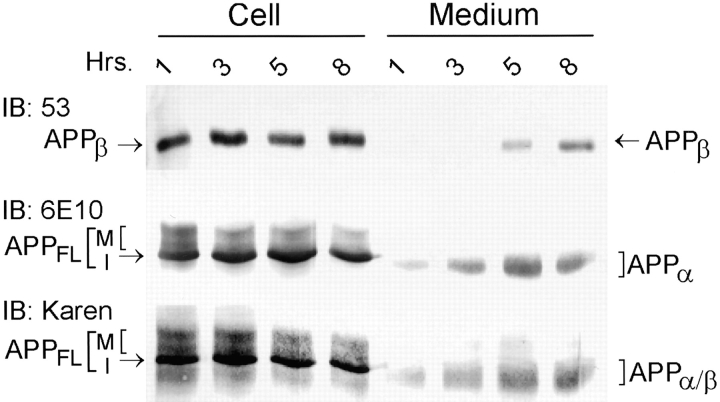
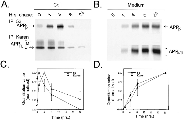
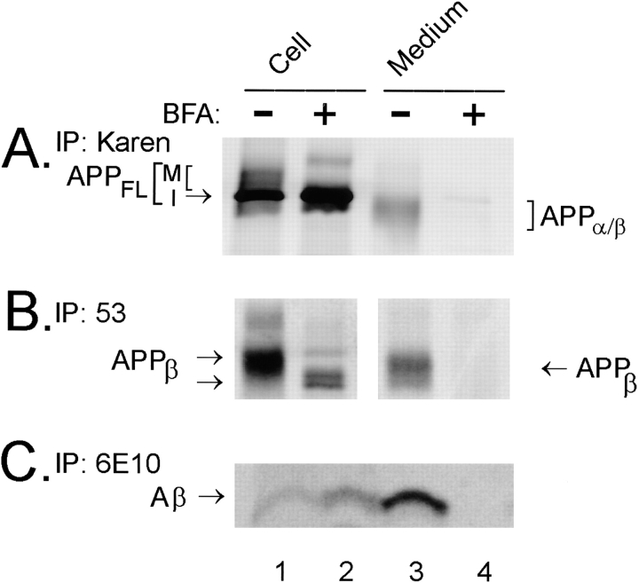
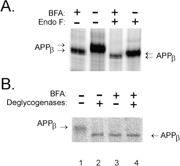
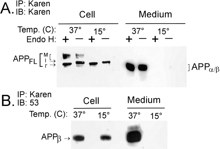
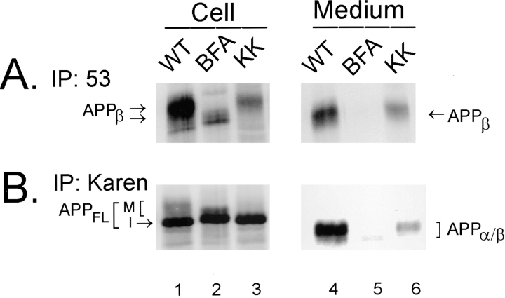
Previous studies have demonstrated that NT2N neurons derived from a human embryonal carcinoma cell line (NT2) constitutively process the endogenous wild-type beta-amyloid precursor protein (APP) to amyloid beta peptide in an intracellular compartment. These studies indicate that other proteolytic fragments generated by intracellular processing must also be present in these cells. Here we show that the NH2-terminal fragment of APP generated by beta-secretase cleavage (APPbeta) is indeed produced from the endogenous full length APP (APPFL). Pulse-chase studies demonstrated a precursor-product relationship between APPFL and APPbeta as well as intracellular and secreted APPbeta fragments. In addition, trypsin digestion of intact NT2N cells at 4 degrees C did not abolish APPbeta recovered from the cell lysates. Furthermore, the production of intracellular APPbeta from wild-type APP appears to be a unique characteristic of postmitotic neurons, since intracellular APPbeta was not detected in several non-neuronal cell lines. Significantly, production of APPbeta occurred even when APP was retained in the ER/ intermediate compartment by inhibition with brefeldin A, incubation at 15 degrees C, or by expression of exogenous APP bearing the dilysine ER retrieval motif.


Figures

Figure 1
Figure 1
Proteolytic processing of APPFL. The diagram depicts APP fragments generated by both the α- and β-secretase pathways. A large, secreted ectodomain called APPα is generated by the putative α-secretase(s) that cleaves APPFL within the Aβ domain. A second cleavage by the γ-secretase(s) releases a subfragment of Aβ known as p3. Alternative cleavage by the β-secretase(s) generates a similarly large ectodomain fragment known as APPβ. After the subsequent γ-secretase cleavage, Aβ is released. This schematic also shows the epitope location of the antibodies used in this study to identify the different proteolytic fragments.
Figure 2
Figure 2
NT2N neurons produce intracellular APPβ and Aβ. To demonstrate the presence of APPβ, samples of cell lysate and medium were collected from NT2N cultures and processed for immunoprecipitation (IP) with Karen, a polyclonal antibody that recognizes epitopes within the large ectodomain of APP. The presence of APPβ, APPα, APPα/β, and APPFL was detected by immunoblotting (IB) with the corresponding antibodies (A). To show that Aβ but not p3 is produced intracellularly, NT2N neurons were radiolabeled with [35S]methionine for 16 h. The cell lysate and the medium were then processed for immunoprecipitation with 4G8, a mAb that binds to both Aβ and p3, or Ban50, a mAb that recognizes only Aβ (B). Immunoprecipitates of Aβ and p3 were separated by electrophoresis in 10/16.5% step-gradient Tris-tricine gels. M, mature APPFL; I, immature APPFL.
Figure 3
Figure 3
APPβ is produced intracellularly in NT2N neurons. Culture dishes containing >99% pure NT2N cells were metabolically labeled with [35S]methionine for 16 h. Cells were rinsed twice with PBS and then incubated on ice for 20 min with PBS alone (lane 1), with 10 μg/ml trypsin (lane 2), or with 10 μg/ml trypsin and 0.1% Triton X-100 (lane 3). The cells were processed for immunoprecipitation with the anti-APPβ antibody 53, as described in Materials and Methods.
Figure 4
Figure 4
Intracellular APPβ is observed only in NT2N neurons. Samples of cell lysate and medium collected from cultures of NT2N, NT2, M17, and CHO cells stably expressing APP695 (CHO695) were processed for immunoprecipitation with the antibody Karen. The immunoprecipitates were separated by SDS-PAGE gels and transferred onto nitrocellulose replicas. APPβ present in the cell lysates and the media were detected by immunoblotting with the anti-APPβ antibody 53 (A). After stripping the nitrocellulose replica in A with 0.1% SDS, the blot was reprobed with Karen to detect all APP ectodomain species (B).
Figure 5
Figure 5
NT2N neurons produce intracellular APPβ before secretion. Cultures of NT2N neurons were washed and fresh medium was replenished before measuring the amount of intracellular and secreted APPβ over an 8-h period. Cell lysate and medium collected at the times indicated were immunoprecipitated with Karen. The immunoprecipitates were separated by SDS-PAGE and then transferred onto nitrocellulose membranes. APPβ was identified in immunoblots using the antibody 53. APPα was detected using the antibody 6E10. APPFL and APPα/β were recognized by Karen.
Figure 6
Figure 6
Pulse–chase labeling demonstrates that intracellular APPβ is produced in an intracellular compartment before secretion in NT2N neurons. NT2N neurons were pulse labeled with [35S]methionine for 1 h and chased for 0, 1, 4, 8, and 24 h. Radiolabeled cell lysates (A) or media (B) were immunoprecipitated sequentially with antibody 53 (for APPβ) followed by Karen (for APPFL in the cell lysates and APPα/β in the media). Radiolabeled immunoprecipitates were used to expose PhosphorImager plates (72 h) or X-ray film (3 wk) for visualization. C and D summarize the quantitation of experiments shown in A and B. Counts from three different experiments were normalized to percentage of maximum and plotted as shown (mean ± standard error).
Figure 7
Figure 7
Intracellular β and γ cleavages occur in a pre-Golgi compartment in NT2N neurons. Cultures of NT2N cells were first preincubated with 20 μg/ml BFA for 1 h before radiolabeling with [35S]methionine for 16 h in the continuous presence of 20 μg/ml BFA. Control cultures were processed similarly, except that BFA was absent in the medium. Radiolabeled proteins from BFA-treated and untreated cell lysates and media were immunoprecipitated with Karen (for APPFL in the cell lysates and APPα/β in the media as shown in A), with antibody 53 (for APPβ in B), and with the mAb 6E10 (for Aβ in C). Note that APPβ and Aβ were recovered in the cell lysate but not in the medium of BFA-treated cells. (M, mature APPFL; I, immature APPFL.
Figure 8
Figure 8
APPβ generated in the presence of BFA is partially glycosylated. Cultures of NT2N neurons were metabolically labeled as in Fig. 7 in the presence or absence of 20 μg/ml BFA. The cell lysates were then immunoprecipitated with the antibody 53. (A) Samples in lanes 3 and 4 were treated with Nglyc F for 16 h to remove N-linked sugars, whereas immunoprecipitates in lanes 1 and 2 were treated with the vehicle. (B) Samples in lanes 2 and 4 were deglycosylated with a combination of Nglyc F, neuraminidase, and O-glycosidase for 16 h to remove both N- and O-linked chains (lanes 2 and 4); lanes 1 and 3 represent samples that were mock digested.
Figure 9
Figure 9
APPβ is generated in the ER/IC of NT2N neurons. Approximately 6 × 10⁶ NT2N neurons were incubated at either 15° or 37°C for 16 h. The cell lysates and media were harvested and immunoprecipitated with Karen. (A) The immunoprecipitates were then split, and half of the samples was treated with Endo H for 18 h, while the other half was mock digested. Subsequent to this step, the immunoprecipitates were separated by SDS-PAGE, transferred onto nitrocellulose replicas, and probed with the antibody Karen. The following observations serve to verify the effectiveness of the temperature block: (a) immature forms of APPFL (I and I′) in the cell lysate retain Endo H sensitivity at 15°C; (b) mature glycosylated forms of APPFL (M) in the cell lysate are not detected at 15°C; and (c) secreted fragments are not detected in the conditioned medium at 15°C. (B) Immunoprecipitates were separated by SDS-PAGE, transferred onto nitrocellulose replicas, and probed with antibody 53. APPβ continued to be produced intracellularly despite the effective temperature block. However, secreted APPβ was not detected in the medium at 15°C. Note that splitting intracellular APPβ samples recovered at 15°C for Endo H digestion decreased the yield to below the level of detection by this assay (data not shown). M, mature APPFL; I, immature APPFL; I′, immature APPFL demonstrating a mobility shift due to Endo H sensitivity.
Figure 10
Figure 10
APPβ is generated from APPFL that is concentrated in the ER. NT2N cultures of ∼1 × 10⁶ cells were infected with recombinant SFV containing either wild-type APP695 or APP695ΔKK constructs. The dilysine motif concentrates APPFL to the ER by an efficient retrieval mechanism. Duplicate cultures infected with wild-type APP695 were treated with 20 μg/ml BFA for comparison. Under these conditions, the cells were metabolically labeled with [35S]methionine for 16 h. Radiolabeled cell lysates and media were then immunoprecipitated with antibody 53 (for APPβ, A) and Karen (for APPFL and APPα/β, B). Radiolabeled immunoprecipitates were used to expose PhosphorImager plates (72 h) for visualization of bands. Unlike APPβ produced under BFA inhibition, APPβ derived from APP695ΔKK was modified and secreted into the medium.

Similar articles

Cited by

References

    1. Arai H, Lee VM-Y, Messinger M, Greenberg BD, Lowery DE, Trojanowski JQ. Expression patterns of β-amyloid precursor protein (βAPP) in neural and nonneural human tissues from Alzheimer's disease and control subjects. Ann Neurol. 1991;30:686–693. - PubMed
    1. Busciglio J, Gabuzda DH, Matsudaira P, Yankner BA. Generation of β-amyloid in the secretory pathway in neuronal and nonneuronal cells. Proc Natl Acad Sci USA. 1993;90:2092–2096. - PMC - PubMed
    1. Cai X-D, Golde TE, Younkin SG. Release of excess amyloid β protein from a mutant amyloid β protein precursor. Science (Wash DC) 1993;259:514–516. - PubMed
    1. Citron M, Oltersdorf T, Haass C, McConlogue L, Hung AY, Seubert P, Vigo-Pelfrey C, Lieberburg I, Selkoe DJ. Mutation of the β-amyloid precursor protein in Alzheimer's disease increases β-protein production. Nature (Lond) 1992;360:672–674. - PubMed
    1. Cook DG, Sung JC, Golde TE, Felsenstein KM, Wojczyk RE, Tanzi RE, Trojanowski JQ, Lee VM-Y, Doms RW. Expression and analysis of presenilin 1 in a human neuronal system: localization in cell bodies and dendrites. Proc Natl Acad Sci USA. 1996;93:9223–9228. - PMC - PubMed

Publication types