Comparative Study
. 1997 Aug 15;17(16):6165-78.

Establishment of a cell-free system of neuronal apoptosis: comparison of premitochondrial, mitochondrial, and postmitochondrial phases

Affiliations
Comparative Study

Establishment of a cell-free system of neuronal apoptosis: comparison of premitochondrial, mitochondrial, and postmitochondrial phases

H M Ellerby et al. J Neurosci. .

Expression of concern in

Abstract

Apoptosis is a fundamental process required for normal development of the nervous system and is triggered during neurodegenerative disease. To dissect the molecular events leading to neuronal cell death, we have developed a cell-free model of neuronal apoptosis. The model faithfully reproduces key elements of apoptosis, including chromatin condensation, DNA fragmentation, caspase activation/processing, and selective substrate cleavage. We report that cell-free apoptosis is activated in premitochondrial, mitochondrial, and postmitochondrial phases by tamoxifen, mastoparan, and cytochrome c, respectively, allowing a functional ordering of these proapoptotic modulators. Furthermore, this is the first report of mitochondrial-mediated activation of cell-free apoptosis in a cell extract. Although Bcl-2 blocks activation at the premitochondrial and mitochondrial levels, it does not affect the postmitochondrial level. The cell-free system described here provides a valuable tool to elucidate the molecular events leading to neuronal cell death.


Figures
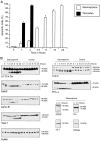
Fig. 1.
Fig. 1.
Staurosporine and tamoxifen-activated neural apoptosis. A, Percentage of apoptotic cells versus time in hours. CSM-25 cells were incubated with staurosporine (10 μm; white rectangles) or tamoxifen (100 μm; black rectangles) for the indicated times (34°C). Apoptotic cells were judged morphologically. Data are mean values ± SD as given by error bars (number of independent experiments, n = 3). No significant apoptosis was observed in control cells during the 24 hr period. B, Proteolytic profile of protein substrates selectively cleaved during staurosporine-initiated neural apoptosis. CSM-25 cells were incubated with staurosporine for the indicated times (10 μm, 34°C). Cell lysates were made at the indicated time points and subjected to Western blot analysis. The data in B are representative of at least three independent experiments, depending on substrate.
Fig. 2.
Fig. 2.
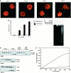
Tamoxifen-primed extract activates neural cell-free apoptosis. A, Nuclear morphological changes in CSM nuclei incubated in a 16,000 × g extract made from tamoxifen-primed NSC-19 cells at 34°C. B, Percentage of apoptotic nuclei incubated as in A in either normal or primed extract. Data are mean values ± SD as given by error bars (n = 3). No significant apoptotic changes were observed in control nuclei during the 2 hr incubation. C, Agarose gel electrophoresis of internucleosomal DNA fragmentation of rat liver nuclei incubated in a 16,000 × g extract made from tamoxifen-primed CSM-25 cells (2 hr, 34°C). D, Selective proteolytic cleavage of key substrates from a cell-free reaction of HeLa nuclei incubated for the indicated times in a 16,000 × gextract made from tamoxifen-primed CSM-25 cells at 37°C. Cleavage was prevented by Ac-DEVD-CHO, but not by Ac-YVAD-CHO (each at 1 μm). E, The activity of CPP32-like caspases as measured by DEVD-pNA hydrolysis. The CPP32-like caspase activity of a tamoxifen-primed NSC-19 extract after a 2 hr incubation at 37°C is given by the top line. The DEVD activity of a normal NSC-19 extract and the YVAD activity of a tamoxifen-primed NSC-19 extract fall at or below the bottom line. In each case the data in A, C–E are representative of at least three independent experiments.
Fig. 5.
Fig. 5.
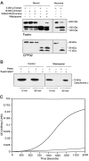
Mastoparan activates neural and neuronal cell-free apoptosis. A, Fodrin cleavage and CPP32 processing in a neural cell-free system composed of a 3000 × gextract (containing mitochondria) made from CSM-25 cells in a neural cell-free system of mouse liver mitochondria incubated in a 16,000 × g extract from NT2 cells and in a neuronal cell-free system of rat neuronal mitochondria in a 16,000 ×g extract from primary cerebellar neurons (2–4 hr at 37°C; 50 μm). Mastoparan did not prime a 16,000 ×g extract without mitochondria. B, Mastoparan induced release of cytochrome c from mitochondria. Mouse liver mitochondria incubated with mastoparan under the conditions in A led to the release of cytochromec, as measured by Western blot of the supernatant from the mitochondrial pellet. C, The processing of DEVD-pNA substrate by a 16,000 × g normal NT2 extract activated by the concentrated supernatant from B is shown in the top curve (37°C). The activity of a 16,000 × g normal NT2 extract, of a 16,000 ×g normal NT2 extract incubated with mastoparan, and mastoparan in buffer was less than or equal to the activity shown by the bottom curve. In each case the data given inA–C are representative of at least three independent experiments.
Fig. 6.
Fig. 6.
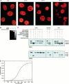
Cytochrome c and dATP activate neural and neuronal cell-free apoptosis. A, Nuclear fragmentation of HeLa nuclei incubated in a 16,000 ×g NT2 extract with horse heart cytochromec and dATP. B, DNA fragmentation of CSM nuclei incubated in a 16,000 × g CSM extract with horse heart cytochrome c and dATP. C, Proteolysis of fodrin and the processing of CPP32 in extracts. Although horse heart cytochrome c activated both 16,000 ×g NT2 extracts and 16,000 × gextracts from rat primary cerebellar neurons, yeast and acetylated horse cytochrome c did not activate extracts. A 16,000 × g CSM extract made from Bcl-2-overexpressing cells, with or without mitochondria from Bcl-2-overexpressing cells, and a 3000 × g extract from such cells is activated by cytochrome c/dATP. A 30–60 min preincubation of 16,000 × g extract at 37°C renders the extract incapable of activation by cytochromec/dATP. Furthermore, the peptide inhibitor zVAD-fmk prevents the activation. Incubation conditions for A–Care 10 μm cytochrome c and 1 mm dATP for 1.5 hr at 37°C. D, Activation of CPP32-like caspase in a 16,000 × g NSC-34 extract incubated with dATP and cytochrome c, shown by the top curve, as measured by hydrolysis of DEVD-pNA (10 μm cytochrome c and 1 mmdATP at 37°C). The activity of extract alone is shown by the bottom curve.The activities of yeast and partially acetylated cytochrome c in the above system and cytochrome c/dATP in buffer lie at or below the activity of normal extract. In each case the data given inA–D are representative of at least three independent experiments.
Fig. 3.
Fig. 3.
Atractyloside activates neural cell-free apoptosis. Atractyloside (5 mm) was incubated in a 3000 × g extract made from CSM-25 cells (2–4 hr, 37°C). The activation of apoptosis was measured by fodrin cleavage. Atractyloside also induced cell-free apoptosis in a system composed of rat liver mitochondria and 16,000 × g extract from CSM-25 cells. However, atractyloside incubated in a 16,000 ×g extract alone did not lead to cell-free apoptosis. The data given in this figure are representative of three independent experiments.
Fig. 4.
Fig. 4.
Mastoparan activates neural apoptosis. Mastoparan induces apoptosis in cultured rat cerebellar neuron precursors (the R2 cell line) as measured by cell death, using propidium iodide staining of DNA in cells with a compromised plasma membrane (see Rabizadeh et al., 1993). Data are mean values ± SD as given by error bars (n = 3). No death was observed in control cells (6 hr).
Fig. 8.
Fig. 8.
CPP32 processing in apoptosis and cell-free apoptosis. As indicated by the processing of CPP32, Tamoxifen (Tam) induces apoptosis in whole cells (Pre-mito) but does not induce cell-free apoptosis in extract with added mitochondria (Mito) or extract alone (Post-mito). Similar results were obtained for staurosporine (data not shown). Mastoparan (Mast) induces apoptosis at the Pre-mito level and cell-free apoptosis at the Mito level but does not induce cell-free apoptosis at the Post-mito level. Similar results were obtained for atractyloside (data not shown). Cytochromec/dATP (Cytc) activates cell-free apoptosis at the Mito and Post-mitolevels but does not induce apoptosis at the Pre-mitolevel. For the premitochondrial level, neural cells (CSM, NSC, or NT2) were incubated with tamoxifen (2 hr), mastoparan (6 hr), or cytochromec/dATP (8 hr). For the mitochondrial level, a cell-free system composed of 16,000 × g neural extract and added rat liver mitochondria was incubated with tamoxifen (4 hr), mastoparan (4 hr), or cytochrome c/dATP (1 hr). For the postmitochondrial level, a cell-free system composed of 16,000 ×g neural extract was incubated with tamoxifen (4 hr), mastoparan (4 hr), or cytochrome c/dATP (1 hr). All incubations were run under the following conditions: 100 μm tamoxifen; 50 μm mastoparan; 10 μm cytochrome c; 1 mm dATP; 37°C). The data given in this figure are representative of at least three independent experiments.
Fig. 7.
Fig. 7.
Sequence alignment of horse and yeast (Iso-1) cytochrome c. Yeast cytochrome c differs from horse heart cytochrome c in the number and distribution of lysines. The open rectangles highlight lysine residues found in horse, but not in yeast cytochromec. The shaded rectangle highlights lysine 72, which is naturally trimethylated in yeast cytochromec (Clements et al., 1989).

Similar articles

Cited by

References

    1. Ajmani AK, Satoh M, Reap E, Cohen PL, Reeves WH. Absence of autoantigen Ku in mature human neutrophils and human promyelocytic leukemia line (HL-60) cells and lymphocytes undergoing apoptosis. J Exp Med. 1995;181:2049–2058. - PMC - PubMed
    1. Alnemri ES, Livingston DJ, Nicholson DW, Salvesen G, Thornberry NA, Wong WW, Yuan J. Human ICE/CED-3 protease nomenclature. Cell. 1996;87:171. - PubMed
    1. Arnett TR, Lindsay R, Kilb JM, Moonga BS, Spowage M, Dempster DW. Selective toxic effects of tamoxifen on osteoclasts: comparison with the effects of estrogen. J Endocrinol. 1996;149:503–508. - PubMed
    1. Azi A, Montecucco C, Richter C. The use of acetylated ferricytochrome c for the detection of superoxide radicals in biological membranes. Biochem Biophys Res Commun. 1975;65:597–603. - PubMed
    1. Bravo R, Frank R, Blundelli PA, McDonald-Bravo H. Cyclin-PCNA is the auxiliary protein of DNA polymerase delta. Nature. 1987;326:515–517. - PubMed

Publication types

MeSH terms

LinkOut - more resources