. 1997 Jun 30;137(7):1495-509.
doi: 10.1083/jcb.137.7.1495.

Sec2p mediates nucleotide exchange on Sec4p and is involved in polarized delivery of post-Golgi vesicles

Affiliations

Sec2p mediates nucleotide exchange on Sec4p and is involved in polarized delivery of post-Golgi vesicles

C Walch-Solimena et al. J Cell Biol. .

Abstract

The small GTPase Sec4p is required for vesicular transport at the post-Golgi stage of yeast secretion. Here we present evidence that mutations in SEC2, itself an essential gene that acts at the same stage of the secretory pathway, cause Sec4p to mislocalize as a result of a random rather than a polarized accumulation of vesicles. Sec2p and Sec4p interact directly, with the nucleotide-free conformation of Sec4p being the preferred state for interaction with Sec2p. Sec2p functions as an exchange protein, catalyzing the dissociation of GDP from Sec4 and promoting the binding of GTP. We propose that Sec2p functions to couple the activation of Sec4p to the polarized delivery of vesicles to the site of exocytosis.


Figures
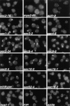
Figure 1
Figure 1
Immunofluorescence localization of Sec4p in sec mutants. Cells were grown overnight in YP dextrose medium at 25°C and then shifted to 37°C for 1 h, fixed, and labeled with αSec4 antibody. The effect of cycloheximide (cyc) and azide treatment on wild-type cells was tested by adding either agent to the culture immediately before shift to 37°C. sec2-78, NY1529; myo2-66, NY1005; act1-3, NY278; sec1-1, NY8; sec3-2, NY45; sec4-8, NY28; sec5-24, NY30; sec6-4, NY18; sec8-9, NY43; sec9-4, NY32; sec10-2, NY36; sec15-1, NY39; wild-type, NY10; sec12-4, NY738; sec16-2, NY416; bos1-1, NY1282 (see Table I for strain genotypes). Wild-type cells and most late-acting sec mutants show immunolabeling preferentially in the bud. In sec2-78, myo2-66, and act1-3, diffuse staining for Sec4p is observed. Also, in sec4-8, ER to Golgi sec mutants (sec12-4, sec16-2, bos1-1), and after treatment of wild-type cells with cycloheximide or azide, no concentration of immunolabeling in the bud is found.
Figure 2
Figure 2
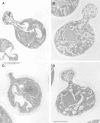
Electron microscopic analysis of sec2-78, wild-type, and sec6-4 cells labeled for Sec4p with immunogold. Cells were grown overnight at 25°C, shifted to 37°C for 30 min, and then prepared for postembedding immunogold labeling as described in Materials and Methods. (A) A sec2-78 (NY1529) cell grown at the permissive temperature where some immunogold labeling is found on vesicles in the bud. Both wild-type (NY10, C) and sec6-4 (NY1294, D) cells show a concentration of immunogold labeling on vesicles in the bud after shift to the restrictive temperature. In contrast, an average sec2-78 cell in B has immunogold-labeled vesicles randomly distributed in the bud and mother cell. At 25°C (A) 3 ± 2 gold particles were observed on vesicles while 3 ± 2 gold particles were found independent of vesicles in the bud. In the mother cell were 1 ± 1 gold particle on vesicles and 10 ± 3 gold particles not on vesicles. At 37°C (B) we found 3 ± 2 gold particles on vesicles and 3 ± 4 gold particles not on vesicles in the bud. In the mother cell were 4 ± 3 gold particles on vesicles and 15 ± 9 gold particles not on vesicles. The error represents the SD for n = 12 at 25°C and n = 24 at 37°C. Bar, 1 μm.
Figure 3
Figure 3
Immunofluorescence localization of Sec4p in different sec2 alleles and a sec2-78,6-4 double mutant after a short shift to the restrictive temperature. Cells were grown overnight in YP dextrose medium at 25°C and then shifted to 37°C for 10 min, fixed, and labeled with αSec4 antibody. sec12-4, NY738; sec2-41, NY132; sec2-59, NY27; sec2-78, NY1529; wild-type, NY10; sec6-4, NY779; sec2-78,6-4, NY1530. In sec2-41, sec2-59, and sec2-78, Sec4p, immunostaining was concentrated in the bud at 25°C but was diffuse already after 10-min shift to the restrictive temperature. In the ER-to-Golgi mutant sec12-4, diffuse immunolabeling was also already observed after 10 min at 37°C. Both in wild-type and sec6-4, Sec4p immunolabeling was concentrated at the bud both at 25°C and after 10 min at 37°C. The double mutant was constructed as described in Materials and Methods. sec2-78, sec2-78,6-4 shows labeling in the buds at the permissive temperature that is no longer detected after 10 min at the restrictive temperature.
Figure 4
Figure 4
Differential centrifugation of lysates derived from wild-type (NY10), sec6-4 (NY779), sec2-41 (NY132), and sec2-78 (NY1529). Cells were grown overnight in YP dextrose and then shifted to 37°C. After 2 h at the restrictive temperature, cells were harvested, and lysates (S1) were prepared as described in Materials and Methods. S1 supernatants were centrifuged at 10,000 g to generate supernatant S2 and pellet P2. The S2 supernatants were centrifuged at 100,000 g to obtain supernatant S3 and pellet P3. Equal volumes of samples were prepared for electrophoresis, separated on SDS-PAGE gels, and transferred to nitrocellulose. Western blots were probed with αSec4p polyclonal antibodies and iodinated protein A. No difference in the relative distribution of Sec4p between S3 and P3 in sec2-41 and sec2-78 compared to sec6-4 is seen. The shift of Sec4p from P2 to P3 in sec6-4 compared to wild-type is caused by vesicle accumulation.
Figure 5
Figure 5
Fractionation of P3 pellets from sec6-4 and sec2-41 cells shifted to 37°C for 2 h on sucrose density gradients. The P3 pellets were generated by differential centrifugation and loaded in 0.32 M sorbitol on top of 0.4–1.75 M sucrose gradients. After centrifugation to equilibrium, the gradients were fractionated from the bottom (fraction 2) in 17 fractions (0.75 ml), and the residual pellets were resuspended in 0.75 ml (fraction 1). Equal volumes of each fraction were analyzed by SDS-PAGE and immunoblotting (affinity-purified polyclonal antibodies to Pma1p and Sec2p detected by chemoluminescence; antisera to Sec4p and Sncp detected by iodinated protein A). The Western blots were quantified by densitometry and for each antigen and gradient normalized to a total integrated optical density of 1. In sec2-41 cells (NY132), Sec4p peaks at the same density as in sec6-4 (NY779). Sec2p partially cofractionates with Sec4p and Sncp in sec6-4 cells and completely in sec2-41 cells.
Figure 6
Figure 6
EM of sec2-78, wild-type, and sec6-4 cells shifted to 37°C for 10 min. Cells were synchronized with α factor, released from G1 arrest, and allowed to form small buds at 25°C, and then incubated at 37°C for 10 min. After this, cells were fixed and prepared for EM, as described in Materials and Methods. (A) Representative sec2-78 cell (NY1529) at 25°C that shows some vesicle accumulation. (B) Representative sec2-78 cell after 10 min at 37°C that accumulated vesicles both in bud and mother cell. In comparison, a typical wild-type cell (NY13; C) does not accumulate secretory vesicles, and in sec6-4 (NY17; D), vesicles accumulate mainly in the bud. Bar, 1 μm.
Figure 7
Figure 7
In vitro association of Sec2p with Sec4p. The binding assays were performed on amylose resin using recombinant MBP-Sec2-59 or MBP alone, which was incubated with 35S-labeled Sec4p produced in an in vitro transcription-coupled translation reaction. (A) The SEC4 mutations are indicated above each lane. (B) Wild-type Sec4p was diluted in buffer with EDTA before being gel filtered and associated with nucleotide as indicated. Input was directly loaded into the well and represents 40% of the 35S-labeled protein used in each binding reaction.
Figure 8
Figure 8
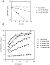
Sec2-59p–stimulated guanine nucleotide exchange on Sec4p. (A) Aliquots containing 1 μM Sec4p-[3H]GDP were incubated with the indicated levels of Sec2-59p or buffer in the presence of excess unlabeled GDP. At the time intervals indicated, Sec4p-bound radioactivity was determined by the filter-binding assay. To retard the intrinsic off rate of Sec4p, this experiment was performed at 14°C. (B) Aliquots containing 1 μM Sec4p were incubated with Sec2-59p or buffer, as indicated in the presence of [35S]GTPγS at 14°C. At the time intervals indicated, Sec4p-bound radioactivity was determined by the filter-binding assay. For each assay, the relative values correspond to the nanomoles of GTPγS retained on the filter.
Figure 9
Figure 9
Sequence of Sec2p and comparison with Rabin3 and expressed sequence tags. Comparison of the Rab-binding portion of the protein sequence of Sec2p with Rabin3 and the predicted protein sequence of ESTs from human liver (H73617), human brain (T09468/N24211), and Caenorhabditis elegans (D37628) homologues. The sequences were aligned using the program MegAlign and visual inspection. EST sequences, which are incomplete cDNA sequences, are shown only for the predicted open reading frames corresponding to the SEC2 sequence. Amino acid residues are numbered according to the protein or DNA sequence. Gaps to improve the alignment are indicated by hyphens. Amino acids that are identical to the yeast sequence in at least two of the five sequences are shaded black. Residues that are similar in class are shaded grey and are grouped according to acidic DE, basic HKR, hydrophobic MPVWAFIL, and polar CGNQSTY.
Figure 10
Figure 10
Differential centrifugation of lysates derived from wild-type (NY10), wild-type overproducing Sec4p on low copy (NY1535), sec6-4 (NY779), sec6-4 overproducing Sec4p (NY1536), and sec4-8 (NY28). Cells were grown overnight in YP dextrose and then shifted to 37°C. After 2 h at the restrictive temperature, cells were harvested and lysates (S1) were prepared as described in Materials and Methods. Differential centrifugation and analysis of the resulting fractions was performed as described in Fig. 4. Western blots were probed with αSec4p polyclonal antibodies and iodinated protein A or affinity-purified αSec2p polyclonal antibodies detected with chemoluminescence. The amount of Sec2p in the P3 fractions parallels the amount of Sec4p. The increase of Sec4p in P3 of strains overproducing the protein is correlated with an increase of Sec2p, and the decrease of Sec4p in P3 in sec4-8 cells is correlated with a decrease of Sec2p in this fraction. The Sec4p blots for wild-type and sec6-4 cells are identical to those shown in Fig. 4 to allow for direct comparison.

Similar articles

Cited by

References

    1. Aalto MK, Ronne H, Keränen S. Yeast syntaxins Sso1p and Sso2p belong to a family of related membrane proteins that function in vesicular transport. EMBO (Eur Mol Biol Organ) J. 1993;12:4095–4104. - PMC - PubMed
    1. Altschul SF, Gish W, Miller W, Myers EW, Lipman DJ. Basic local alignment search tool. J Mol Biol. 1990;215:403–410. - PubMed
    1. Araki S, Kaibuchi K, Sasaki T, Hata Y, Takai Y. Role of the C-terminal region of smg p25A in its interaction with membranes and the GDP/GTP exchange protein. Mol Cell Biol. 1991;11:1438–1447. - PMC - PubMed
    1. Bourne HR, Sanders DA, McCormick F. The GTPase superfamily: a conserved switch for diverse cell functions. Nature (Lond) 1990;348:125–132. - PubMed
    1. Bowser R, Novick P. Sec15 protein, an essential component of the exocytotic apparatus, is associated with the plasma membrane and with a soluble 19.5 S particle. J Cell Biol. 1991;112:1117–1131. - PMC - PubMed

Publication types

MeSH terms

Substances