. 1997 Jul 1;17(13):5046-61.
doi: 10.1523/JNEUROSCI.17-13-05046.1997.

Cellular composition and three-dimensional organization of the subventricular germinal zone in the adult mammalian brain

Affiliations

Cellular composition and three-dimensional organization of the subventricular germinal zone in the adult mammalian brain

F Doetsch et al. J Neurosci. .

Abstract

The adult mammalian subventricular zone (SVZ) contains stem cells that give rise to neurons and glia. In vivo, SVZ progeny migrate 3-8 mm to the olfactory bulb, where they form neurons. We show here that the SVZ of the lateral wall of the lateral ventricles in adult mice is composed of neuroblasts, glial cells, and a novel putative precursor cell. The topographical organization of these cells suggests how neurogenesis and migration are integrated in this region. Type A cells had the ultrastructure of migrating neuronal precursors. These cells were arranged as chains parallel to the walls of the ventricle and were polysialylated neural adhesion cell molecule- (PSA-NCAM), TuJ1- (beta-tubulin), and nestin-positive but GFAP- and vimentin-negative. Chains of Type A cells were ensheathed by two ultrastructurally distinct astrocytes (Type B1 and B2) that were GFAP-, vimentin-, and nestin-positive but PSA-NCAM- and TuJ1-negative. Type A and B2 (but not B1) cells incorporated [3H]thymidine. The most actively dividing cell in the SVZ corresponded to Type C cells, which had immature ultrastructural characteristics and were nestin-positive but negative to the other markers. Type C cells formed focal clusters closely associated with chains of Type A cells. Whereas Type C cells were present throughout the SVZ, they were not found in the rostral migratory stream that links the SVZ with the olfactory bulb. These results suggest that chains of migrating neuroblasts in the SVZ may be derived from Type C cells. Our results provide a topographical model for the adult SVZ and should serve as a basis for the in vivo identification of stem cells in the adult mammalian brain.


Figures
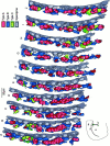
Fig. 5.
Fig. 5.
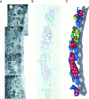
Serial section (separated by 2 μm) reconstruction of the dorsolateral aspect of the lateral wall of the lateral ventricle at the level indicated by the arrow in the coronal hemisection (bottom left). Cell types were identified in photo montages on the basis of characteristics described in the text. The contours and processes of cells were drawn and transferred into drawing programs as described under Materials and Methods and illustrated in Figure 4. Colorscorresponding to the different cell types are indicated in thekey. In this portion of the ventricle the chains of migrating cells (Type A cells) are very abundant. Type A cells (red) form continuous chains that branch and converge. These chains are covered mainly by Type B cell processes (blue) and frequently are associated with clusters of Type C cells (green). Empty spaces are areas of dense neuropil. Nc, Neocortex; cc, corpus callosum; St, striatum; LV, lateral ventricle. Scale bar, 20 μm.
Fig. 4.
Fig. 4.
Steps in the extraction of information about SVZ cell arrangement. A, Contiguous electron micrographs of the SVZ were assembled into a photo montage. B, The contours and nuclei of the different cell types were traced in different colors. C, This information was transferred into a computer, and cells were filled as described in Materials and Methods. This SVZ representation allowed identification of cell types at a glance and the interpretation of serial section reconstructions presented in Figure 5.
Fig. 1.
Fig. 1.
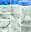
Cell types in the SVZ of adult mice. The identification of the different cell types was made in serial reconstructions, and not all ultrastructural features are present in each photomicrograph. A, Three Type A cells (a) showing scant, dark cytoplasm with many free ribosomes, a small Golgi apparatus (arrow), and dense heterochromatin. The surface of Type A cells is relatively smooth. Their elongated shape is not visible in this cross section. Type A cells frequently are separated by open extracellular spaces (asterisks) and are joined by specialized junctional complexes (see Fig. 2). Magnification, 10,500×. B, Type B1 (b) cells have light cytoplasm, dense bodies, thick bundles of intermediate filaments (arrow), and irregular contours that penetrate the intercellular space around them. Type B1 cells are located at the interface between ependymal cells (e) and overlying tissue. Magnification, 13,000×. C, Type B2 cells have light cytoplasm, irregular contours, and dense bodies in their cytoplasm (arrows). The cytoplasm of Type B2 cells contains few free ribosomes. Type B2 cells (b) are localized basally at the interface of the striatal parenchyma and Type A cells (a). Notice contacts of Type B2 cells with myelinated and unmyelinated axons of the striatal parenchyma. Magnification, 8000×. D, Type C cells (c) have irregular nuclei with deep invaginations, mostly lax chromatin, and a large, reticulated nucleolus. The cytoplasm is more electron-dense than that of Type B cells (b) and contains a clear Golgi apparatus (arrow). Magnification, 6500×. E, Ependymal cells (e) line the ventricle and frequently are ciliated. They are heavily interdigitated and contain apical junctional complexes. Their nuclei are round and their chromatin unclumped. Their cytoplasm is very light and contains many basal mitochondria and a few free ribosomes. Lipid droplets (arrow) are unique to ependymal cells. Note two Type B cells (b). Magnification, 3800×. F, G, Tanycytes (d) are dark, unciliated cells that contact the ventricle. They have irregular nuclei and an electron-dense cytoplasm containing many mitochondria and a large Golgi apparatus. They exhibit lateral branches (arrows) that interdigitate with ependymal cells (e) and Type B cells. Magnification: 6000× in F; 4500× in G.
Fig. 3.
Fig. 3.
Relationships among the major cell types in the SVZ. Identification of the individual cell types was made in serial ultrathin sections. A, Frontal section through the SVZ in the lateral wall revealing the topographical relationships of the different cell types to one another. A transversely cut chain of Type A (a) cells is separated from ependymal cells (e) by the electron-lucent lateral expansions of Type B1 cells (b1), which are found adjacent to the ependymal layer. In contrast, the processes of Type B2 cells (b2), which are localized basally adjacent to the striatal parenchyma, isolate the chain of Type A cells from the surrounding neuropil. Type B cells are never found inside chains. Notice the clumped chromatin typical of Type B2 cells and the unclumped chromatin of Type B1 cells. A Type C cell (c) with its typical reticulated nucleolus is located close to the chain of Type A cells. Magnification, 6700×. B, A thin lamina (arrows) formed by individual or multiple processes of Type B1 cells separate Type A cells (a) from ependymal cells (e). Magnification, 16,800×. C, Tangential section through the SVZ showing the chain organization of Type A cells at the light microscope in a semithin section. Thearrows indicate the presence of two chains of dark cells corresponding to Type A cells. Magnification, 360×. D, Tangential section through the SVZ showing the elongated shape of Type A cells in this plane and their close association with Type C cells. The larger size of Type C cells is clearly visible here, as are the differences in cytoplasmic electron density of Type A, B, and C cells.a, Type A cell; b, Type B cell;c, Type C cell. Magnification, 3500×.
Fig. 2.
Fig. 2.
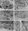
Selected sections of a serial reconstruction of small zonula adherens-like contacts between Type A cells. The junctions between two Type A cells were photographed in 39 ultrathin serial sections; section number is indicated in thelower right corner. Four junctions (I, II, III, IV) appear in this reconstruction. The junctions are not continuous but are disk-like in shape with a diameter of ∼0.5–1 μm. Endocytic vesicles (arrows) frequently were associated with these junctions. Magnification, 12,500×.
Fig. 8.
Fig. 8.
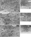
Cell types in the adult mouse SVZ that incorporate [3H]thymidine. A, Pair of Type C cells (c) labeled 1 hr after injection of [3H]thymidine. Silver grains over the nuclei of these two cells are shown in a semithin section to theright. Magnification, 4500×. B, Two labeled cells (arrows in semithin section to theright and in electron micrograph to left) 1 hr after [3H]thymidine injection correspond to a Type C cell (c) and a Type A cell (a). Magnification, 3400×. C, Type B2 cells also were labeled by [3H]thymidine 1 hr after injection. In this electron micrograph an elongated Type B2 cell (b2, arrow) is labeled with silver grains (semithin autoradiogram on the right,arrow). Ependymal cells (e) and Type B1 cells (b1) were not observed to be labeled with [3H]thymidine. Magnification, 3500×.
Fig. 6.
Fig. 6.
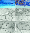
Immunocytochemical characterization of different cell types in the SVZ: PSA–NCAM and TuJ1. The chains of Type A cells are immuno-positive for both PSA–NCAM and TuJ1. Other cell types are immunonegative for these markers. A, PSA–NCAM staining in toluidine blue-stained coronal semithin section showing a chain of immunopositive cells flanked by immunonegative cells. Magnification, 400×. B, C, Immunostaining for PSA–NCAM at the ultrastructural level. Type A cells are immunopositive for PSA–NCAM, whereas Type B cells are immunonegative (arrows in B). PSA–NCAM staining is continuous along the plasma membrane of Type A cells (a); nuclear membranes of Type A cells are unstained (arrow in C). Magnification: 8000× inB; 11,500× in C. D, TuJ1 immunostaining in toluidine blue-stained coronal semithin section. Magnification, 500×. E, F, Immunostaining for TuJ1 at the ultrastructural level. The cytoplasm of Type A cells is darkly stained by anti-TuJ1 antibodies. Type B cells (b) and ependymal cells (e) are immunonegative for this marker. Magnification: 6800× inE; 12,000× in F.
Fig. 7.
Fig. 7.
Immunocytochemical characterization of different cell types in the SVZ: GFAP, vimentin, and nestin. Chains of Type A cells are immunonegative for GFAP and vimentin but are immunopositive for nestin. Type B cells, which ensheath the chains of Type A cells, are immunopositive for GFAP, vimentin, and nestin. Ependymal cells are stained very strongly by nestin and vimentin antibodies but more weakly by GFAP antibodies. Shown is immunostaining of GFAP (A), vimentin (E), and nestin (H) in toluidine blue-stained coronal semithin sections of the SVZ. A, Clusters of cells in chains are surrounded by processes immunopositive for GFAP. Ependymal cells also are stained by GFAP antibodies. E, Ependymal cells are stained strongly by anti-vimentin antibodies. The processes of astrocytes are also vimentin-positive. H, Ependymal cells are very darkly stained by nestin antibodies. Staining is also visible in astrocytes and Type A cells (darker nuclei). Magnification: 300× in A; 400× in E; 400× inH. B, At the electron microscope the processes and cell bodies of Type B cells (b) are stained by GFAP antibodies. These immunopositive processes surround a chain of Type A cells (a) that are immunonegative for GFAP. Ependymal cells (e) are lightly stained. Magnification, 3000×. C, Type D cells (tanycytes,arrow) are very darkly stained with GFAP antibodies. Magnification, 2800×. D, Thin lamellar processes (arrows; see also Fig. 2E) of Type B1 cells that separate Type A cells (a) from ependymal cells (e) are GFAP-positive. Magnification, 26,000×.F, Ependymal cells (e) are strongly immunopositive for vimentin. Type B cells are also positive to vimentin but do not stain as darkly as ependymal cells. Magnification, 3000×.G, Higher magnification of vimentin-immunonegative Type A cells adjacent to a vimentin-positive Type B cell (b) with dark immunoreactive precipitate in the cytoplasm. Magnification, 24,500×. I, Nestin immunoreactivity in ependymal cells (e), Type A cells (a), and Type C cells (c). The underlying striatal neuropil and neurons were not stained by the nestin antibody. The cytoplasm of ependymal cells is stained homogeneously, whereas staining is in clumps in Type A and C cells. Magnification, 5000×. J, Clumps of nestin immunoreactivity in Type A cells (a) concentrate in the perinuclear cytoplasm (arrows; see alsoI). Magnification, 6000×. K, Type B cells (b) also contain nestin immunoreactive material in their cytoplasm. Magnification, 8500×.
Fig. 9.
Fig. 9.
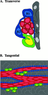
Summary diagram of the organization of the adult SVZ. A, Schematic cross section through a chain of migrating neuroblasts (red) ensheathed by two types of glial cells (B1, B2, blue) that separate the migrating cells from the striatum (left) and ependymal cells (gray). Type C cells (green, putative precursor) are not ensheathed by glia and are associated closely with the chains of migrating neuroblasts. B, Schematic en face view of the SVZ viewed from the striatum. The red channelsrepresent the chains of migrating neuroblasts (Type A cells) with tangentially elongated nuclei (light red). Theblue blocks represent the ensheathing glial cells (Type B1 and B2). These cells form tunnel-like structures through which the Type A cells migrate. Putative precursors (Type C cells,green) are closely associated with—and speckled in small clusters along—chains of migrating neuroblasts. The underlying ependymal cells (gray) form a sheet lining the ventricular surface.

Similar articles

Cited by

References

    1. Allen E. The cessation of mitosis in the central nervous system of the albino rat. J Comp Neurol. 1912;22:547–568.
    1. Altman J. Autoradiographic and histological studies of postnatal neurogenesis. IV. Cell proliferation and migration in the anterior forebrain, with special reference to persisting neurogenesis in the olfactory bulb. J Comp Neurol. 1969;137:433–458. - PubMed
    1. Alvarez-Buylla A, Lois C. Neuronal stem cells in the brain of adult vertebrates. Stem Cells. 1995;13:263–272. - PubMed
    1. Alvarez-Buylla A, Buskirk DR, Nottebohm F. Monoclonal antibody reveals radial glia in adult avian brain. J Comp Neurol. 1987;264:159–170. - PubMed
    1. Alvarez-Buylla A, Theelen M, Nottebohm F. Proliferation “hot spots” in adult avian ventricular zone reveal radial cell division. Neuron. 1990;5:101–109. - PubMed

Publication types