Peyer's patch organogenesis is intact yet formation of B lymphocyte follicles is defective in peripheral lymphoid organs of mice deficient for tumor necrosis factor and its 55-kDa receptor
- PMID: 9177215
- PMCID: PMC21047
- DOI: 10.1073/pnas.94.12.6319
Peyer's patch organogenesis is intact yet formation of B lymphocyte follicles is defective in peripheral lymphoid organs of mice deficient for tumor necrosis factor and its 55-kDa receptor
Erratum in
- Proc Natl Acad Sci U S A 1997 Aug 19;94(17):9510
Abstract
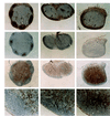
Targeted inactivation of genes in the tumor necrosis factor (TNF)/lymphotoxin (LT) ligand and receptor system has recently revealed essential roles for these molecules in lymphoid tissue development and organization. Lymphotoxin-alphabeta (LTalphabeta)/lymphotoxin-beta receptor (LTbeta-R) signaling is critical for the organogenesis of lymph nodes and Peyer's patches and for the structural compartmentalization of the splenic white pulp into distinct B and T cell areas and marginal zones. Moreover, an essential role has been demonstrated for TNF/p55 tumor necrosis factor receptor (p55TNF-R) signaling in the formation of splenic B lymphocyte follicles, follicular dendritic cell networks, and germinal centers. In contrast to a previously described essential role for the p55TNF-R in Peyer's patch organogenesis, we show in this report that Peyer's patches are present in both TNF and p55TNF-R knockout mice, demonstrating that these molecules are not essential for the organogenesis of this lymphoid organ. Furthermore, we show that in the absence of TNF/p55TNF-R signaling, lymphocytes segregate normally into T and B cell areas and a normal content and localization of dendritic cells is observed in both lymph nodes and Peyer's patches. However, although B cells are found to home normally within Peyer's patches and in the outer cortex area of lymph nodes, organized follicular structures and follicular dendritic cell networks fail to form. These results show that in contrast to LTalphabeta signaling, TNF signaling through the p55TNF-R is not essential for lymphoid organogenesis but rather for interactions that determine the cellular and structural organization of B cell follicles in all secondary lymphoid tissues.
Figures


Similar articles
-
Peyer's patch organogenesis--cytokines rule, OK?Gut. 1997 Nov;41(5):707-9. doi: 10.1136/gut.41.5.707. Gut. 1997. PMID: 9414984 Free PMC article. Review.
-
Tumor necrosis factor and the p55TNF receptor are required for optimal development of the marginal sinus and for migration of follicular dendritic cell precursors into splenic follicles.Cell Immunol. 2000 Apr 10;201(1):33-41. doi: 10.1006/cimm.2000.1636. Cell Immunol. 2000. PMID: 10805971
-
Defective Peyer's patch organogenesis in mice lacking the 55-kD receptor for tumor necrosis factor.J Exp Med. 1996 Jul 1;184(1):259-64. doi: 10.1084/jem.184.1.259. J Exp Med. 1996. PMID: 8691140 Free PMC article.
-
Lymphotoxin-alpha-deficient and TNF receptor-I-deficient mice define developmental and functional characteristics of germinal centers.Immunol Rev. 1997 Apr;156:137-44. doi: 10.1111/j.1600-065x.1997.tb00965.x. Immunol Rev. 1997. PMID: 9176705 Review.
-
Effects of tumor necrosis factor and lymphotoxin on peripheral lymphoid tissue development.Int Immunol. 1998 Jun;10(6):727-41. doi: 10.1093/intimm/10.6.727. Int Immunol. 1998. PMID: 9678753
Cited by
-
Protective and pathologic roles of the immune response to mouse hepatitis virus type 1: implications for severe acute respiratory syndrome.J Virol. 2009 Sep;83(18):9258-72. doi: 10.1128/JVI.00355-09. Epub 2009 Jul 1. J Virol. 2009. PMID: 19570864 Free PMC article.
-
Lymphotoxin alpha/beta and tumor necrosis factor are required for stromal cell expression of homing chemokines in B and T cell areas of the spleen.J Exp Med. 1999 Jan 18;189(2):403-12. doi: 10.1084/jem.189.2.403. J Exp Med. 1999. PMID: 9892622 Free PMC article.
-
Role of tumor necrosis factor alpha in Helicobacter pylori gastritis in tumor necrosis factor receptor 1-deficient mice.Infect Immun. 2002 Jun;70(6):3149-55. doi: 10.1128/IAI.70.6.3149-3155.2002. Infect Immun. 2002. PMID: 12011009 Free PMC article.
-
Role of an intact splenic microarchitecture in early lymphocytic choriomeningitis virus production.J Virol. 2002 Mar;76(5):2375-83. doi: 10.1128/jvi.76.5.2375-2383.2002. J Virol. 2002. PMID: 11836415 Free PMC article.
-
TNF-α acts as an immunoregulator in the mouse brain by reducing the incidence of severe disease following Japanese encephalitis virus infection.PLoS One. 2013 Aug 5;8(8):e71643. doi: 10.1371/journal.pone.0071643. Print 2013. PLoS One. 2013. PMID: 23940775 Free PMC article.
References
-
- Vassalli P. Annu Rev Immunol. 1992;10:411–452. - PubMed
-
- Paul N L, Ruddle N H. Annu Rev Immunol. 1988;6:407–438. - PubMed
-
- Vandenabeele P, Declercq W, Beyaert R, Fiers W. Trends Cell Biol. 1995;5:392–399. - PubMed
-
- Grell M, Douni E, Wajant H, Lohden M, Clauss M, Maxeiner B, Georgopoulos S, Lesslauer W, Kollias G, Pfizenmaier K, Scheurich P. Cell. 1995;83:793–802. - PubMed
-
- Browning J L, Ngam ek A, Lawton P, DeMarinis J, Tizard R, Chow E P, Hession C, O’Brine-Greco B, Foley S F, Ware C F. Cell. 1993;72:847–856. - PubMed
Publication types
MeSH terms
Substances
LinkOut - more resources
Full Text Sources
Molecular Biology Databases

