An in vitro tubulogenesis system using cell lines derived from the embryonic kidney shows dependence on multiple soluble growth factors
- PMID: 9177208
- PMCID: PMC21040
- DOI: 10.1073/pnas.94.12.6279
An in vitro tubulogenesis system using cell lines derived from the embryonic kidney shows dependence on multiple soluble growth factors
Abstract
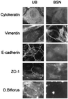
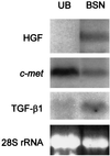
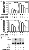
Interactions between the ureteric bud (UB) and metanephric mesenchyme are crucial for tubulogenesis during kidney development. Two immortalized cell lines derived from the day 11.5 embryonic kidney, UB cells, which appear to be epithelial (cytokeratin-positive, E-cadherin-positive, and ZO-1-positive by immunostaining) and BSN cells, which are largely mesenchymal (vimentin-positive, but negative for cytokeratin, cell surface E-cadherin, and cell surface ZO-1), were used to establish an in vitro tubulogenesis system. BSN cells expressed hepatocyte growth factor (HGF) and transforming growth factor-beta1 mRNAs, and its conditioned medium (BSN-CM) contained factors capable of activating the epidermal growth factor (EGF) receptor (EGFR). When UB cells were cultured in an extracellular matrix gel in the presence of the embryonic kidney or BSN-CM, the UB cells underwent morphogenetic changes characteristic of early in vitro branching tubulogenesis. These changes were largely inhibited by a combination of neutralizing anti-HGF antibodies and the EGFR inhibitor tyrphostin AG1478, suggesting that EGFR ligands, together with HGF, account for much of this early morphogenetic activity. Nevertheless, there was a significant fraction of tubulogenic activity that could not be inhibited, suggesting the existence of other soluble factors. Whereas HGF, EGF, transforming growth factor alpha, basic fibroblast growth factor (bFGF), and insulin-like growth factor 1 (IGF-1), or a mixture of these growth factors, induced epithelial processes for up to 3 days, only IGF-1, possibly bFGF, and the mixture were able to sustain morphogenesis for longer periods, though not nearly to the same degree as BSN-CM. Moreover, only BSN-CM induced branching tubular structures with clear lumens, consistent with the existence of other soluble factors crucial for the formation and/or maintenance of branching tubular structures with lumens in vitro.
Figures


Similar articles
-
Differential tubulogenic and branching morphogenetic activities of growth factors: implications for epithelial tissue development.Proc Natl Acad Sci U S A. 1995 May 9;92(10):4412-6. doi: 10.1073/pnas.92.10.4412. Proc Natl Acad Sci U S A. 1995. PMID: 7753820 Free PMC article.
-
EGF receptor ligands are a large fraction of in vitro branching morphogens secreted by embryonic kidney.Am J Physiol. 1997 Sep;273(3 Pt 2):F463-72. doi: 10.1152/ajprenal.1997.273.3.F463. Am J Physiol. 1997. PMID: 9321921
-
Transforming growth factor-beta selectively inhibits branching morphogenesis but not tubulogenesis.Am J Physiol. 1997 Jan;272(1 Pt 2):F139-46. doi: 10.1152/ajprenal.1997.272.1.F139. Am J Physiol. 1997. PMID: 9039060
-
Development of the tubular nephron.Semin Nephrol. 1995 Jul;15(4):315-26. Semin Nephrol. 1995. PMID: 7569411 Review.
-
Epithelial tubulogenesis through branching morphogenesis: relevance to collecting system development.J Am Soc Nephrol. 1995 Oct;6(4):1151-9. doi: 10.1681/ASN.V641151. J Am Soc Nephrol. 1995. PMID: 8589281 Review.
Cited by
-
Identification of matrix physicochemical properties required for renal epithelial cell tubulogenesis by using synthetic hydrogels.J Cell Sci. 2019 Oct 21;132(20):jcs226639. doi: 10.1242/jcs.226639. J Cell Sci. 2019. PMID: 31558679 Free PMC article.
-
TIMP-1 induces an EMT-like phenotypic conversion in MDCK cells independent of its MMP-inhibitory domain.PLoS One. 2012;7(6):e38773. doi: 10.1371/journal.pone.0038773. Epub 2012 Jun 11. PLoS One. 2012. PMID: 22701711 Free PMC article.
-
Developmental tumourigenesis: NCAM as a putative marker for the malignant renal stem/progenitor cell population.J Cell Mol Med. 2009 Aug;13(8B):1792-1808. doi: 10.1111/j.1582-4934.2008.00607.x. J Cell Mol Med. 2009. PMID: 20187302 Free PMC article.
-
Role of extracellular matrix components and structure in new renal models in vitro.Front Physiol. 2022 Dec 7;13:1048738. doi: 10.3389/fphys.2022.1048738. eCollection 2022. Front Physiol. 2022. PMID: 36569770 Free PMC article. Review.
-
Receptor tyrosine kinases in kidney development.J Signal Transduct. 2011;2011:869281. doi: 10.1155/2011/869281. Epub 2011 Mar 3. J Signal Transduct. 2011. PMID: 21637383 Free PMC article.
References
-
- Saxen L. Organogenesis of the Kidney. Cambridge, U.K.: Cambridge Univ. Press; 1987.
-
- Nigam S K, Aperia A, Brenner B M. In: The Kidney. Brenner B M, editor. Philadelphia: Saunders; 1996. pp. 72–98.
-
- Stark K, Vainio S, Vassileva G, McMahon A P. Nature (London) 1994;372:679–683. - PubMed
-
- Luo G, Hofmann C, Bronckers A L, Sohocki M, Bradley A, Karsenty G. Genes Dev. 1995;9:2808–2820. - PubMed
-
- Dudley A T, Lyons K M, Robertson E J. Genes Dev. 1995;9:2795–2807. - PubMed
Publication types
MeSH terms
Substances
LinkOut - more resources
Full Text Sources
Other Literature Sources
Research Materials
Miscellaneous

