Degradation of T cell receptor (TCR)-CD3-zeta complexes after antigenic stimulation
- PMID: 9151711
- PMCID: PMC2196323
- DOI: 10.1084/jem.185.10.1859
Degradation of T cell receptor (TCR)-CD3-zeta complexes after antigenic stimulation
Abstract
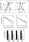
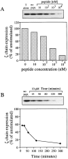
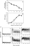
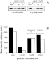
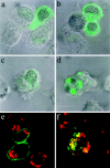
T cell activation by specific antigen results in a rapid and long-lasting downregulation of triggered T cell receptors (TCRs). In this work, we investigated the fate of downregulated TCR- CD3-zeta complexes. T cells stimulated by peptide-pulsed antigen-presenting cells (APCs) undergo an antigen dose-dependent decrease of the total cellular content of TCR-beta, CD3-epsilon, and zeta chains, as detected by FACS(R) analysis on fixed and permeabilized T-APC conjugates and by Western blot analysis on cell lysates. The time course of CD3-zeta chain consumption overlaps with that of TCR downregulation, indicating that internalized TCR-CD3 complexes are promptly degraded. Inhibitors of lysosomal function (bafilomycin A1, folimycin) markedly reduced zeta chain degradation, leading to the accumulation of zeta chain in large Lamp1(+) vesicles. These results indicate that in T cell-APC conjugates, triggered TCRs are rapidly removed from the cell surface and are degraded in the lysosomal compartment.
Figures
Comment in
-
Understanding the mechanisms of sustained signaling and T cell activation.J Exp Med. 1997 May 19;185(10):1717-9. doi: 10.1084/jem.185.10.1717. J Exp Med. 1997. PMID: 9198667 Free PMC article. Review. No abstract available.
Similar articles
-
Degradation of ZAP-70 following antigenic stimulation in human T lymphocytes: role of calpain proteolytic pathway.J Immunol. 1999 Jul 1;163(1):50-6. J Immunol. 1999. PMID: 10384098
-
Qualitative and quantitative contributions of the T cell receptor zeta chain to mature T cell apoptosis.J Exp Med. 1996 May 1;183(5):2109-17. doi: 10.1084/jem.183.5.2109. J Exp Med. 1996. PMID: 8642321 Free PMC article.
-
Functionally active T cell receptor/CD3 complexes are present at the surface of cloned cytotoxic T cells without fluorescence-immunological detectability.Cell Immunol. 1996 Jul 10;171(1):62-7. doi: 10.1006/cimm.1996.0173. Cell Immunol. 1996. PMID: 8660838
-
The T-cell antigen receptor: a complex signal-transducing molecule.Princess Takamatsu Symp. 1988;19:87-104. Princess Takamatsu Symp. 1988. PMID: 2978621 Review.
-
Internalization and intracellular fate of TCR-CD3 complexes.Crit Rev Immunol. 2000;20(4):325-46. Crit Rev Immunol. 2000. PMID: 11100805 Review.
Cited by
-
Phosphorylation of CDK9 at Ser175 enhances HIV transcription and is a marker of activated P-TEFb in CD4(+) T lymphocytes.PLoS Pathog. 2013;9(5):e1003338. doi: 10.1371/journal.ppat.1003338. Epub 2013 May 2. PLoS Pathog. 2013. PMID: 23658523 Free PMC article. Clinical Trial.
-
HLA-restricted epitope identification and detection of functional T cell responses by using MHC-peptide and costimulatory microarrays.Proc Natl Acad Sci U S A. 2005 Mar 8;102(10):3744-9. doi: 10.1073/pnas.0407019102. Epub 2005 Feb 23. Proc Natl Acad Sci U S A. 2005. PMID: 15728728 Free PMC article.
-
Suboptimal engagement of the T-cell receptor by a variety of peptide-MHC ligands triggers T-cell anergy.Immunology. 2010 Jan;129(1):1-7. doi: 10.1111/j.1365-2567.2009.03206.x. Epub 2009 Dec 2. Immunology. 2010. PMID: 20002785 Free PMC article. Review.
-
Qa-1 restriction of CD8+ suppressor T cells.J Clin Invest. 2004 Nov;114(9):1218-21. doi: 10.1172/JCI23152. J Clin Invest. 2004. PMID: 15520850 Free PMC article. Review.
-
Effector CD8+CD45RO-CD27-T cells have signalling defects in patients with squamous cell carcinoma of the head and neck.Br J Cancer. 2003 Jan 27;88(2):223-30. doi: 10.1038/sj.bjc.6600694. Br J Cancer. 2003. PMID: 12610507 Free PMC article.
References
-
- Weiss A, Littman DR. Signal transduction by lymphocyte antigen receptors. Cell. 1994;76:263–274. - PubMed
-
- Klausner RD, Lippincott J, Schwartz, Bonifacino JS. The T cell antigen receptor: insights into organelle biology. Annu Rev Cell Biol. 1990;6:403–431. - PubMed
-
- Donnadieu E, Cefai D, Tan YP, Paresys G, Bismuth G, Trautmann A. Imaging early steps of human T cell activation by antigen-presenting cells. J Immunol. 1992;148:2643–2653. - PubMed
-
- Valitutti S, Müller S, Cella M, Padovan E, Lanzavecchia A. Serial triggering of many T-cell receptors by a few peptide–MHC complexes. Nature (Lond) 1995;375:148–151. - PubMed
Publication types
MeSH terms
Substances
LinkOut - more resources
Full Text Sources
Other Literature Sources
Miscellaneous

