

Bipolar localization of a chromosome partition protein in Bacillus subtilis
- PMID: 9114058
- PMCID: PMC20791
- DOI: 10.1073/pnas.94.9.4721
Bipolar localization of a chromosome partition protein in Bacillus subtilis
Abstract
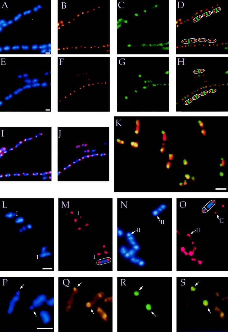
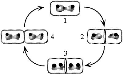
We have determined the subcellular localization of the chromosome partition protein Spo0J of Bacillus subtilis by immunofluorescence microscopy and visualizing fluorescence of a Spo0J-GFP fusion protein. Spo0J was associated with a region of the nucleoid proximal to the cell pole, both in growing cells dividing symmetrically and in sporulating cells dividing asymmetrically. Additional experiments indicated that Spo0J was bound to sites in the origin-proximal third of the chromosome. These results show that the replicating chromosomes are oriented in a specific manner during the division cycle, with the Spo0J binding region positioned toward the cell poles. Experiments characterizing cells at different stages of the cell cycle showed that chromosome orientation is established prior to the initiation of cell division. Our results indicate that there is a mechanism for orienting the chromosomes and that the chromosome partition protein Spo0J might be part of a bacterial mitotic-like apparatus.
Figures

Similar articles
-
Effects of the chromosome partitioning protein Spo0J (ParB) on oriC positioning and replication initiation in Bacillus subtilis.J Bacteriol. 2003 Feb;185(4):1326-37. doi: 10.1128/JB.185.4.1326-1337.2003. J Bacteriol. 2003. PMID: 12562803 Free PMC article.
-
RacA and the Soj-Spo0J system combine to effect polar chromosome segregation in sporulating Bacillus subtilis.Mol Microbiol. 2003 Sep;49(6):1463-75. doi: 10.1046/j.1365-2958.2003.03643.x. Mol Microbiol. 2003. PMID: 12950914
-
Cell division protein DivIB influences the Spo0J/Soj system of chromosome segregation in Bacillus subtilis.Mol Microbiol. 2005 Jan;55(2):349-67. doi: 10.1111/j.1365-2958.2004.04399.x. Mol Microbiol. 2005. PMID: 15659156
-
Regulation of initiation of Bacillus subtilis chromosome replication.Plasmid. 1999 Jan;41(1):17-29. doi: 10.1006/plas.1998.1381. Plasmid. 1999. PMID: 9887303 Review.
-
Generating and exploiting polarity in bacteria.Science. 2002 Dec 6;298(5600):1942-6. doi: 10.1126/science.1072163. Science. 2002. PMID: 12471245 Review.
Cited by
-
Spatio-temporal organization of replication in bacteria and eukaryotes (nucleoids and nuclei).Cold Spring Harb Perspect Biol. 2012 Aug 1;4(8):a010389. doi: 10.1101/cshperspect.a010389. Cold Spring Harb Perspect Biol. 2012. PMID: 22855726 Free PMC article. Review.
-
A single parS sequence from the cluster of four sites closest to oriC is necessary and sufficient for proper chromosome segregation in Pseudomonas aeruginosa.PLoS One. 2015 Mar 20;10(3):e0120867. doi: 10.1371/journal.pone.0120867. eCollection 2015. PLoS One. 2015. PMID: 25794281 Free PMC article.
-
Phosphorylation of Mycobacterium tuberculosis ParB participates in regulating the ParABS chromosome segregation system.PLoS One. 2015 Mar 25;10(3):e0119907. doi: 10.1371/journal.pone.0119907. eCollection 2015. PLoS One. 2015. PMID: 25807382 Free PMC article.
-
Identification and characterization of a negative regulator of FtsZ ring formation in Bacillus subtilis.Proc Natl Acad Sci U S A. 1999 Aug 17;96(17):9642-7. doi: 10.1073/pnas.96.17.9642. Proc Natl Acad Sci U S A. 1999. PMID: 10449747 Free PMC article.
-
Rhodoccoccus erythropolis Is Different from Other Members of Actinobacteria: Monoploidy, Overlapping Replication Cycle, and Unique Segregation Pattern.J Bacteriol. 2019 Nov 20;201(24):e00320-19. doi: 10.1128/JB.00320-19. Print 2019 Dec 15. J Bacteriol. 2019. PMID: 31570531 Free PMC article.
References
-
- Murray A, Hunt T. The Cell Cycle: An Introduction. New York: Freeman; 1993.
-
- Hyman A A, Sorger P K. Annu Rev Cell Dev Biol. 1995;11:471–495. - PubMed
-
- Hiraga S. Annu Rev Biochem. 1992;61:283–306. - PubMed
-
- Rothfield L I. Cell. 1994;77:963–966. - PubMed
-
- Wake R G, Errington J. Annu Rev Genet. 1995;29:41–67. - PubMed
Publication types
MeSH terms
Substances
Grants and funding
LinkOut - more resources
Full Text Sources
Other Literature Sources
Molecular Biology Databases
