. 1997 May 1;17(9):2967-79.
doi: 10.1523/JNEUROSCI.17-09-02967.1997.

Second messengers, trafficking-related proteins, and amino acid residues that contribute to the functional regulation of the rat brain GABA transporter GAT1

Affiliations

Second messengers, trafficking-related proteins, and amino acid residues that contribute to the functional regulation of the rat brain GABA transporter GAT1

M W Quick et al. J Neurosci. .

Abstract

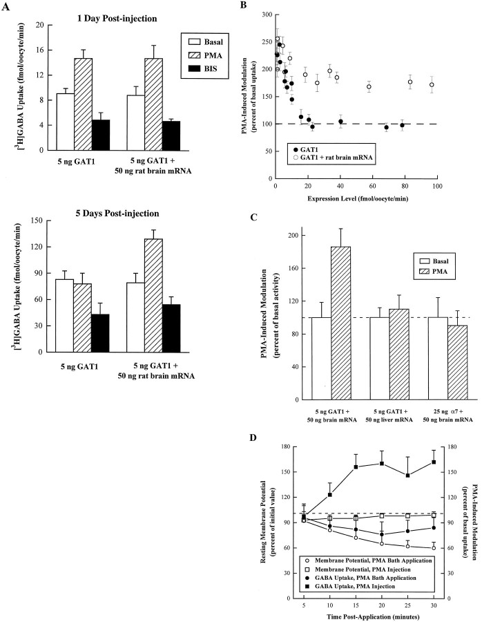
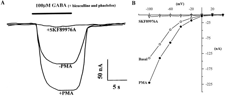
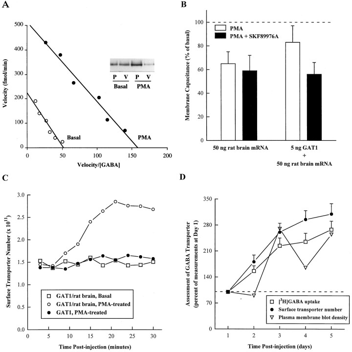
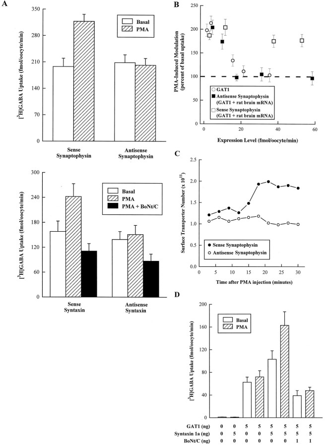
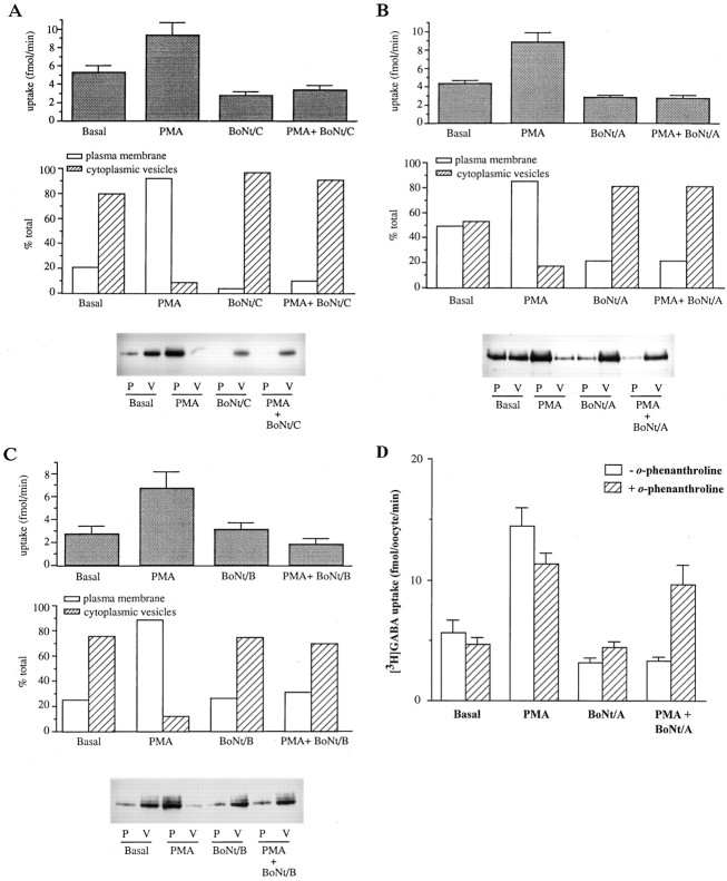
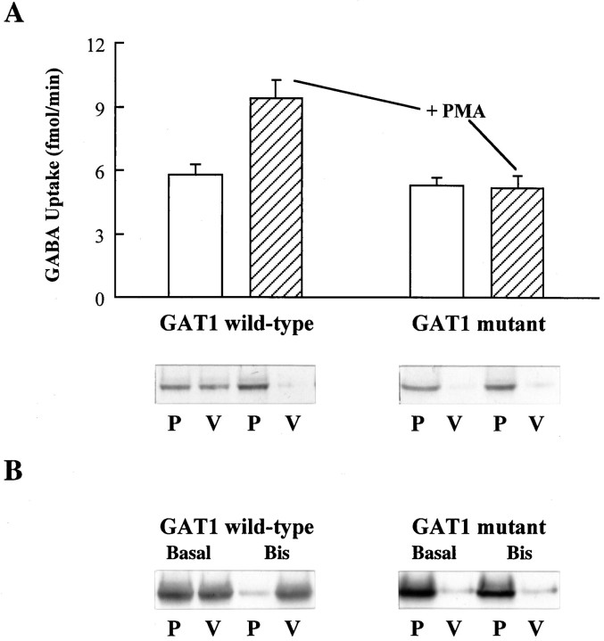
Recent evidence indicates that several members of the Na+-coupled transporter family are regulated, and this regulation in part occurs by redistribution of transporters between intracellular locations and the plasma membrane. We elucidate components of this process for both wild-type and mutant GABA transporters (GAT1) expressed in Xenopus oocytes using a combination of uptake assays, immunoblots, and electrophysiological measurements of membrane capacitance, transport-associated currents, and GAT1-specific charge movements. At low GAT1 expression levels, activators of protein kinase C (PKC) induce redistribution of GAT1 from intracellular vesicles to the plasma membrane; at higher GAT1 expression levels, activators of PKC fail to induce this redistribution. However, coinjection of total rat brain mRNA with GAT1 permits PKC-mediated modulation at high transporter expression levels. This effect of brain mRNA on modulation is mimicked by coinjection of syntaxin 1a mRNA and is eliminated by injecting synaptophysin or syntaxin antisense oligonucleotides. Additionally, botulinum toxins, which inactivate proteins involved in vesicle release and recycling, reduce basal GAT1 expression and prevent PKC-induced translocation. Mutant GAT1 proteins, in which most or all of a leucine heptad repeat sequence was removed, display altered basal distribution and lack susceptibility to modulation by PKC, delineating one region of GAT1 necessary for its targeting. Thus, functional regulation of GAT1 in oocytes occurs via components common to transporters and to trafficking in both neural and non-neural cells, and suggests a relationship between factors that control neurotransmitter secretion and the components necessary for neurotransmitter uptake.


Figures

Fig. 1.
Fig. 1.
PMA-induced modulation of GAT1 uptake. A, Coinjection of rat brain mRNA allows PMA-induced GAT1 modulation at higher expression levels. Oocytes were injected with 5 ng of GAT1 cRNA alone or in combination with 50 ng of rat brain mRNA, then assayed by [3H]GABA uptake. The assay time was 15 min. Data represent an experiment (5–7 oocytes per condition, mean ± SEM) for oocytes from a single batch, examined either at 1 d (low expression; top) or at 5 d (high expression;bottom) after RNA injection. Before the assay, oocytes were injected with 25 nl of a vehicle solution (Basal,open bars) or a solution containing 400 nmPMA (15 min before assay, hatched bars) or 100 nm bisindolylmaleimide (BIS; 10 min before assay, filled bars). Note that the topand bottom panels have different vertical scales.B, Summary of data for the effect of rat brain mRNA coinjection. Expression level is quantified as the amount of [3H]GABA uptake in GAT1 RNA-injected oocytes (filled circles) or GAT1 and rat brain RNA-injected oocytes (open circles) injected with vehicle solution 15 min before assay (abscissa). The change in uptake induced by 400 nm PMA (ordinate) is plotted as a percentage of vehicle-only injected oocytes. Each data point represents the mean ± SEM for three to seven oocytes. Expression levels were varied by injecting different amounts of GAT1 cRNA and by performing the assay at different translation times after RNA injection. C, PMA modulation shows specificity for GAT1 and rat brain mRNA. Oocytes from the same frog were injected with 5 ng of GAT1 cRNA in combination with either 50 ng of rat brain mRNA or 50 ng of rat liver mRNA; or, oocytes were injected with 25 ng of cRNA encoding the rat homomeric nicotinic acetylcholine receptor α7 in combination with 50 ng of rat brain cRNA. For the GAT1 cRNA-injected oocytes, GABA transport was assessed as described in A. For oocytes expressing α7, peak currents induced by perfusion of 200 μm nicotine were measured using a two-electrode voltage clamp. The holding potential was −70 mV. The change in function induced by 400 nm PMA (hatched bars) is plotted as a percentage of vehicle-only treated oocytes (open bars). Each data point represents the mean ± SEM for seven oocytes. D, Differential effects of PMA administration on GAT1 uptake. Oocytes were injected with 5 ng of GAT1 cRNA and assayed (5 oocytes per condition) for [3H]GABA uptake after bath application (filled circles) or injection (filled squares) with 400 nm PMA. The change in uptake (right ordinate) is plotted as a percentage of vehicle-only treated oocytes. The resting membrane potential for subsets (5 each) of PMA-incubated (open circles) and PMA-injected (open squares) oocytes was monitored electrophysiologically. The results are plotted as a percentage of their resting membrane potential before PMA application (left ordinate).
Fig. 2.
Fig. 2.
Electrophysiological characterization of PMA-induced GAT1 modulation and block by SKF89976A. A, Two-electrode voltage-clamp analysis of GABA-induced currents for a GAT1/rat brain mRNA-injected oocyte. The holding potential was −80 mV. The solid bar above the traces represents the application of 100 μm GABA. Bicuculline (20 μm) and phaclofen (10 μm) were included to block potential responses attributable to GABA receptor stimulation. PMA (400 nm) was injected after measurement of the basal condition; the PMA-induced trace was recorded 20 min later. The response in the presence of 10 μm SKF89976A was recorded 5 min later. B, Current–voltage relationship for another oocyte recorded as described in A and subjected to voltage ramps from −100 mV to +40 mV over 1 sec.
Fig. 3.
Fig. 3.
PMA-induced modulation occurs by increasing surface transporter expression. Oocytes were injected with 5 ng of GAT1 cRNA and/or 50 ng of rat brain mRNA and assayed 4 d postinjection. PMA (25 nl; 400 nm) was injected 15 min before assay.A, Eadie–Hofstee transformation of dose–response [3H]GABA uptake data for basal (open circles) and PMA (filled circles) conditions (5 oocytes per condition). The assay time was 15 min. GABA concentrations were 0.5, 1, 2, 4, 8, and 16 μm. The average SEM across all conditions was 16.4% of mean values.Inset shows PMA-induced translocation of GAT1 as assessed by Western blot. B, Changes in cell capacitance associated with PMA injection. Membrane potential was held at −40 mV and stepped to −100 mV for 1 sec in the presence (filled bars) and absence (open bars) of 10 μm SKF89976A. Capacitance data are presented as a percentage of the basal capacitance measured before PMA injection. Five oocytes were tested in each of the injection conditions; each oocyte was tested in the absence and presence of SKF89976A. C, PMA-induced (circles) changes in oocyte plasma membrane transporter number as assessed by measurement of charge movements. Calculations are described in Materials and Methods. Oocytes were injected either with 5 ng of GAT1 cRNA alone (filled symbols) or GAT1 cRNA plus 50 ng of rat brain mRNA (open symbols). Oocytes in the basal condition (squares) were control-injected with 25 nl of water. The data presented are from individual oocytes with comparable initial charge movements and are representative of five oocytes/condition.D, Comparison of GABA uptake, surface GABA transporter number, and plasma membrane protein levels. Oocytes from a single frog were injected with 5 ng of GAT1 cRNA and 50 ng of rat brain mRNA and assayed for 5 consecutive d. [3H]GABA uptake (squares) assays were 15 min (5 oocytes/data point). Surface transporter number (circles) was calculated from GAT1-specific charge movements as described in Materials and Methods (5 oocytes/data point). Subcellular fractionation was performed on parallel oocyte samples (25 oocytes/sample) and analyzed by immunoblot as described in Materials and Methods. GAT1 protein present in plasma membrane (triangles) was measured by densitometry (nominal units). All data are plotted relative to the values obtained on day 1.
Fig. 4.
Fig. 4.
Synaptophysin and syntaxin 1a alter the function and expression of GAT1. A, Coinjection of synaptophysin or syntaxin 1a antisense oligonucleotides in oocytes injected with GAT1 and total rat brain mRNA eliminates PMA-induced modulation. Oocytes were coinjected with 5 ng of GAT1 and 50 ng of rat brain RNA. In addition, oocytes were also injected with 25 ng of either sense (control) or antisense oligonucleotides to synaptophysin or syntaxin 1a at the time of RNA injection, and again 24 hr later. Oocytes were assayed 72 hr after RNA injection. The PMA concentration was 400 nm. For oocytes injected with syntaxin 1a oligonucleotides, a subset of oocytes was injected with 1 ng of BoNt/C. Data are mean ± SEM. Oocytes (6 oocytes/condition) were assayed for [3H]GABA uptake 15 min after injection of a control solution (open bars), PMA (hatched bars), or PMA and BoNt/C (filled bars). Assay time was 15 min. B, Summary data for the effect of sense and antisense synaptophysin oligonucleotide injection. Expression level is quantified as the amount of [3H]GABA uptake in GAT1/rat brain-injected oocytes additionally injected with antisense (filled squares) or sense (open squares) oligonucleotides (abscissa). The change in uptake induced by 400 nm PMA (ordinate) is plotted as a percentage of vehicle-only injected oocytes. For comparison, the results for oocytes injected with only GAT1 are included (open circles). Each data point represents the mean ± SEM for three to seven oocytes. Different expression levels were obtained by performing the assay at different times after RNA injection. C, PMA-induced changes in oocyte plasma membrane number as assessed by measurement of charge movements. Calculations are described in Materials and Methods. Measurements were made on GAT1/rat brain mRNA-expressing oocytes injected with either sense (filled circles) or antisense (open circles) oligonucleotides. The data presented are from individual oocytes with comparable initial charge movements and are representative of five oocytes/condition. D, Coexpression of syntaxin 1a with GAT1 permits PMA-induced modulation at higher expression levels. Oocytes were assayed 48 hr after RNA injection. Data are mean ± SEM. Oocytes (5 oocytes/condition) were assayed for [3H]GABA uptake 15 min after injection of a control solution (open bars), 400 nmPMA (hatched bars), or PMA and 1 ng of BoNt/C (filled bars).
Fig. 5.
Fig. 5.
Effect of botulinum toxins on basal GABA transport and on PMA-induced translocation of GAT1. A–C, GAT1-expressing oocytes were untreated (Basal) or injected with 400 nm PMA, botulinum toxin, or both. A subset of these oocytes was assayed 50 min later for [3H]GABA uptake (top). The uptake data represent the mean ± SEM for five oocytes/condition. The assay time was 15 min. Subcellular fractionation was performed on parallel oocyte samples (25–40 oocytes/sample) and analyzed by equilibrium sedimentation on discontinuous sucrose gradients as described in Materials and Methods. The membrane pellets were resuspended, subjected to SDS-PAGE, and transferred to nitrocellulose, and GAT1 protein present in plasma membrane (P) and cytoplasmic vesicle (V) fractions was visualized by immunoblotting (bottom). Densitometry was performed on the immunoblots, and band densities for the plasma membrane and cytoplasmic vesicle fractions are plotted as a percentage of the total density within each treatment group (P + V) (middle). These results are a representative example from two separate experiments. A, Results using BoNt/A (50 ng/oocyte).B, Results using BoNt/C (1 ng/oocyte). C, Results using BoNt/B (1 ng/oocyte). D, Inhibition of the botulinum toxin effects on basal and PMA-induced GABA transport byo-phenanthroline. GAT1-expressing oocytes were untreated (Basal) or injected with 400 nm PMA, 50 ng of BoNt/A, or both. One-half of each group were additionally injected with o-phenanthroline to a final concentration of 1 mm.
Fig. 6.
Fig. 6.
Elimination of a leucine heptad repeat in GAT1 alters transporter regulation. A, Oocytes were injected with wild-type GAT1 cRNA or cRNA encoding a mutant GAT1 in which four leucines were changed to alanine (at positions 83, 90, 97, and 104). [3H]GABA uptake assays were performed 48 hr later on both basal (open bars) and PMA-treated (hatched bars) oocytes. The PMA concentration was 400 nm. The uptake data represent the mean ± SEM for 15 oocytes/condition. Subcellular fractionation was performed on parallel oocyte samples (25 oocytes/sample) and analyzed by equilibrium sedimentation on discontinuous sucrose gradients as described in Materials and Methods. The membrane pellets were resuspended, subjected to SDS-PAGE, and transferred to nitrocellulose, and GAT1 protein present in plasma membrane (P) and cytoplasmic vesicle (V) fractions was visualized by immunoblotting.B, Immunoblots after subcellular fractionation of oocytes injected with cRNA either for wild-type GAT1 or for the quadruple leucine-to-alanine mutant described in A. Fifteen minutes before fractionation, oocytes were injected with either 50 nl of vehicle solution (Basal) or 100 nm bisindolylmaleimide (Bis).

Similar articles

Cited by

References

    1. Ahn J, Mundigl O, Muth TR, Rudnick G, Caplan MJ. Polarized expression of GABA transporters in Madin-Darby canine kidney cells and cultured hippocampal neurons. J Biol Chem. 1996;271:6917–6924. - PubMed
    1. Alder J, Lu B, Valtorta F, Greengard P, Poo M. Calcium-dependent transmitter secretion reconstituted in Xenopus oocytes: requirement for synaptophysin. Science. 1992;257:657–661. - PubMed
    1. Asano T, Takata K, Katagiri H, Tsukuda K, Lin J-L, Ishihara H, Inukai K, Hirano H, Yazaki Y, Oka Y. Domains responsible for the differential targeting of glucose transporter isoforms. J Biol Chem. 1992;267:19636–19641. - PubMed
    1. Bennett MK, Scheller RH. The molecular machinery for secretion is conserved from yeast to neurons. Proc Natl Acad Sci USA. 1993;90:2559–2563. - PMC - PubMed
    1. Bennett MK, Calakos N, Scheller RH. Syntaxin: a synaptic protein implicated in docking of synaptic vesicles at presynaptic active zones. Science. 1992;257:255–259. - PubMed

Publication types

LinkOut - more resources