. 1997 Mar 24;136(6):1169-83.
doi: 10.1083/jcb.136.6.1169.

Chromosomal proteins and cytokinesis: patterns of cleavage furrow formation and inner centromere protein positioning in mitotic heterokaryons and mid-anaphase cells

Affiliations

Chromosomal proteins and cytokinesis: patterns of cleavage furrow formation and inner centromere protein positioning in mitotic heterokaryons and mid-anaphase cells

D M Eckley et al. J Cell Biol. .

Abstract

After the separation of sister chromatids in anaphase, it is essential that the cell position a cleavage furrow so that it partitions the chromatids into two daughter cells of roughly equal size. The mechanism by which cells position this cleavage furrow remains unknown, although the best current model is that furrows always assemble midway between asters. We used micromanipulation of human cultured cells to produce mitotic heterokaryons with two spindles fused in a V conformation. The majority (15/19) of these cells cleaved along a single plane that transected the two arms of the V at the position where the metaphase plate had been, a result at odds with current views of furrow positioning. However, four cells did form an additional ectopic furrow between the spindle poles at the open end of the V, consistent with the established view. To begin to address the mechanism of furrow assembly, we have begun a detailed study of the properties of the chromosome passenger inner centromere protein (INCENP) in anaphase and telophase cells. We found that INCENP is a very early component of the cleavage furrow, accumulating at the equatorial cortex before any noticeable cortical shape change and before any local accumulation of myosin heavy chain. In mitotic heterokaryons, INCENP was detected in association with spindle midzone microtubules beneath sites of furrowing and was not detected when furrows were absent. A functional role for INCENP in cytokinesis was suggested in experiments where a nearly full-length INCENP was tethered to the centromere. Many cells expressing the chimeric INCENP failed to complete cytokinesis and entered the next cell cycle with daughter cells connected by a large intercellular bridge with a prominent midbody. Together, these results suggest that INCENP has a role in either the assembly or function of the cleavage furrow.


Figures
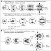
Figure 1
Figure 1
Relationship between astral position and furrow assembly in embryonic and somatic cells. (A) Top: A sand dollar zygote was manipulated into a toroidal shape by pressing with a blunt needle (Rappaport, 1961). Cytokinesis produced a tubular binucleate cell. During the following mitosis, this single cell contained two mitotic spindles arranged in tandem. During the subsequent cytokinesis, furrows formed above each spindle midzone. An additional ectopic furrow formed between the two adjacent asters that were oriented “back-to-back” and not connected by a spindle midzone (hollow arrow). Bottom: Cytokinesis was reversed in a fertilized sea urchin egg by the application of hydrostatic pressure (Salmon and Wolniak, 1990). The egg subsequently traversed interphase as a dikaryon, which formed two independent spindles during the next mitosis. After completion of anaphase, two orthogonal cleavage furrows formed. In this case, an ectopic furrow formed between two pairs of adjacent asters that were oriented “side-by-side” and not connected by a spindle midzone (hollow arrows). The outcome in both cases was cleavage giving rise to four diploid progeny cells. (B) Two adjacent mitotic U2OS cells were punctured with a microneedle at the point of contact. These cells fused to form a heterokaryon containing two mitotic spindles that fused to make a V-shaped unit with a single bent metaphase plate. These cells initiated anaphase normally with the chromatids segregating into three masses. At cytokinesis, one furrow always formed across the middle of the V above the position where the metaphase plate had been (solid arrows). In most cells only this furrow formed; however, in four cells a second furrow formed between the adjacent asters at the open end of the V. Thus, although furrows can form between asters that are oriented “side-by-side” and not connected by a spindle midzone, this is not the most common outcome in these cells.
Figure 2
Figure 2
Adjacent asters usually do not direct cleavage furrow formation in somatic heterokaryons. Neighboring metaphase U2OS cells were microinjected at their point of contact and allowed to fuse, as depicted in Fig. 1. The heterokaryons were then observed as they progressed through the subsequent mitosis. (0 min) Chromosomes congress to a bent metaphase plate. (12, 20 min) Anaphase initiates with the metaphase plate resolving into three separate masses of chromatids. (16, 24 min) Separation of the chromatids is completed. A phase-lucent zone is visible along the axis of chromosome separation for both heterokaryons. An additional phase-lucent zone developed in 4 of 19 heterokaryons (right; 22, 24 min, white arrow). (18 min; left) In 15 of 19 cells followed, cytokinesis involved a single furrow over the spindle midzones at the location of the bent metaphase plate. (24, 26 min; right) In four cells, cytokinesis involved a furrow at the location of the bent metaphase plate plus an ectopic furrow located between the two distal spindle poles (white arrow).
Figure 2
Figure 2
Adjacent asters usually do not direct cleavage furrow formation in somatic heterokaryons. Neighboring metaphase U2OS cells were microinjected at their point of contact and allowed to fuse, as depicted in Fig. 1. The heterokaryons were then observed as they progressed through the subsequent mitosis. (0 min) Chromosomes congress to a bent metaphase plate. (12, 20 min) Anaphase initiates with the metaphase plate resolving into three separate masses of chromatids. (16, 24 min) Separation of the chromatids is completed. A phase-lucent zone is visible along the axis of chromosome separation for both heterokaryons. An additional phase-lucent zone developed in 4 of 19 heterokaryons (right; 22, 24 min, white arrow). (18 min; left) In 15 of 19 cells followed, cytokinesis involved a single furrow over the spindle midzones at the location of the bent metaphase plate. (24, 26 min; right) In four cells, cytokinesis involved a furrow at the location of the bent metaphase plate plus an ectopic furrow located between the two distal spindle poles (white arrow).
Figure 3
Figure 3
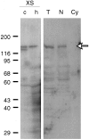
A single human INCENP is detected in purified chromosomes and cell nuclei. (Lanes c and h) Purified chromosomes from DU249 cells (chicken; lane c) or U2OS cells (human; lane h) were resolved on a 7.5% polyacrylamide gel before transfer to nitrocellulose. (Lanes T, N, and Cy) Human U2OS cells were lysed in SDS sample buffer and total protein was resolved on a 7.5% polyacrylamide gel (lane T) followed by blotting to nitrocellulose. Cells were fractionated by Dounce homogenization into nuclei (lane N) and cytoplasm (lane Cy) before gel electrophoresis and transfer to nitrocellulose. Blots were probed with cross-reactive polyclonal antiserum raised against recombinant chicken INCENP and detected using 125I-labeled protein A. The positions of molecular weight standards on Ponceau S–stained membranes are indicated, as is the position of the major human immunoreactive band (arrow).
Figure 4
Figure 4
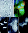
INCENP distribution correlates with sites of furrowing in a mitotic heterokaryon undergoing a triradial mitosis without an ectopic furrow. Human U2OS cells were fused by needle puncture, allowed to progress through mitosis until the commencement of furrowing, and then fixed and stained to localize INCENP. (A) Phase contrast image of the fixed cell. (B) Simultaneous localization of INCENP (red), tubulin (green), and the chromosomes (blue). (C) The separated sister chromatids. Arrows indicate the direction of chromatid movement. (D) The V-shaped spindle. Arrows indicate the axis of cleavage. (E) Localization of INCENP, with staining limited to the region above the original spindle midzone.
Figure 5
Figure 5
INCENP distribution correlates with sites of furrowing in mitotic heterokaryon undergoing a triradial mitosis with an ectopic furrow. (A) Phase contrast image of the fixed cell. This cell shows three sites of furrowing. These are numbered in order of the degree of microtubule bundling (1 = strongest, 3 = weakest). (B) Simultaneous localization of INCENP (red), tubulin (green), and the chromosomes (blue). (C) The separated sister chromatids. Arrows indicate the direction of chromatid movement. (D) The distribution of microtubules. (E) Localization of INCENP. All furrows have associated INCENP staining.
Figure 6
Figure 6
INCENP accumulates on organized spindle midzone microtubules but not on overlapping astral microtubules. Untreated U2OS cells with three masses of chromatids were fixed and stained for DNA, tubulin, and INCENP. (A) An anaphase cell with three masses of chromatids. (B) Microtubules formed a large and well-organized spindle midzone. (C) INCENP concentrated at the spindle midzone. (D) A telophase cell with three reforming nuclei. (E) Spindle midzone microtubules are collapsed under the advancing cleavage furrow into a single midbody structure. In the upper portion of the cell, microtubules are overlapping and unorganized, and no additional cleavage furrow is seen. (F) INCENP concentrated at the midbody but not on the disorganized microtubules at the top.
Figure 6
Figure 6
INCENP accumulates on organized spindle midzone microtubules but not on overlapping astral microtubules. Untreated U2OS cells with three masses of chromatids were fixed and stained for DNA, tubulin, and INCENP. (A) An anaphase cell with three masses of chromatids. (B) Microtubules formed a large and well-organized spindle midzone. (C) INCENP concentrated at the spindle midzone. (D) A telophase cell with three reforming nuclei. (E) Spindle midzone microtubules are collapsed under the advancing cleavage furrow into a single midbody structure. In the upper portion of the cell, microtubules are overlapping and unorganized, and no additional cleavage furrow is seen. (F) INCENP concentrated at the midbody but not on the disorganized microtubules at the top.
Figure 7
Figure 7
INCENP localizes to the cleavage furrow before cytoplasmic myosin in early anaphase cells. Mitotic cells were fixed and stained for INCENP, myosin, and DNA. Separate stages of anaphase were ordered by cell shape, the distance between the separating sister chromatids, and the relative distribution of INCENP and myosin. The stages are defined as follows: STAGE I, round cell with <2-μm separation between sister chromatids and with neither INCENP nor myosin at the equatorial cortex; STAGE II, round cell with 2–4-μm separation between sister chromatids. INCENP is concentrated at the equatorial cortex while myosin is still diffuse or in patches; STAGE III, oval cell with 3–5-μm separation between sister chromatids. Both INCENP and myosin are concentrated in the equatorial cortex; STAGE IV, elongated, furrowing cell with >4-μm separation between sister chromatids. Both INCENP and myosin are concentrated in the advancing furrow. Bar, 5 μm.
Figure 8
Figure 8
Radixin concentrates at the equatorial cortex only when noticeable furrowing has occurred. Mitotic cells were fixed and stained for INCENP, radixin, and DNA. Separate stages of anaphase were ordered by cell shape and the distance between separating sister chromatids, as described in the legend to Fig. 7. INCENP first concentrates at the equatorial cortex in stage II cells. In contrast, radixin is found in patches throughout the cortex of the cell in stages I–III and does not concentrate at the equatorial cortex until furrowing is well established (STAGE IV). Bar, 5 μm.
Figure 9
Figure 9
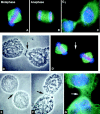
A dominant negative INCENP mutant disrupts the completion of cytokinesis. (A–C) Association of CENP-B1–158:INCENP43–839 with centromeres during mitosis. In these images of transfected cells, taken 24 h after transfection, CENP-B1–158:INCENP43–839 is red, DNA is blue, and microtubules are green. The chimeric protein remains associated with centromeres at all stages of the mitotic cycle. (D and D′) Paired metaphases observed 48 h after transfection. In the phase contrast image (D), two cells are seen to be joined by a prominent intracellular bridge with a midbody (arrow). In the immunofluorescence image (D′), both cells are seen to be in metaphase of the next cell cycle. Neither tubulin nor INCENPs are detected in the midbody. (E and F) Two further examples of paired metaphase cells joined by prominent intercellular bridges with midbodies. (G) Dikaryon with a prominent intercellular bridge containing an interphase array of microtubules.

Similar articles

Cited by

References

    1. Andreassen PR, Palmer DK, Wener MH, Margolis RL. Telophase disk: a new mammalian mitotic organelle that bisects telophase cells with a possible function in cytokinesis. J Cell Sci. 1991;99:523–534. - PubMed
    1. Aubin JE, Osborn M, Weber K. Inhibition of cytokinesis and altered contractile ring morphology induced by cytochalasins in synchronized PtK2 cells. Exp Cell Res. 1981;136:63–79. - PubMed
    1. Ausubel, F.M., R. Brent, R.E. Kingston, D.D. Moore, J.G. Seidman, J.A. Smith, and K. Struhl. 1991. Current Protocols in Molecular Biology. John Wiley & Sons, New York.
    1. Bray D, White JG. Cortical flow in animal cells. Science (Wash DC) 1988;239:883–888. - PubMed
    1. Buck RC, Tisdale JM. The fine structure of the mid-body of the rat erythroblast. J Cell Biol. 1962;13:109–115. - PMC - PubMed

Publication types