The HIV coreceptors CXCR4 and CCR5 are differentially expressed and regulated on human T lymphocytes
- PMID: 9050881
- PMCID: PMC20019
- DOI: 10.1073/pnas.94.5.1925
The HIV coreceptors CXCR4 and CCR5 are differentially expressed and regulated on human T lymphocytes
Abstract
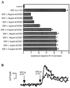
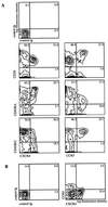
The chemokine receptors CXCR4 and CCR5 function as coreceptors for HIV-1 entry into CD4+ cells. During the early stages of HIV infection, viral isolates tend to use CCR5 for viral entry, while later isolates tend to use CXCR4. The pattern of expression of these chemokine receptors on T cell subsets and their regulation has important implications for AIDS pathogenesis and lymphocyte recirculation. A mAb to CXCR4, 12G5, showed partial inhibition of chemotaxis and calcium influx induced by SDF-1, the natural ligand of CXCR4. 12G5 stained predominantly the naive, unactivated CD26(low) CD45RA+ CD45R0- T lymphocyte subset of peripheral blood lymphocytes. In contrast, a mAb specific for CCR5, 5C7, stained CD26(high) CD45RA(low) CD45R0+ T lymphocytes, a subset thought to represent previously activated/memory cells. CXCR4 expression was rapidly up-regulated on peripheral blood mononuclear cells during phytohemagglutinin stimulation and interleukin 2 priming, and responsiveness to SDF-1 increased simultaneously. CCR5 expression, however, showed only a gradual increase over 12 days of culture with interleukin 2, while T cell activation with phytohemagglutinin was ineffective. Taken together, the data suggest distinct functions for the two receptors and their ligands in the migration of lymphocyte subsets through lymphoid and nonlymphoid tissues. Furthermore, the largely reciprocal expression of CXCR4 and CCR5 among peripheral blood T cells implies distinct susceptibility of T cell subsets to viral entry by T cell line-tropic versus macrophage-tropic strains during the course of HIV infection.
Figures


Comment in
-
Expression pattern of HIV-1 coreceptors on T cells: implications for viral transmission and lymphocyte homing.Proc Natl Acad Sci U S A. 1997 Mar 4;94(5):1615-8. doi: 10.1073/pnas.94.5.1615. Proc Natl Acad Sci U S A. 1997. PMID: 9050826 Free PMC article. No abstract available.
Similar articles
-
CXCR4 and CCR5 on human thymocytes: biological function and role in HIV-1 infection.J Immunol. 1998 Sep 15;161(6):3103-13. J Immunol. 1998. PMID: 9743377
-
The lymphocyte chemoattractant SDF-1 is a ligand for LESTR/fusin and blocks HIV-1 entry.Nature. 1996 Aug 29;382(6594):829-33. doi: 10.1038/382829a0. Nature. 1996. PMID: 8752280
-
Expression pattern of HIV-1 coreceptors on T cells: implications for viral transmission and lymphocyte homing.Proc Natl Acad Sci U S A. 1997 Mar 4;94(5):1615-8. doi: 10.1073/pnas.94.5.1615. Proc Natl Acad Sci U S A. 1997. PMID: 9050826 Free PMC article. No abstract available.
-
[Deep lung--cellular reaction to HIV].Rev Port Pneumol. 2007 Mar-Apr;13(2):175-212. Rev Port Pneumol. 2007. PMID: 17492233 Review. Portuguese.
-
Chemokines as natural HIV antagonists.Curr Mol Med. 2002 Dec;2(8):691-702. doi: 10.2174/1566524023361862. Curr Mol Med. 2002. PMID: 12462390 Review.
Cited by
-
Effects of HIV-1 genotype on baseline CD4+ cell count and mortality before and after antiretroviral therapy.Sci Rep. 2020 Sep 28;10(1):15875. doi: 10.1038/s41598-020-72701-4. Sci Rep. 2020. PMID: 32985559 Free PMC article.
-
HIV-MTB Co-Infection Reduces CD4+ T Cells and Affects Granuloma Integrity.Viruses. 2024 Aug 21;16(8):1335. doi: 10.3390/v16081335. Viruses. 2024. PMID: 39205309 Free PMC article.
-
Immunophenotypic alterations in acute and early HIV infection.Clin Immunol. 2007 Dec;125(3):299-308. doi: 10.1016/j.clim.2007.08.011. Epub 2007 Oct 3. Clin Immunol. 2007. PMID: 17916441 Free PMC article. Clinical Trial.
-
CXCR4-Using HIV Strains Predominate in Naive and Central Memory CD4+ T Cells in People Living with HIV on Antiretroviral Therapy: Implications for How Latency Is Established and Maintained.J Virol. 2020 Feb 28;94(6):e01736-19. doi: 10.1128/JVI.01736-19. Print 2020 Feb 28. J Virol. 2020. PMID: 31852784 Free PMC article.
-
Exosomal transmission of viruses, a two-edged biological sword.Cell Commun Signal. 2023 Jan 23;21(1):19. doi: 10.1186/s12964-022-01037-5. Cell Commun Signal. 2023. PMID: 36691072 Free PMC article. Review.
References
Publication types
MeSH terms
Substances
Grants and funding
LinkOut - more resources
Full Text Sources
Other Literature Sources
Research Materials
Miscellaneous

