

Intestinal tumorigenesis is suppressed in mice lacking the metalloproteinase matrilysin
- PMID: 9037065
- PMCID: PMC19803
- DOI: 10.1073/pnas.94.4.1402
Intestinal tumorigenesis is suppressed in mice lacking the metalloproteinase matrilysin
Abstract
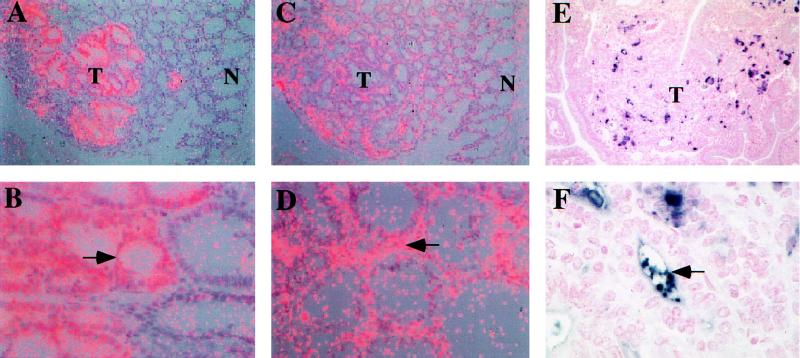
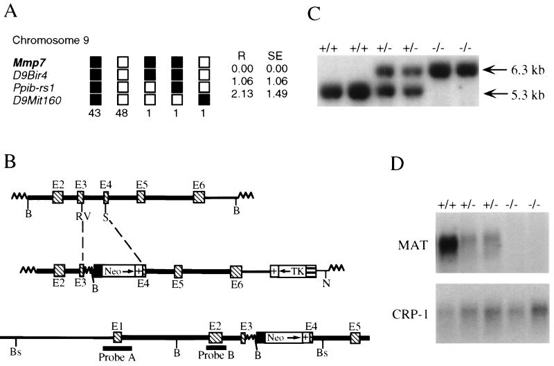
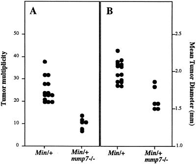
Matrix metalloproteinases (MMPs) classically have been implicated in basement membrane destruction associated with late-stage tumor cell invasion and metastasis. However, recent studies have demonstrated that one MMP family member, matrilysin, is expressed in a high percentage of early-stage human colorectal tumors. We analyzed matrilysin expression in benign intestinal tumors from mice heterozygous for the ApcMin allele (Min/+) and found that the mRNA was induced in the majority (88%) of these adenomas. Protein was detected in the tumor cells, where, surprisingly, it was predominantly immunolocalized to the lumenal surface of dysplastic glands rather than the basement membrane or extracellular matrix. To address the role of matrilysin in Min intestinal tumorigenesis, we generated Min/+ mice deficient in this MMP by gene targeting and homologous recombination. The absence of matrilysin resulted in a reduction in mean tumor multiplicity in Min/+ animals of approximately 60% and a significant decrease in the average tumor diameter. Based on these findings, we conclude that matrilysin is a suppressor of the Min phenotype, possibly by functioning in a capacity independent of matrix degradation. These results argue for the use of MMP inhibitors in the treatment and prevention of early-stage colon cancer.
Figures
Similar articles
-
Differential expression of matrilysin and cyclooxygenase-2 in intestinal and colorectal neoplasms.Mol Carcinog. 1999 Mar;24(3):177-87. Mol Carcinog. 1999. PMID: 10204802
-
Matrilysin gene expression in sporadic and familial colorectal adenomas.Mol Carcinog. 1997 Aug;19(4):225-9. Mol Carcinog. 1997. PMID: 9290698
-
The metalloproteinase matrilysin is a target of beta-catenin transactivation in intestinal tumors.Oncogene. 1999 May 6;18(18):2883-91. doi: 10.1038/sj.onc.1202627. Oncogene. 1999. PMID: 10362259
-
[Genetic diagnosis of colorectal cancer].Hokkaido Igaku Zasshi. 1996 Jan;71(1):9-14. Hokkaido Igaku Zasshi. 1996. PMID: 8727369 Review. Japanese.
-
Matrix-degrading metalloproteinases in tumor progression.Princess Takamatsu Symp. 1994;24:152-61. Princess Takamatsu Symp. 1994. PMID: 8983072 Review.
Cited by
-
Molecular Basis of Acute Cystitis Reveals Susceptibility Genes and Immunotherapeutic Targets.PLoS Pathog. 2016 Oct 12;12(10):e1005848. doi: 10.1371/journal.ppat.1005848. eCollection 2016 Oct. PLoS Pathog. 2016. PMID: 27732661 Free PMC article.
-
Resistance of young gelatinase B-deficient mice to experimental autoimmune encephalomyelitis and necrotizing tail lesions.J Clin Invest. 1999 Dec;104(11):1507-15. doi: 10.1172/JCI6886. J Clin Invest. 1999. PMID: 10587514 Free PMC article.
-
Severe polyposis in Apc(1322T) mice is associated with submaximal Wnt signalling and increased expression of the stem cell marker Lgr5.Gut. 2010 Dec;59(12):1680-6. doi: 10.1136/gut.2009.193680. Epub 2010 Oct 6. Gut. 2010. PMID: 20926645 Free PMC article.
-
Matrilysin (matrix metalloproteinase-7) mediates E-cadherin ectodomain shedding in injured lung epithelium.Am J Pathol. 2003 Jun;162(6):1831-43. doi: 10.1016/S0002-9440(10)64318-0. Am J Pathol. 2003. PMID: 12759241 Free PMC article.
-
The adenomatous polyposis coli-associated exchange factors Asef and Asef2 are required for adenoma formation in Apc(Min/+)mice.EMBO Rep. 2009 Dec;10(12):1355-62. doi: 10.1038/embor.2009.233. Epub 2009 Nov 6. EMBO Rep. 2009. PMID: 19893577 Free PMC article.
References
-
- MacDougall J R, Matrisian L M. Cancer Metastasis Rev. 1995;14:351–362. - PubMed
-
- Powell W C, Matrisian L M. In: Attempts to Understand Metastasis Formation: I. Metastasis-Related Molecules. Gunthert W, Birchmeier W, editors. Heidelberg: Springer; 1996. pp. 1–22.
-
- Wilson C L, Matrisian L M. Int J Biochem Cell Biol. 1996;28:123–136. - PubMed
-
- Saarialho-Kere U K, Crouch E C, Parks W C. J Invest Dermatol. 1995;105:190–196. - PubMed
Publication types
MeSH terms
Substances
Grants and funding
LinkOut - more resources
Full Text Sources
Other Literature Sources
Molecular Biology Databases
Research Materials
