

Extracellular fibrillar structure of latent TGF beta binding protein-1: role in TGF beta-dependent endothelial-mesenchymal transformation during endocardial cushion tissue formation in mouse embryonic heart
- PMID: 9008713
- PMCID: PMC2132455
- DOI: 10.1083/jcb.136.1.193
Extracellular fibrillar structure of latent TGF beta binding protein-1: role in TGF beta-dependent endothelial-mesenchymal transformation during endocardial cushion tissue formation in mouse embryonic heart
Abstract
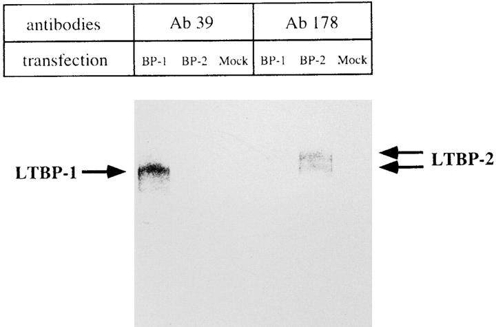
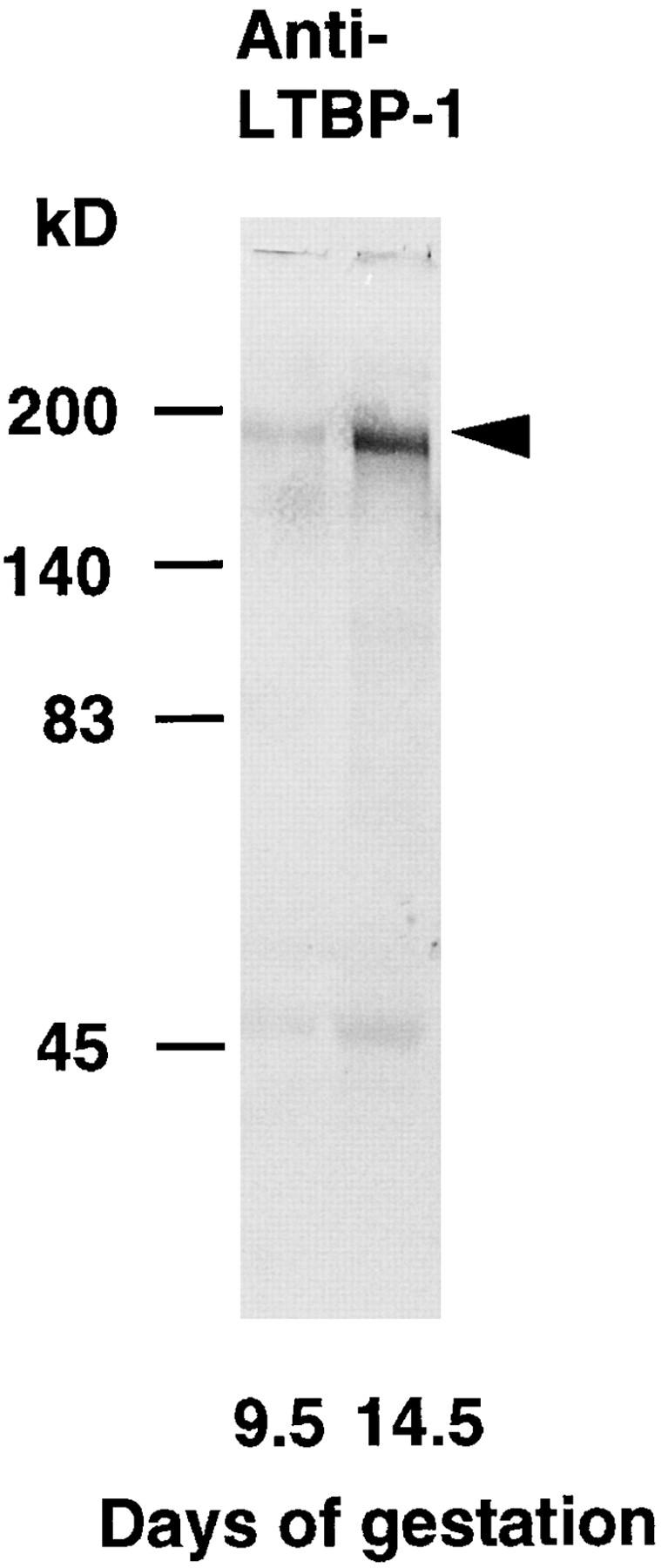
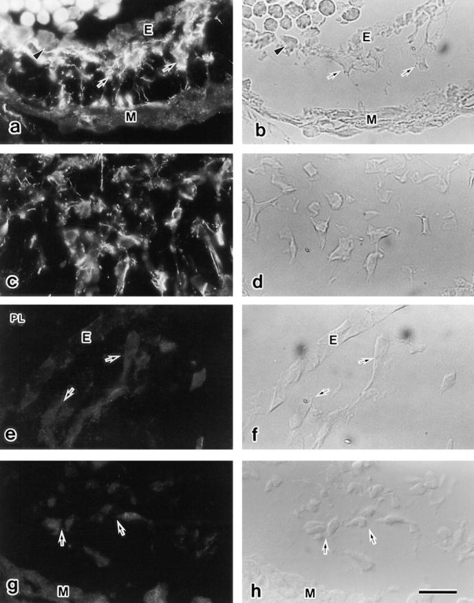
Transforming growth factor-beta (TGF beta) is a dimeric peptide growth factor which regulates cellular differentiation and proliferation during development. Most cells secrete TGF beta as a large latent TGF beta complex containing mature TGF beta, latency associated peptide, and latent TGF beta-binding protein (LTBP)-1. The biological role of LTBP-1 in development remains unclear. Using a polyclonal antiserum specific for LTBP-1 (Ab39) and three-dimensional collagen gel culture assay of embryonic heart, we examined the tissue distribution of LTBP-1 and its functional role during the formation of endocardial cushion tissue in the mouse embryonic heart. Mature TGF beta protein was required at the onset of the endothelial-mesenchymal transformation to initiate endocardial cushion tissue formation. Double antibody staining showed that LTBP-1 colocalized with TGF beta 1 as an extracellular fibrillar structure surrounding the endocardial cushion mesenchymal cells. Immunogold electronmicroscopy showed that LTBP-1 localized to 40-100 nm extracellular fibrillar structure and 5-10-nm microfibrils. The anti-LTBP-1 antiserum (Ab39) inhibited the endothelial-mesenchymal transformation in atrio-ventricular endocardial cells cocultured with associated myocardium on a three-dimensional collagen gel lattice. This inhibitory effect was reversed by administration of mature TGF beta proteins in culture. These results suggest that LTBP-1 exists as an extracellular fibrillar structure and plays a role in the storage of TGF beta as a large latent TGF beta complex.
Figures


Similar articles
-
Latent transforming growth factor-beta 1 and its binding protein are components of extracellular matrix microfibrils.J Histochem Cytochem. 1996 Aug;44(8):875-89. doi: 10.1177/44.8.8756760. J Histochem Cytochem. 1996. PMID: 8756760
-
The latent transforming growth factor-beta-binding protein-1 promotes in vitro differentiation of embryonic stem cells into endothelium.Mol Biol Cell. 2000 Dec;11(12):4295-308. doi: 10.1091/mbc.11.12.4295. Mol Biol Cell. 2000. PMID: 11102524 Free PMC article.
-
Dual role for the latent transforming growth factor-beta binding protein in storage of latent TGF-beta in the extracellular matrix and as a structural matrix protein.J Cell Biol. 1995 Oct;131(2):539-49. doi: 10.1083/jcb.131.2.539. J Cell Biol. 1995. PMID: 7593177 Free PMC article.
-
Latent transforming growth factor-beta binding proteins (LTBPs)--structural extracellular matrix proteins for targeting TGF-beta action.Cytokine Growth Factor Rev. 1999 Jun;10(2):99-117. doi: 10.1016/s1359-6101(99)00010-6. Cytokine Growth Factor Rev. 1999. PMID: 10743502 Review.
-
Mechanisms involved in valvuloseptal endocardial cushion formation in early cardiogenesis: roles of transforming growth factor (TGF)-beta and bone morphogenetic protein (BMP).Anat Rec. 2000 Feb 1;258(2):119-27. doi: 10.1002/(SICI)1097-0185(20000201)258:2<119::AID-AR1>3.0.CO;2-U. Anat Rec. 2000. PMID: 10645959 Review.
Cited by
-
Loss of muscleblind-like 1 promotes invasive mesenchyme formation in endocardial cushions by stimulating autocrine TGFβ3.BMC Dev Biol. 2012 Aug 6;12:22. doi: 10.1186/1471-213X-12-22. BMC Dev Biol. 2012. PMID: 22866814 Free PMC article.
-
Latent transforming growth factor-beta-binding protein-4 regulates transforming growth factor-beta1 bioavailability for activation by fibrogenic lung fibroblasts in response to bleomycin.Am J Pathol. 2009 Jan;174(1):21-33. doi: 10.2353/ajpath.2009.080620. Epub 2008 Dec 4. Am J Pathol. 2009. PMID: 19056849 Free PMC article.
-
Isoform-specific effects of transforming growth factor β on endothelial-to-mesenchymal transition.J Cell Physiol. 2018 Nov;233(11):8418-8428. doi: 10.1002/jcp.26801. Epub 2018 Jun 1. J Cell Physiol. 2018. PMID: 29856065 Free PMC article.
-
Noonan syndrome cardiac defects are caused by PTPN11 acting in endocardium to enhance endocardial-mesenchymal transformation.Proc Natl Acad Sci U S A. 2009 Mar 24;106(12):4736-41. doi: 10.1073/pnas.0810053106. Epub 2009 Feb 27. Proc Natl Acad Sci U S A. 2009. PMID: 19251646 Free PMC article.
-
Transforming growth factor-beta signaling in epithelial-mesenchymal transition and progression of cancer.Proc Jpn Acad Ser B Phys Biol Sci. 2009;85(8):314-23. doi: 10.2183/pjab.85.314. Proc Jpn Acad Ser B Phys Biol Sci. 2009. PMID: 19838011 Free PMC article. Review.
References
-
- Akhurst, R.J. 1994. The transforming growth factor β family in vertebrate embryogenesis. In Growth Factors and Signal Transduction in Development. M. Nilsen-Hamilton, editor. Wiley-Liss, Inc. New York. pp. 97–122.
-
- Akhurst RJ, Lehnert SA, Faissner A, Duffie E. TGF β in murine morphogenetic processes: the early embryo and cardiogenesis. Development. 1990;108:645–656. - PubMed
-
- Appella E, Weber IT, Blasi F. Structure and function of epidermal growth factor-like regions in proteins. FEBS Lett. 1988;231:1–4. - PubMed
-
- Barnard JA, Lyons RM, Moses HL. The cell biology of transforming growth factor β. Biochem Biophys Acta. 1990;1032:79–87. - PubMed
-
- Bernanke DH, Markwald RR. Migratory behavior of cardiac cushion tissue cells in a collagen-lattice culture system. Dev Biol. 1982;91:235–245. - PubMed
Publication types
MeSH terms
Substances
LinkOut - more resources
Full Text Sources
Molecular Biology Databases
Research Materials
