. 1997 Feb 1;17(3):904-16.
doi: 10.1523/JNEUROSCI.17-03-00904.1997.

Kinetics, Ca2+ dependence, and biophysical properties of integrin-mediated mechanical modulation of transmitter release from frog motor nerve terminals

Collaborators, Affiliations

Kinetics, Ca2+ dependence, and biophysical properties of integrin-mediated mechanical modulation of transmitter release from frog motor nerve terminals

B M Chen et al. J Neurosci. .

Abstract

Neurotransmitter release from frog motor nerve terminals is strongly modulated by change in muscle length. Over the physiological range, there is an approximately 10% increase in spontaneous and evoked release per 1% muscle stretch. Because many muscle fibers do not receive suprathreshold synaptic inputs at rest length, this stretch-induced enhancement of release constitutes a strong peripheral amplifier of the spinal stretch reflex. The stretch modulation of release is inhibited by peptides that block integrin binding of natural ligands. The modulation varies linearly with length, with a delay of no more than approximately 1-2 msec and is maintained constant at the new length. Moreover, the stretch modulation persists in a zero Ca2+ Ringer and, hence, is not dependent on Ca2+ influx through stretch activated channels. Eliminating transmembrane Ca2+ gradients and buffering intraterminal Ca2+ to approximately normal resting levels does not eliminate the modulation, suggesting that it is not the result of release of Ca2+ from internal stores. Finally, changes in temperature have no detectable effect on the kinetics of stretch-induced changes in endplate potential (EPP) amplitude or miniature EPP (mEPP) frequency. We conclude, therefore, that stretch does not act via second messenger pathways or a chemical modification of molecules involved in the release pathway. Instead, there is direct mechanical modulation of release. We postulate that tension on integrins in the presynaptic membrane is transduced mechanically into changes in the position or conformation of one or more molecules involved in neurotransmitter release, altering sensitivity to Ca2+ or the equilibrium for a critical reaction leading to vesicle fusion.


Figures
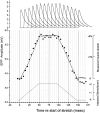
Fig. 4.
Fig. 4.
Kinetics of stretch enhancement of EPP amplitude in a representative sartorius junction in low Ca2+ Ringer. The muscle was attached at either end to a pair of arms that moved simultaneously in opposite directions in response to a command signal from the computer, beginning to lengthen at 0 msec, reaching 2 mm (6%) stretch at 50 msec, holding that length for 45 msec, and shortening in another 50 msec. The actual measured change in length of the muscle (noisy solid line) and corresponding EPP amplitudes at 36 time points during the cycle (filled circles) are shown in the middle records, and sample EPPs taken at different times are shown at the top. Each point is the average of 50 or more EPPs, each evoked in a single lengthening–shortening cycle.
Fig. 5.
Fig. 5.
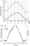
Kinetics of stretch enhancement of EPP amplitude. Average of six experiments like that shown in Figure 4. Note the relatively large arrow bars at the peak of the lengthening phase and the beginning of shortening. Muscle length is shown by the noisy line, where the noise arose primarily in the light source used to measure length (see Materials and Methods). Except at the beginning of the shortening phase, there was virtually no delay between change in muscle length and change in release efficacy.
Fig. 7.
Fig. 7.
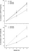
Suppression of the stretch enhancement by RGD peptides. The graphs show that enhancement of mEPP frequency and EPP amplitude was approximately linear with length and that 0.2 mm RGD, but not the inactive control RGE, suppressed the effect. EPPs were recorded in 3–6 μm curare.
Fig. 8.
Fig. 8.
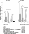
Effects of manipulations affecting Ca2+ levels on the magnitude of the increase in mEPP frequency with stretch. Zero Ca2+ Ringer reduced the enhancement by ∼50% (column 2), but the enhancement was still highly significant. Mn2+ (0.5 mm) restored much of the enhancement (column 3), which was then essentially totally blocked by 0.2 mm RGD, applied both before and after addition of Mn2+ (column 4). Loading of terminals with a strong Ca2+buffer by exposure to DM-BAPTA-AM in zero Ca2+ Ringer strongly suppressed the enhancement (column 5), but again 0.5 mm Mn2+ mostly restored it (column 6); 0.2 mm RGD, applied both in the zero Ca2+ Ringer and during subsequent addition of Mn2+, totally eliminated the enhancement (column 7). These data show that Ca2+ influx is not necessary for the enhancement and that the enhancement can be totally eliminated by agents that interfere with integrin binding. Absolute mEPP frequencies at rest length are also indicated for each condition, and asterisks indicate the probability of the indicated matches (*p < 0.05, **p < 0.005).
Fig. 1.
Fig. 1.
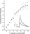
Average change in EPP amplitude as a function of stretch of the sartorius muscle. Mean ± SE for six junctions.Inset, EPPs obtained from a sensitive preparation at 95, 100, 110, and 120% of rest length. All in low Ca2+ Ringer containing 0.56 mm Ca2+ and 4 mmMg2+.
Fig. 2.
Fig. 2.
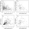
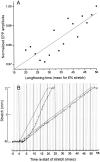
Variability and sensitivity of different junctions to 6% muscle stretch. A1 andA2, scattergraphs showing percentage increase in mEPP frequency and EPP amplitude for different junctions, plotted against their release at rest length. The vast majority of junctions showed an increase in release with stretch, but it varied between no change and a two- to threefold increase. The dashed lines represent linear best fits. B1 andB2, absolute values of change in mEPP frequency and EPP amplitude in the same junctions. Note that there was little or no correlation between degree of enhancement and resting release levels in the stronger junctions. EPP recordings were in 3 μmcurare.
Fig. 3.
Fig. 3.
Effects of stretch on response in NFR.A, intracellular records from an unblocked junction in which stretch of the muscle elicited only an EPP at rest length but a full action potential after 1 mm (3%) muscle stretch.B, Records of twitch tension at rest length and after 1 and 2 mm (6%) stretch in a 30 mm sartorius in response to nerve stimulation (left) and direct muscle stimulation (right). The increase in tension with stretch in the directly stimulated muscle is predicted from the length–tension relationship of individual fibers. The much greater enhancement with indirect stimulation reflects the recruitment of large populations of fibers that were not excited above threshold at rest length. That indirect stimulation produced a larger twitch than direct stimulation at 6% stretch suggests that the direct stimulus did not evoke action potentials in all fibers in the muscle.
Fig. 6.
Fig. 6.
A, EPP amplitudes in low Ca2+ Ringer evoked by stimulation at the moment of reaching the 2 mm stretch command voltage, when the stretch was accomplished at different rates between 2 mm/20 msec and 2 mm/50 msec. The enhancement after the most rapid lengthening (20 msec) was ∼3% less than after a lengthening that took 50 msec. B, Optical measurement of muscle length during 2 mm stretches in 20 and 50 msec. The greater lag between command voltage and actual muscle stretch at 2 mm/20 msec can explain the slightly reduced enhancement. The muscle length was 30 mm, hence 2 mm represented 6% stretch.
Fig. 9.
Fig. 9.
Stretch enhancement of release with 3 and 6% stretch in preparations in which [Ca2+]iwas “clamped” at ∼100 nm by loading the terminal with BAPTA, disabling the main intrinsic Ca2+ buffers with 10 μm thapsigargin, and reducing or eliminating the electrochemical gradient for Ca2+ across the terminal membrane by immersion in a Ringer containing 1 nmCa2+ and 2 μm of the Ca2+ionophore A23187. Under these conditions, stretch enhancement of release was still about one-half that found in NFR and very close to that obtained in zero Ca2+ Ringer (see Fig. 8).
Fig. 10.
Fig. 10.
Effects of change in temperature on stretch enhancement of the EPP in low Ca2+ Ringer.A, Average of records from five lengthening–shortening cycles at a single representative junction at 22.2° and at 13°, showing the change in absolute EPP amplitude with stretch and shortening [using a stretching regime similar to that of Figs. 4 and 5but with stretch maintained at +2 mm (6%) for only 25 msec]. The absolute amplitude was much decreased at 13°, but the percentage change with stretch was actually increased at the lower temperature.B, Normalized plots of the lengthening and shortening phases at the two temperatures. The kinetics of the change in amplitude with change in length were essentially unaffected by temperature in this range.
Fig. 11.
Fig. 11.
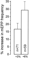
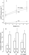
Effects of temperature on sensitivity of mEPP frequency to stretch. A, mEPP frequency as a function of muscle length at 10° and 23° in NFR, and at 10° in NFR plus 10 mm K+. B, Percentage change in mEPP frequency at 3 and 6% mm stretch under each of these conditions. Cooling reduced the resting mEPP frequency but had little effect on the enhancement by stretch. When the resting mEPP frequency was increased by K+ depolarization to approximately the level in NFR at 23°, the effect of stretch was indistinguishable from that at 23°C.
Fig. 12.
Fig. 12.
A schematic model suggesting, generically, how extracellularly applied tension on integrins might increase the probability of vesicle fusion. The fusion process is pictured as resulting when a docked vesicle (V) is allowed to be pulled into contact with the plasma membrane (P) at a specific site. This process is normally triggered by Ca2+interaction with a Ca2+-sensing molecule that can, through an unknown number of steps, activate the fusion process, perhaps by displacing a molecule (B) that physically blocks contact of the two membranes. Tension on integrins (I) might alter the Ca2+ binding affinity of the Ca2+ sensor (1) or physically exert force contributing to the removal of the block of fusion (2a) or pulling of the vesicle down toward fusion-promoting molecules in the plasma membrane (2b). Another interpretation of alternative (2b) might be an increased probability of vesicle–plasma membrane fusion, the result somehow of a direct effect of mechanical stress on the conformation of the plasma membrane lipid. RGD inhibits the modulation by blocking integrin binding to ligands in the ECM.

Similar articles

Cited by

References

    1. Adams BA. Temperature and synaptic efficacy in frog skeletal muscle. J Physiol (Lond) 1989;408:443–455. - PMC - PubMed
    1. Andreu R, Barrett EF. Calcium dependence of evoked transmitter release at very low quantal contents at the frog neuromuscular junction. J Physiol (Lond) 1980;308:79–97. - PMC - PubMed
    1. Barrett EF, Magleby KL. Physiology of cholinergic transmission. In: Goldberg AM, Hanin I, editors. Biology of cholinergic function. Raven; New York: 1976. pp. 29–100.
    1. Chen BM, Grinnell AD. Integrins and modulation of transmitter release from motor nerve terminals by stretch. Science. 1995;269:1578–1580. - PubMed
    1. Cruz LJ, Gray WR, Olivera BM, Zeikus RD, Kerr L, Yoshikami D, Moczydlowski E. Conus geographus toxins that discriminate between neuronal and muscle sodium channels. J Biol Chem. 1985;260:9280–9288. - PubMed

LinkOut - more resources