. 1997 Feb 1;17(3):882-90.
doi: 10.1523/JNEUROSCI.17-03-00882.1997.

The seizure locus encodes the Drosophila homolog of the HERG potassium channel

Affiliations

The seizure locus encodes the Drosophila homolog of the HERG potassium channel

X J Wang et al. J Neurosci. .

Abstract

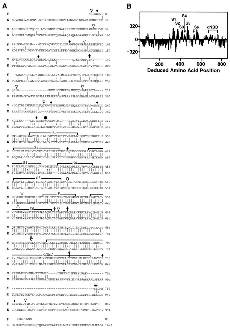
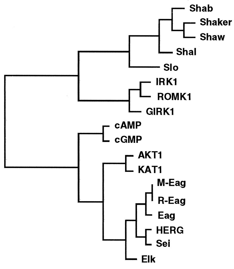
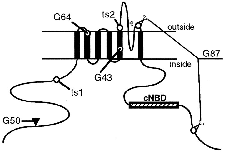
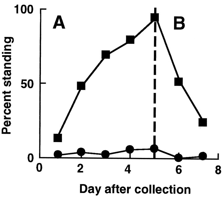
Mutations in the seizure (sei) locus cause temperature-induced hyperactivity, followed by paralysis. Gene cloning studies have established that the seizure gene product is the Drosophila homolog of HERG, a member of the eag family of K+ channels implicated in one form of hereditary long QT syndrome in humans. A series of five null alleles with premature stop codons are all recessive, but viable. A missense mutation in the sei gene, which changes the charge at a conserved glutamate residue near the outer mouth of the pore, has a semidominant phenotype, suggesting that the mutant seizure protein acts as a poison in a multimeric complex. Transformation rescue of a null allele with a cDNA under the control of an inducible promoter demonstrates that induced expression of seizure potassium channels in adults rescues the paralytic phenotype. This rescue decays with a t1/2 of approximately 1-1.5 d after gene induction is discontinued, providing the first estimate of ion channel stability in an intact, multicellular animal.


Figures

Fig. 3.
Fig. 3.
Structural features of the seizure protein.A, The sei (S) amino acid sequence was compared with the HERG (H) potassium channel subunit. The alignment was generated using the GAP program in GCG software (Devereux et al., 1984). Vertical lines connect identical amino acids; two vertical dots represent conservative substitutions; dashes indicate gaps introduced to facilitate alignment. Conserved groups were defined as (M, I, L, V); (A, G); (S, T); (Q, N); (K, R); (E, D); and (F, Y, W). The six hydrophobic domains (S1S6), the pore (P), and the cyclic nucleotide binding domain (cNBD) are overlined. Predicted N-glycosylation sites (Asn 515 in sei, Asn 598 in HERG) are marked with Ψ. The position of the premature stop codon inseits1 is indicated by aclosed circle; the missense mutation inseits2 is indicated by an open circle. Potential phosphorylation sites are indicated as follows: CaM kinase,downward-pointing arrows; protein kinase C, inverted open triangles; protein kinase A,upside down caret; and casein kinase, filled diamonds. The C terminus (1045–1159) of HERG is not shown.B, The hydropathy plot for the deduced sei protein was determined by the method of Kyte and Doolittle (1982). Regionsabove the line are hydrophobic.
Fig. 1.
Fig. 1.
Localization of the seizuregene. A, Deletion mapping. Heterozygotes forseits1 and each deficiency (Df) were tested for temperature-induced paralysis at 38°C. OnlyDf(2R)orBR6and Df(2R)orBR11 failed to complement sei, locating the gene between 60A8 and B10. Deleted regions are indicated as black bars with regions of uncertainty shown as open bars. Other genes shown are eye color genes (bw, or), cloned genes for a muscarinic acetylcholine receptor (mAChR) (Onai et al., 1989), and a sodium channel homolog (DSC1) (Salkoff et al., 1987). The regions deleted are as follows:Df(2R)orBR6, 59D5–D10 to 60B3–B8;Df(2R)orBR11, 59F6–F8 to 60A8–A16; andDf(2R)G10BR27, 59F3 to 60A8–A16 (breakpoints defined in Reed, 1992);Df(2R)bw-S46, 59D8–D11 to 60A7 (Simpson, 1983);Df(2R)bw-D23, 59D4–D5 to 60A1–A2;Df(2R)Px, 60B8–B10 to 60D1–D2; andDf(2R)Px4, 60B8–B10 to 60D1 (breakpoints defined in Lindsley and Zimm, 1992); andDf(2R)spMP, 60B8–B13 to 60D3–D8 (Parisi and Hall, unpublished results). B–D, RFLP mapping and chromosome walking. Twenty recombinants betweenor49h andseits1 were analyzed for six RFLPs that distinguish theor49h chromosome from theseits1 chromosome. The positions of these RFLPs are indicated by an asterisk on the genomic DNA restriction maps (C). H,HpaI; X, XbaI;E, EcoRI; B,BglII; O, XhoI. The number of recombinants with the or49hRFLP is plotted versus RFLP location on genomic DNA inB. The X intercept, determined by linear regression analysis, shows the approximate physical location ofseizure. The small black bar inC shows the region in which genomic DNA aberrations were detected in seizure alleles. The cosmid clones from the chromosome walk are shown in D, relative to the end of deletion Df(2R)G10BR27. The heavy black bar represents the known end of this deletion, whereas the open bar shows a region of uncertainty. The dotted line indicates that the deletion extends to the left beyond the limits of this figure.
Fig. 2.
Fig. 2.
Northern blot of seizure alleles. Poly(A+) RNA (∼10 μg) from wild-type (CS) and sei mutant adult flies was loaded into each lane, as indicated. The blot was probed with aseizure cDNA, including bases +127 to +2668 of the ORF. (Northern blots of wild type probed with single-stranded riboprobes or with the 0.8 kb genomic fragment originally used to probe the cDNA library each identify only the same 3.4 and 3.0 kb transcripts shown in this blot.) Later the blot was reprobed with Drosophilaribosomal protein 49 (rp49) cDNA (O’Connell and Rosbash, 1984) to standardize for mRNA recovery (bottom panel).
Fig. 4.
Fig. 4.
Dendrogram of the potassium channel family. This tree shows the relationship between sei and other potassium channel family members, using the hydrophobic cores for comparison. Similarity is inversely proportional to the horizontal distance of any two sequences from a branch point. The GrowTree program in the Wisconsin Genetics Computer Group (GCG) sequence analysis software (version 8.0) was used to produce this diagram from a distance matrix created by Distances, using UPGMA. The potassium channels (and their GenBank accession numbers) are the Drosophila Shaker family of voltage-gated channels Shab (M32659),Shaker (M17211), Shaw (M32661),Shal (M32660); Drosophilacalcium-activated channel Slo (M96840); inwardly rectifying channels IRK1 (X73052), ROMK1(X72341) GIRK1 (L25264); cyclic nucleotide-gated channels cAMP (X55519) and cGMP (X51604); plant inward rectifiers AKT1 (X62907) andKAT1 (M86990); and eag family of channels mouseM-Eag (U04294), rat R-Eag (Z34264),Drosophila Eag (M61157), Elk (U04246), and human HERG (U04270).
Fig. 5.
Fig. 5.
Location of mutations in the sei protein. The predicted membrane topology of the sei protein is shown. Theheavy black vertical lines indicate the six transmembrane domains. The postulated pore-forming domain dips into the membrane between transmembrane domains 5 and 6. A potential cyclic nucleotide binding domain (cNBD) is shown as ahatched box in the C terminus. Ψ shows the position of a predicted N-glycosylation site. The filled trianglerepresents the site of a 0.5 kb insertion containing an in-frame stop codon in seiG50. Thescissors indicate the 779 bp deletion inseiG87. This causes a +1 frameshift leading to a premature stop codon that follows the seven altered amino acids after N548. The positions of changes in the other four mutant alleles are shown by open circles.G64 has a single nucleotide deletion leading to five changed amino acids that follow P363 and end with a premature stop codon at amino acid 369. G43 has a TG to ATTT change leading to a +2 frameshift. The 49 amino acids that follow I473 are changed, and there is a premature stop codon in place of I522. The nature of the ts1 and ts2 point mutations is discussed in the text.
Fig. 6.
Fig. 6.
Paralysis rescue (A) and estimation of apparent channel half-life (B) using an induciblesei+ transgene. Adult flies homozygous for the seits1 null allele and carrying the wild-type sei transgene under the control of a heat–shock promoter were grown at 21°C and collected within 18 hr of adult eclosion. The transgene was induced by heat treatments at 35°C for 1 hr per day. A, Induction began within 2 hr after collection and continued for a total of 5 d. Each day, ∼24 hr after the last induction, a group of flies was scored for percentage standing (not paralyzed) after 2 min at 38°C. Under these conditions, 100% of wild-type flies would be standing (data not shown). Flies were discarded after a single paralysis test. Data from multiple experiments have been combined. Filled squaresrepresent transgenic flies given the inducing heat treatment (109–226 flies per point); filled circles show their uninduced sibs (102–204 flies per point). B, Flies tested on day 6 (after 5 d of induction) showed 94% rescue of the paralytic phenotype. After day 5 no further inducing heat pulses were given, but paralysis testing on different batches of flies continued until day 7.

Similar articles

Cited by

References

    1. Brodie C, Brody M, Sampson SR. Characterization of the relation between sodium channels and electrical activity in cultured rat skeletal myotubes: regulatory aspects. Brain Res. 1989;488:186–194. - PubMed
    1. Cai H, Kiefel P, Yee J, Duncan I. A yeast artificial chromosome clone map of the Drosophila genome. Genetics. 1994;136:1385–1399. - PMC - PubMed
    1. Catterall WA. Cellular and molecular biology of voltage-gated sodium channels. Physiol Rev. 1992;72:s15–s48. - PubMed
    1. Chandy KG, Gutman GA. Voltage-gated potassium channel genes. In: North RA, editor. Ligand- and voltage-gated ion channels. CRC; Boca Raton, FL: 1995. pp. 1–71.
    1. Chouinard SW, Wilson GF, Schlimgen AK, Ganetzky B. A potassium channel beta subunit related to the aldo-keto reductase superfamily is encoded by the Drosophila Hyperkinetic locus. Proc Natl Acad Sci USA. 1995;92:6763–6767. - PMC - PubMed

Publication types

MeSH terms

Associated data

LinkOut - more resources