

The mouse Pax2(1Neu) mutation is identical to a human PAX2 mutation in a family with renal-coloboma syndrome and results in developmental defects of the brain, ear, eye, and kidney
- PMID: 8943028
- PMCID: PMC19453
- DOI: 10.1073/pnas.93.24.13870
The mouse Pax2(1Neu) mutation is identical to a human PAX2 mutation in a family with renal-coloboma syndrome and results in developmental defects of the brain, ear, eye, and kidney
Abstract
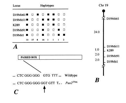
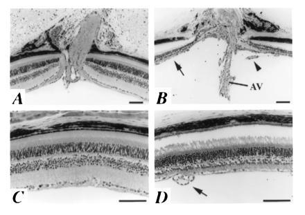
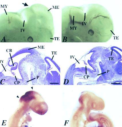
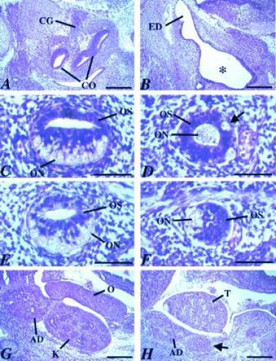
We describe a new mouse frameshift mutation (Pax2(1Neu)) with a 1-bp insertion in the Pax2 gene. This mutation is identical to a previously described mutation in a human family with renal-coloboma syndrome [Sanyanusin, P., McNoe, L. A., Sullivan, M. J., Weaver, R. G. & Eccles, M. R. (1995) Hum. Mol. Genet. 4, 2183-2184]. Heterozygous mutant mice exhibit defects in the kidney, the optic nerve, and retinal layer of the eye, and in homozygous mutant embryos, development of the optic nerve, metanephric kidney, and ventral regions of the inner ear is severely affected. In addition, we observe a deletion of the cerebellum and the posterior mesencephalon in homozygous mutant embryos demonstrating that, in contrast to mutations in Pax5, which is also expressed early in the mid-hindbrain region, loss of Pax2 gene function alone results in the early loss of the mid-hindbrain region. The mid-hindbrain phenotype is similar to Wnt1 and En1 mutant phenotypes, suggesting the conservation of gene regulatory networks between vertebrates and Drosophila.
Figures
Similar articles
-
Homonucleotide expansion and contraction mutations of PAX2 and inclusion of Chiari 1 malformation as part of renal-coloboma syndrome.Hum Mutat. 1999;14(5):369-76. doi: 10.1002/(SICI)1098-1004(199911)14:5<369::AID-HUMU2>3.0.CO;2-E. Hum Mutat. 1999. PMID: 10533062
-
Further delineation of renal-coloboma syndrome in patients with extreme variability of phenotype and identical PAX2 mutations.Am J Hum Genet. 1997 Apr;60(4):869-78. Am J Hum Genet. 1997. PMID: 9106533 Free PMC article.
-
Mutation of the PAX2 gene in a family with optic nerve colobomas, renal anomalies and vesicoureteral reflux.Nat Genet. 1995 Apr;9(4):358-64. doi: 10.1038/ng0495-358. Nat Genet. 1995. PMID: 7795640
-
Renal-coloboma syndrome: a multi-system developmental disorder caused by PAX2 mutations.Clin Genet. 1999 Jul;56(1):1-9. doi: 10.1034/j.1399-0004.1999.560101.x. Clin Genet. 1999. PMID: 10466411 Review.
-
Pax2 in development and renal disease.Int J Dev Biol. 1999;43(5):463-8. Int J Dev Biol. 1999. PMID: 10535325 Review.
Cited by
-
Alport-like glomerular basement membrane changes with renal-coloboma syndrome.Pediatr Nephrol. 2012 Jul;27(7):1189-92. doi: 10.1007/s00467-012-2125-9. Epub 2012 Feb 21. Pediatr Nephrol. 2012. PMID: 22350371
-
Mutations in PAX2 associate with adult-onset FSGS.J Am Soc Nephrol. 2014 Sep;25(9):1942-53. doi: 10.1681/ASN.2013070686. Epub 2014 Mar 27. J Am Soc Nephrol. 2014. PMID: 24676634 Free PMC article.
-
PAX2 polymorphisms and congenital abnormalities of the kidney and urinary tract in a Brazilian pediatric population: evidence for a role in vesicoureteral reflux.Mol Diagn Ther. 2014 Aug;18(4):451-7. doi: 10.1007/s40291-014-0096-1. Mol Diagn Ther. 2014. PMID: 24633556
-
Inhibition of Pax2 Transcription Activation with a Small Molecule that Targets the DNA Binding Domain.ACS Chem Biol. 2017 Mar 17;12(3):724-734. doi: 10.1021/acschembio.6b00782. Epub 2017 Jan 24. ACS Chem Biol. 2017. PMID: 28094913 Free PMC article.
-
Developmental Genetics and Congenital Anomalies of the Kidney and Urinary Tract.J Pediatr Genet. 2016 Mar;5(1):51-60. doi: 10.1055/s-0035-1558423. Epub 2015 Sep 7. J Pediatr Genet. 2016. PMID: 27617142 Free PMC article. Review.
References
-
- Strachan T, Read A P. Curr Opin Genet Dev. 1994;4:427–438. - PubMed
-
- Walther C, Guenet J-L, Smion D, Deutsch U, Jostes B, Goulding M D, Plachov D, Balling R, Gruss P. Genomics. 1991;11:424–434. - PubMed
-
- Wehr R, Gruss P. Int J Dev Biol. 1996;40:369–377. - PubMed
-
- Sanyanusin P, Schimmenti L A, McNoe L A, Ward T A, Pierpont M E M, Sullivan M J, Dobyns W B, Eccles M R. Nat Genet. 1995;9:358–364. - PubMed
-
- Sanyanusin P, McNoe L A, Sullivan M J, Weaver R G, Eccles M R. Hum Mol Genet. 1995;4:2183–2184. - PubMed
Publication types
MeSH terms
Substances
Associated data
- Actions
LinkOut - more resources
Full Text Sources
Molecular Biology Databases
