. 1995 Feb 24;80(4):621-30.
doi: 10.1016/0092-8674(95)90516-2.

Nucleocapsid and glycoprotein organization in an enveloped virus

Affiliations

Nucleocapsid and glycoprotein organization in an enveloped virus

R H Cheng et al. Cell. .

Abstract

Alphaviruses are a group of icosahedral, positive-strand RNA, enveloped viruses. The membrane bilayer, which surrounds the approximately 400 A diameter nucleocapsid, is penetrated by 80 spikes arranged in a T = 4 lattice. Each spike is a trimer of heterodimers consisting of glycoproteins E1 and E2. Cryoelectron microscopy and image reconstruction of Ross River virus showed that the T = 4 quaternary structure of the nucleocapsid consists of pentamer and hexamer clusters of the capsid protein, but not dimers, as have been observed in several crystallographic studies. The E1-E2 heterodimers form one-to-one associations with the nucleocapsid monomers across the lipid bilayer. Knowledge of the atomic structure of the capsid protein and our reconstruction allows us to identify capsid-protein residues that interact with the RNA, the glycoproteins, and adjacent capsid-proteins.


Figures
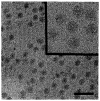
Figure 1
Figure 1
Low Magnification View of Frozen-Hydrated RR Recorded at 1.5 μm Underfocus The inset shows some RR images that were used in the reconstruction (Figure 2) at <1 μm underfocus. Bar is 2000 Å, and inset is 1000 Å.
Figure 2
Figure 2
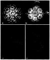
Surface-Shaded and Depth-Cued Representations of the RR Reconstruction The surface-shaded representations are viewed along a twofold axis (a) and a threefold axis (b). The 3D reconstruction was computed from 68 independent particle images. The depth-cued representation (c), viewed along a threefold axis, is overlaid with a T = 4 lattice to show the relative positions of the glycoprotein spikes with respect to the five-, three-, and twofold symmetry axes (labeled with numbers). Bar is 200 Å. Small holes are also observed on the fivefold axes, and even smaller holes are at the base of each spike.
Figure 3
Figure 3
Radial Density Distributions (a) Equatorial cross-section of the virus reconstruction along a twofold axis, with high density in white and low density (solvent and lipid) in black. The right half is a continuous, grey-level representation of the section, whereas the left half is a surface representation, shaded at a contour level of 0.5 standard deviation above the average. The positions of icosahedral two-, three-, fivefold, and quasi-threefold (Q3) axes are shown along with a scale marker. The closed arrows show regions of membrane pinching suggested to be the location of the transmembrane connections. (b) Projected density distributions (twofold view) at specific radii of the front half of the RR reconstruction. High to low densities are represented with a white to black grey scale, respectively. Bar is 200 Å. The juxtapositions of the spike glycoproteins (260 Å) and the nucleocapsid (210 Å) at both surfaces of the lipid bilayer are evident in these views.
Figure 4
Figure 4
Glycoprotein Spike (a) Close-up view of an adjacent pair of spikes at the icosahedral threefold (3) and quasi-threefold axes (Q3), showing nearly identical morphologies. (b) Close-up view at a glancing angle of the two spikes shown in (a). Notice the small holes at the base of the spike. (c) The molecular envelopes of the threefold (blue) and the quasi-threefold (red) spikes are nearly superimposable. (d) Schematic diagrams of the RR spike structure. The side view (A) is a cross-section through an individual spike (virus center towards the bottom), showing the inner cavity that lies directly below the bulk of the spike head and extending ~ 30 Å above the surface of the lipid bilayer. Cross-section B is a slice through the high radius portion of the spike, viewed from outside the viral surface. Cross-section C shows the stem portion of the spike, ~ 25 Å above the top of the lipid bilayer.
Figure 5
Figure 5
Structure of the RR Nucleocapsid Core (a) Surface-shaded, twofold view of the vifion reconstruction after all densities outside a radius of 210 Å were removed. The pentamer and hexamer arrangements of the nucleocapsid subunits are visible. (b) Depth-cued representation of the map in (a) with superimposed T = 4 lattice. One icosahedral asymmetric unit lies within the triangle (red) bounded by two threefold axes and one fivefoid axis. It includes four monomers (A1, B1, C1, and D1). Yellow numbers identify the icosahedral axes, and green symbols label the pseudoaxes. The polar angles are defined (as in Rossmann and Blow, 1962). (c) Placement of the crystallographic SCP dimer Cα structure (yellow) into the observed electron density(blue). Neither the A1C1 nor the B1D2 dimers fit into the observed density. (d) In contrast with (c), the SCP monomer (yellow) fits into the RR nucleocapsid density (blue) quite well. In agreement with cross-linking results, Lys-250 residues (denoted by asterisks) on adjacent capsomeres are ~ 12 Å apart.
Figure 6
Figure 6
Mutilayer Organization of Alphaviruses (a) Left, depth-cued representation of the 3D structure of RR (threefold view), showing the disposition of the spikes relative to the surface lattice (blue lines). Middle, schematic representation of the envelope glycoproteins as they emerge from the base of the spike, traverse the lipid bilayer membrane, and contact the underlying nucleocapsid monomers. Spikes at the icosahedral threefold (3) or quasi-threefold (Q3) are depicted by circles from which dark blue and light blue lines splay outwards at the outer surface of the membrane and then traverse the membrane and contact the underlying nucleocapsid monomers (yellow). Right, depth-cued representation of the 3D structure of the RR nucleocapsid (threefold view) showing the disposition of the core capsomeres relative to the icosahedral lattice (red lines). (b) A 5 Å thick, detailed view of an equatorial slice of the RR image reconstruction that contains one vertical twofold axis and two adjacent threefold axes (yellow lines). Distances from the virus center are marked along the twofold axis. The density is contoured at three levels: blue (low), purple (medium), and red (high). The left half of the diagram is colored to represent different viral components: spikes (blue), phospholipid bilayer (green), core protein (yellow), and the RNA–protein interior (red). The electron density at the highest contour level (red) identifies the most icosahedrally ordered portion of the density (i.e., protein in the spikes and in the nucleocapsid capsomeres). The fit of the Cα backbone of the SCP model (yellow-green) within the density is shown at the right of the diagram.

Similar articles

Cited by

References

    1. Aldroubi A, Trus BL, Unser M, Booy FP, Steven AC. Magnification mismatches between micrographe: corrective procedures and implications for structural analysis. Ultramicroscopy. 1992;46:175–188. - PubMed
    1. Aliperti G, Schlesinger MJ. Evidence for an autoprotease activity of Sindbis virus capsid protein. Virology. 1978;90:366–369. - PubMed
    1. Anthony RP, Brown DT. Protein–protein interactions in an alphavirus membrane. J Virol. 1991;65:1187–1194. - PMC - PubMed
    1. Baker TS, Drak J, Bina M. Reconstruction of the three-dimensional structure of simian virus 40 and visualization of the chromatin core. Proc Natl Acad Sci USA. 1988;85:422–426. - PMC - PubMed
    1. Baker TS, Newcomb WW, Olson NH, Cowsert LM, Olson C, Brow JC. Structures of bovine and human papillomaviruses. Analysis by cryoelectron microscopy and three- dimensional reconstruction. Biophys J. 1991;60:1445–1456. - PMC - PubMed

Publication types

MeSH terms