. 1995 Jun;14(6):1213-22.
doi: 10.1016/0896-6273(95)90268-6.

The integrin receptor alpha 8 beta 1 mediates interactions of embryonic chick motor and sensory neurons with tenascin-C

Affiliations

The integrin receptor alpha 8 beta 1 mediates interactions of embryonic chick motor and sensory neurons with tenascin-C

B Varnum-Finney et al. Neuron. 1995 Jun.

Abstract

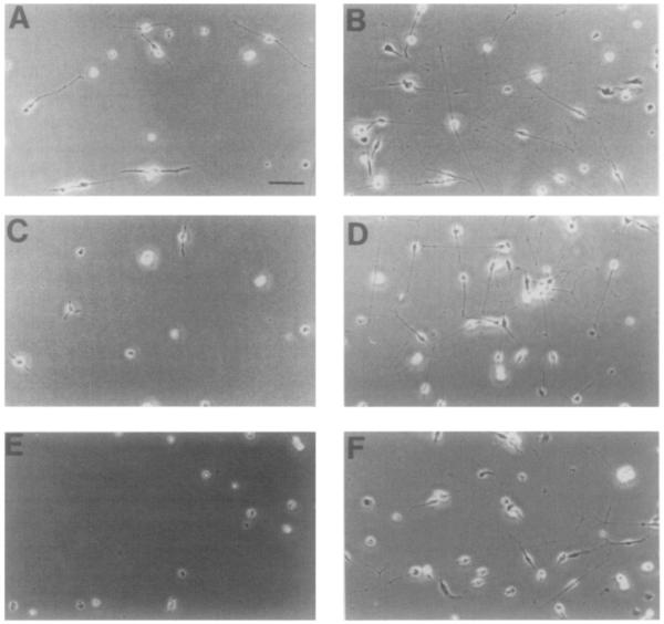
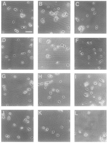
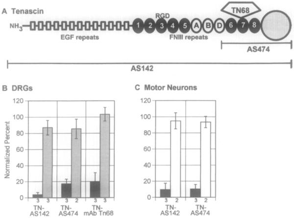
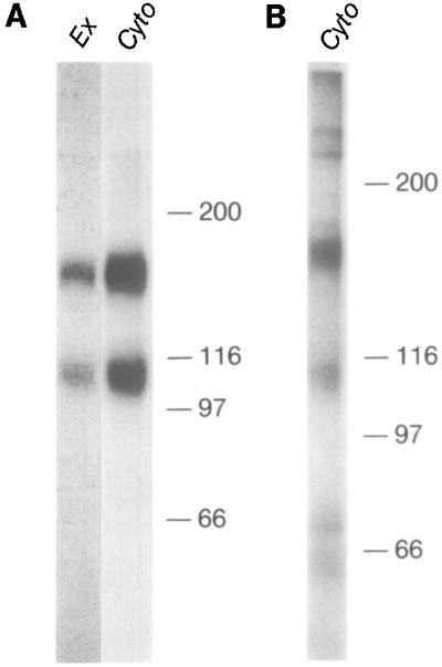
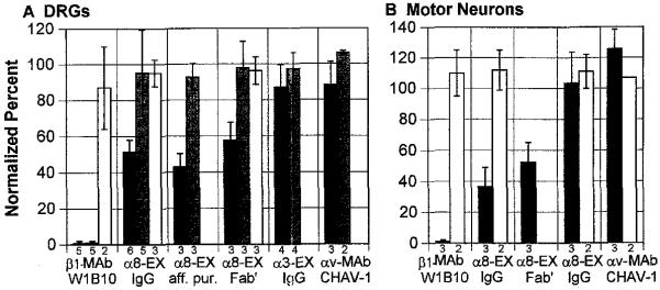
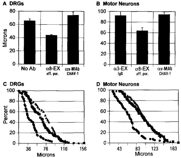
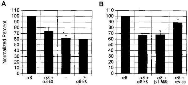
This paper identifies a neuronal receptor for tenascin-C (tenascin/cytotactin), an extracellular matrix protein that has previously been detected in developing sensory and motor neuron pathways and has been shown to regulate cell migration in the developing CNS. Antibodies specific for each subunit of the integrin alpha 8 beta 1 are used to demonstrate that alpha 8 beta 1 mediates neurite outgrowth of embryonic sensory and motor neurons on this extracellular matrix protein. In addition, expression of alpha 8 in K562 cells results in surface expression of alpha 8 beta 1 heterodimers that are shown to promote attachment of this cell line to tenascin. The major domain in tenascin that mediates neurite outgrowth is shown to be localized to fibronectin type III repeats 6-8.


Figures

Figure 1
Figure 1
Effects of Integrin- and Tenascin-Specific Antibodies on DRG Sensory Neuron Cultures Plated on Substrate-Bound Tenascin and Laminin DRGs were dissected from E7 chickens, dissociated, and plated on substrate-bound tenascin (A, C, and E) or laminin (B, D, and F). The following were added at the specified concentration to the culture medium: nothing (control culture; A and B), anti-α8-EX (300 μg/ml; C and D), and MAb Tn68 (100 μg/ml; E and F). Cultures on laminin were incubated for 4 hr, and those on tenascin, for 6 hr. Cultures were then fixed with paraformaldehyde, viewed with phase optics, and photographed. Bar, 30 μm.
Figure 2
Figure 2
Effects of Integrin- and Tenascin-Specific Antibodies on Motor Neuron Cultures Plated on Substrate-Bound Tenascin and N-Cadherin Neural tubes were dissected from stage 19 chick embryos, dissociated, and plated on substrate-bound tenascin (A-F) or N-cadherin (G-L). The following antibodies were added at the specified concentration to the culture medium: anti-αv MAb CHAV-1 (50 μg/ml; A and G), anti-β1 MAb W1B10 (50 μg/ml; B and H), anti-α8-EX (300 μg/ml; C and I), anti-α3-EX (300 μg/ml; D and J), anti-tenascin AS142 (1:100; E and K), and anti-tenascin AS474 (1:100; F and L). Cultures were incubated for 24 hr, fixed, viewed with phase optics, and photographed. Bar, 30 μm.
Figure 3
Figure 3
Schematic Drawing of a Tenascin Subunit Showing the Largest Splice Variant and the Effects of Anti-Tenascin Antibodies on Neurite Outgrowth by Sensory and Motor Neurons (A) Schematic drawing of a tenascin subunit showing the largest splice variant. Open squares, EGF repeats; closed ovals, FNIII repeats present in all tenascin isoforms; open ovals, differentially spliced FNIII repeats not present in all isoforms. The RGD sequence in FNIII domain 3 is illustrated, and the large open circle depicts the domain homologous to a domain in fibrinogen. The domains of tenascin recognized by two polyclonal antibodies (AS142 and AS474) and one MAb (TN68) are also shown. (B and C) Effects of anti-tenascin antibodies on neurite outgrowth by sensory (B) or motor (C) neurons. Neurons were dissected and dissociated as described in Experimental Procedures and plated on substrate-bound tenascin (closed bars), laminin (striped bars), or N-cadherin (open bars) in the presence of the anti-tenascin antibodies. Mean values for 2 wells from each experiment were normalized to mean values for control wells; n is listed below each bar. Values represent means ± SD (for n = 3) or means ± range (for n = 2).
Figure 4
Figure 4
Nonreducing SDS-PAGE of 125I-Surface Labeled Proteins (A) Chick embryo fibroblast lysates immunoprecipitated with anti-α8-EX (Ex) or anti-α8-cyto (Cyto). (B) Dissociated E7.5 chick DRG neurons were cultured, labeled, lysed, and immunoprecipitated with anti-α8-cyto. The positions of standards are indicated. Molecular weights are depicted in kilodaltons.
Figure 5
Figure 5
Normalized Percentages of Neurons Bearing Neurites after Culture in the Presence of Anti-Integrin Subunit-Specific Antibodies (A) E7 DRG sensory neurons from DRG were cultured for 5 hr on substrate-bound tenascin (closed bars), laminin (striped bars), or N-cadherin (open bars). Cultures were incubated in the presence of the the following anti-integrin antibodies at the designated concentrations: anti-β1 MAb W1B10 (50 μg/ml), anti-α8-EX (IgG; 300 μg/ml), anti-α8-EX (affinity-purified; 25 μg/ml), anti-α8-EX (Fab’; 300 μg/ml), anti-α3-EX (IgG; 300 μg/ml), and anti-αv MAb CHAV-1 (100 μg/ml). (B) Motor neurons were cultured on substrate-bound tenascin (closed bars) or N-cadherin (open bars) in the presence of the the following anti-integrin antibodies: anti-β1 MAb W1B10 (50 μg/ml), anti-α8-EX (IgG; 300 μg/ml), anti-α8-EX (Fab’; 300 μg/ml), anti-α3-EX (IgG; 300 μg/ml), and anti-αv MAb CHAV-1 (50 μg/ml). Mean values for 2 wells from each experiment were normalized to mean values for control wells (wells to which no antibody was added). Values in bar graph represent means ± SD (fcr n = 3-6) or means ± range (for n = 2); n is indicated below each bar.
Figure 6
Figure 6
Effect of Integrin-Specific Antibodies on Neurite Lengths in Sensory and Motor Neuron Cultures (A and C) DRG sensory neurons were cultured for 24 hr. Processes longer than 20 μm were measured for 20 neurons in each well, and a mean length was calculated for each well. Mean values (A) represent the means ± SEM for 4 wells from two experiments for the control and anti-α8-EX, and the mean ± range for 2 wells from a single experiment for anti-αv MAb CHAV-1. (C) shows a cumulative frequency distribution plot for the same neurons. The plot shows the percentage of neurons with neurites longer than a given length in micrometers (closed squares, control [no antibody]; closed circles, affinity-purified anti-α8-EX; closed triangles, anti-αv MAb CHAV-1). For sensory neurons, mean lengths (± SEM) were 92 ± 8 μm (control, anti-α3-EX), 64 ± 6 μm (anti-α8-EX), and 94 ± 5 μm (anti-αv). (B and D) Mean length and cumulative frequency distribution plots for motor neurons cultured for 7 hr. Motor neurons were incubated with anti-α3-EX (control; closed squares), anti-α8-EX (closed circles), or anti-αv MAb CHAV-1 (closed triangles). Mean values (B) represent the mean for 6 wells from three experiments. Cumulative frequency distribution plots (D) are for the same neurons as in (B). For motor neurons, mean lengths (± SEM) were 66 ± 3 μm (control; no antibody), 44 ± 1 μm (anti-α8-EX), and 74 ± 5 μm (anti-αv). For both sensory and motor neurons, differences in mean length between control or anti-αv versus anti-α8 cultures were significant, as determined by ANOVA (p < .001; Sokol and Rohlf, 1969).
Figure 7
Figure 7
Percentage Area of Attachment of K562 Cells to Substrate-Bound Tenascin A centrifugation assay was used to measure attachment as described in Experimental Procedures. After centrifugation, the area of cellular attachment to tenascin was quantitated by measuring the diameter of the cell pellet. A mean diameter of the cell-covered area was calculated from duplicate wells coated with tenascin. The mean diameter of the cell-covered area in wells coated with BSA was subtracted. Diameters were normalized, using the area covered by α8-expressing K562 cell transfectants as 100%. (A) Attachment for α8-transfected K562 is compared with control K562 in the presence and absence of anti-α8-EX affinity-purified Fab’ at 70 μg/ml. Each value represents the mean ± SD from three experiments, with the exception of parental K562 plus anti-α8-EX, which is from a single experiment. (B) Attachment for α8-transfected K562 cells was compared in cultures with the following antibodies: affinity-purified anti-α8-EX Fab’ at 70 μg/ml, anti-human β1 MAb A2BII (ascites 1:500), and anti-human integrin αv (serum; 1:500). Values from anti-α8-EX and anti-human β1 MAb represent the mean ± SD from three experiments, and values from antibody to the human integrin αv represent the mean ± range from two experiments.

Similar articles

Cited by

References

    1. Bartsch S, Bartsch U, Dorries U, Faissner A, Weller A, Ekblom P, Schachner M. Expression of tenascin in the developing and adult cerebellar cortex. J. Neurosci. 1992;12:736–748. - PMC - PubMed
    1. Bossy B, Bossy-Wetzel E, Reichardt LF. Characterization of the integrin α8 subunit: a new integrin β1-associated subunit, which is prominently expressed on axons and on cells in contact with basal laminae in chick embryos. EMBO J. 1991;10:2375–2385. - PMC - PubMed
    1. Burns FR, von Kannen S, Guy L, Raper JA, Kamholz J, Chang S. DM-GRASP, a novel immunoglobulin superfamily axonal surface protein that supports neurite extension. Neuron. 1991;7:209–220. - PubMed
    1. Bourdon MA, Ruoslahti E. Tenascin mediates cell attachment through an RGD-dependent receptor. J. Cell Biol. 1989;108:1149–1155. - PMC - PubMed
    1. Chen H, Sedat JW, Agard DA. Manipulation, display and analysis of three dimensional biological images. In: Pawley J, editor. The Handbook of Biological Confocal Microscopy. IMR Press; Madison, Wisconsin: 1989. pp. 127–135.

Publication types

MeSH terms

LinkOut - more resources