. 2024 Nov 15;13(11):bio061697.
doi: 10.1242/bio.061697. Epub 2024 Nov 25.

Feeding a rich diet supplemented with the translation inhibitor cycloheximide decreases lifespan and ovary size in Drosophila

Affiliations

Feeding a rich diet supplemented with the translation inhibitor cycloheximide decreases lifespan and ovary size in Drosophila

Hye Jin Hwang et al. Biol Open. .

Abstract

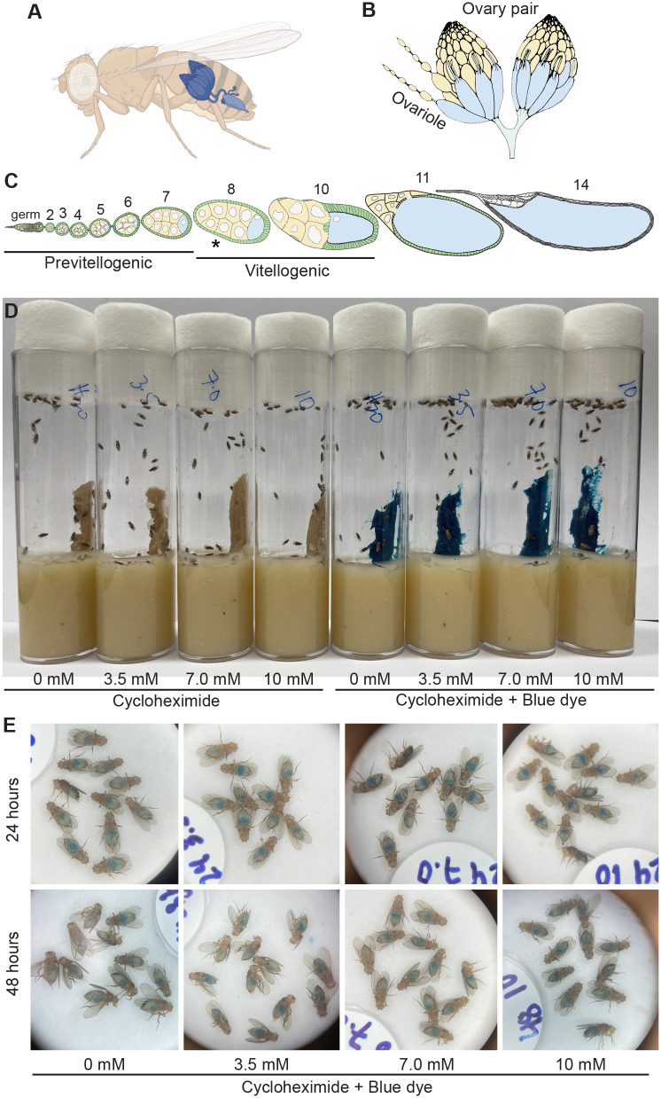
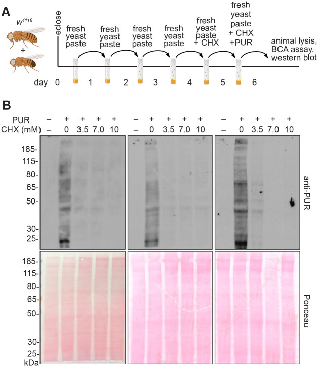
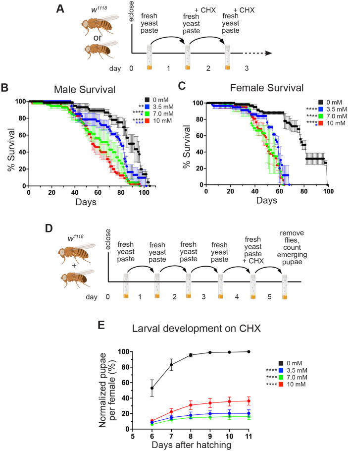
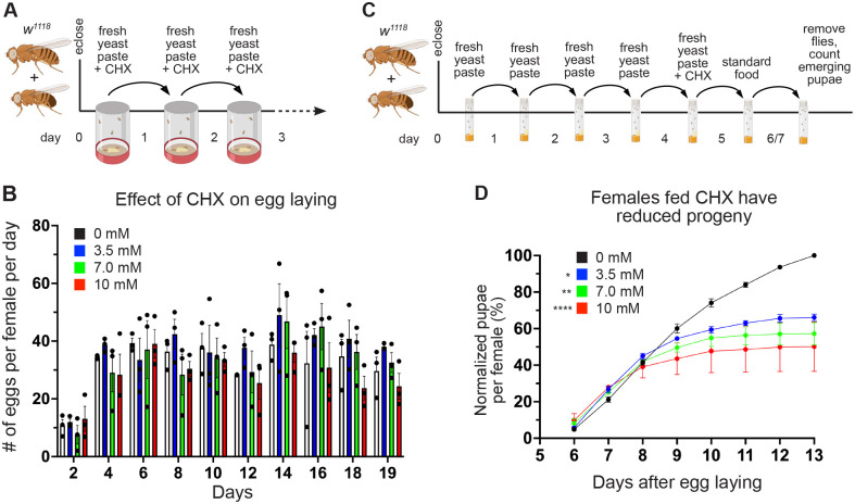
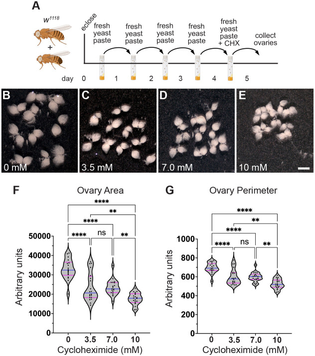
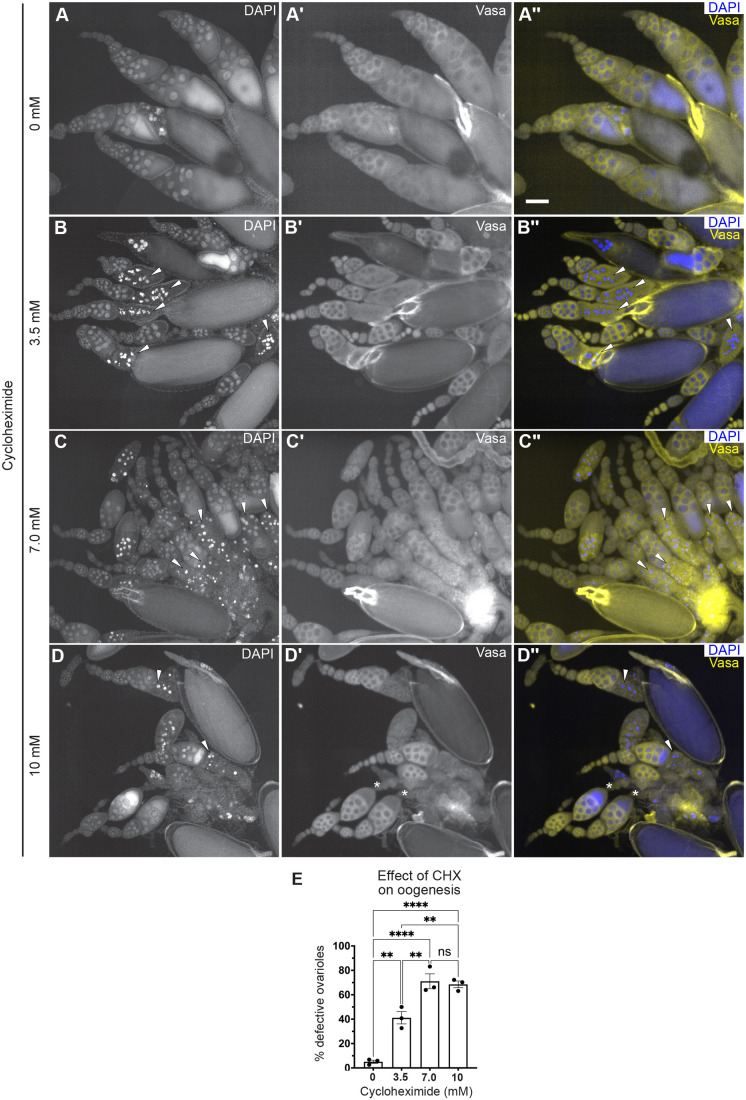
Drosophila oogenesis has long been an important model for understanding myriad cellular processes controlling development, RNA biology and patterning. Flies are easily fed drugs to disrupt various molecular pathways. However, this is often done under poor nutrient conditions that adversely affect oogenesis, thus making analysis challenging. Cycloheximide is a widely used compound that binds to and stalls the ribosome, therefore reducing protein synthesis. As egg production is a highly nutrient-dependent process, we developed a method to feed female Drosophila a rich diet of yeast paste supplemented with cycloheximide to better determine the effect of cycloheximide treatment on oogenesis. We found that flies readily consumed cycloheximide-supplemented yeast paste. Males and females had reduced lifespans when maintained on cycloheximide, with males exhibiting a dose-dependent decrease. Although females did not exhibit decreased egg laying, their ovaries were smaller and the number of progeny reduced, indicating substandard egg quality. Finally, females fed cycloheximide had disrupted oogenesis, with smaller ovaries, missing ovariole stages, and an increase in apoptotic follicles. Together, these data support that reduced protein synthesis adversely affects oogenesis with a rich diet that provides optimal nutrient conditions. In addition, this method could be used more broadly to test the effect of other drugs on Drosophila oogenesis without the confounding effects caused by poor nutrition.

Keywords: Drosophila; Cycloheximide; Lifespan; Nutrition; Oogenesis; Protein synthesis.


Conflict of interest statement

Competing interests The authors declare no competing or financial interests.

Figures

Fig. 1.
Fig. 1.
Flies readily consume wet yeast paste supplemented with CHX. (A-C) Schematics of Drosophila female ovaries. (A) Female flies contain a pair of ovaries. (B) Each ovary is composed of strings of developing follicles called ovarioles. (C) Ovarioles in females fed rich diets contain all stages of follicles (stages 2-14). Stage 8 (asterisk) is a developmental stage that undergoes apoptosis with nutritional stress. (D) Vials of wild-type flies feeding on wet yeast paste with 0, 3.5, 7.0 and 10 mM cycloheximide (CHX). The dye FD&C Blue No. 1 was used to visualize food ingestion by the flies (right). (E) Blue food was visible in the guts of female flies from the vials in D. Thirty animals were tested for each treatment on the same day.
Fig. 2.
Fig. 2.
Flies fed CHX-supplemented yeast paste have reduced protein synthesis. (A) Schematic illustrating the feeding regimen for puromycin (PUR) incorporation in B. Newly eclosed flies fed freshly prepared yeast paste for 4 days were given fresh yeast paste containing cycloheximide (CHX) for 24 h, after which they were transferred to the food vial with fresh yeast paste containing both CHX and PUR. 24 h later, protein was extracted from the female adults and analyzed. (B) Biological triplicate western blots probed with anti-PUR antibodies showing decreased puromycin incorporation with CHX feeding. 40 µg of protein sample was loaded per lane. Ponceau staining (lower blots) were used as a control for gel transfer to the membrane. PUR –, negative control for puromycin feeding.
Fig. 3.
Fig. 3.
A rich diet supplemented with CHX affects lifespan and larval development. (A) Schematic illustrating the feeding regimen for lifespan measurement in B,C. Newly eclosed flies were fed freshly prepared yeast paste for 1 day, after which they were transferred daily to fresh yeast paste containing CHX until they died. (B,C) Lifespan of flies fed with yeast paste containing varying concentrations of CHX at room temperature. Both males (B) and females (C) had significantly shortened lifespans when fed with CHX compared to controls. Males also showed significantly shortened lifespans with increasing CHX concentration. (D) Schematic illustrating the feeding regimen for larval development with CHX in E. Newly eclosed flies were fed freshly prepared yeast paste for 4 days, after which they were fed with fresh yeast paste containing CHX for 24 h, then removed. The progeny from the eggs laid for 24 h were maintained until the pupae emerged. (E) The percentages of pupae per female fed with varying concentrations of CHX normalized to wild type are shown. CHX-fed progenies had significant growth defects compared to the control group. All experimental conditions had three replicates. In B,C,E, error bars show the standard error (s.e.m.). Data were analyzed for significant differences using the log-rank test using the OASIS2 online application with combined triplicates (B,C) or using two-way ANOVA followed by Tukey's post hoc test (E). Asterisks in graphs indicate significance as follows: *P<0.05; **P<0.01; ***P<0.001; ****P<0.0001. Asterisks in E indicate significance on day 6 after egg laying. The colors of asterisks in the graphs denote the concentration of the comparison.
Fig. 4.
Fig. 4.
A rich diet supplemented with CHX affects egg quality but not quantity. (A) Schematic illustrating the feeding regimen for egg laying measurements in B. Newly eclosed flies were fed freshly prepared yeast paste for 1 day, after which they were transferred daily to an egg-laying cup with fresh yeast paste containing CHX. (B) Quantification of the number of eggs laid per female fed with varying concentrations of CHX. The total number of eggs was counted every 2 days. Points on the graph represent each of three replicates. Bars on the graph represent the average of all three replicates. (C) Schematic illustrating the feeding regimen for progeny developmental assays in D. Newly eclosed flies were fed freshly prepared yeast paste daily for 4 days, after which they were fed fresh yeast paste containing CHX for 24 h. Flies were transferred to a standard food vial and maintained for 48 h, after which they were removed. The progenies from the eggs laid for 48 h were maintained until pupae emerged. (D) The normalized percentages of pupae per female fed with varying concentrations of CHX for 24 h are shown. The number of pupae from flies fed with CHX-containing yeast paste was significantly decreased compared to controls. All experimental conditions had three replicates. In B,D, error bars show the s.e.m. Data were analyzed for significant differences by two-way ANOVA followed by Tukey's post hoc test. Asterisks in graphs indicate significance on day 11 after egg laying. *P<0.05; **P<0.01; ****P<0.0001. The colors of asterisks in the graphs denote the concentration of the comparison.
Fig. 5.
Fig. 5.
A rich diet supplemented with CHX affects ovary size. (A) Schematic illustrating the feeding regimen for measuring ovary size in B-G. Newly eclosed flies were fed freshly prepared yeast paste daily for 4 days, then fed with fresh yeast paste containing CHX for 24 h, after which the ovaries were dissected. (B-E) Ten pairs of ovaries from the females fed with varying concentrations of CHX for 24 h are shown. Females fed CHX had smaller ovaries. The pictures shown are representative of triplicates. These ovaries were analyzed using FIJI/Image J for the whole ovary area (F) and the perimeter (G). Scale bar: 1 mm. (F,G) Violin plots for ovary area (F) and perimeter (G) from females fed CHX. Each point on the violin plot represents one ovary. The median and quartiles are represented by blue and magenta horizontal solid lines, respectively. The plots shown are representative of triplicates. All experimental conditions had three replicates. Data were analyzed for significant differences by one-way ANOVA followed by Tukey's post hoc test. Asterisks in graphs indicates significance as follows: ns, no significance; **P<0.01; ****P<0.0001.
Fig. 6.
Fig. 6.
A rich diet supplemented with CHX affects oogenesis. (A-D″) Dissected ovaries labeled with anti-Vasa (Aʹ-Dʹ, yellow in A″-D″) to label the germplasm and DAPI (A-D, blue in A″-D″) to label nuclei. Ovaries were dissected from females fed for 24 h with wet yeast paste containing 0 (A-A″), 3.5 (B-B″), 7.0 (C-C″) or 10 (D-D″) mM CHX. White arrowheads indicate apoptotic cells (B-D″). Asterisks indicate ovarioles with missing follicles stages (D′,D″). Ovarioles from CHX-fed females had an increased number of mid-stage apoptotic follicles as indicated by condensed DAPI-labeled nuclei (B,B″,C,C″,D,D″, arrowheads). Scale bar: 100 µm. (E) The relative number of ovarioles having developmental defects shown in A-D″. Ovarioles with developmental defects contained follicles with apoptotic nuclei and/or missing follicle stages. Approximately 50-90 ovarioles were counted in an individual experimental condition (see Materials and Methods for details.) Error bars show the s.e.m. Points on the graph represent each of three replicates. Bars on the graph represent the average of all three independent experiments and data were analyzed for significant differences by one-way ANOVA followed by Tukey's post hoc test. Asterisks in the graph indicate significance as follows: ns, not significant; **P<0.01; ****P<0.0001.

Update of

Similar articles

References

    1. Armstrong, A. R. (2020). Drosophila melanogaster as a model for nutrient regulation of ovarian function. Reproduction 159, R69-R82. 10.1530/REP-18-0593 - DOI - PubMed
    1. Belozerov, V. E., Ratkovic, S., Mcneill, H., Hilliker, A. J. and McDermott, J. C. (2014). In vivo interaction proteomics reveal a novel p38 mitogen-activated protein kinase/Rack1 pathway regulating proteostasis in Drosophila muscle. Mol. Cell. Biol. 34, 474-484. 10.1128/MCB.00824-13 - DOI - PMC - PubMed
    1. Cervantes-Sandoval, I., Chakraborty, M., MacMullen, C. and Davis, R. L. (2016). Scribble scaffolds a signalosome for active forgetting. Neuron 90, 1230-1242. 10.1016/j.neuron.2016.05.010 - DOI - PMC - PubMed
    1. Darvishi, E. and Woldemichael, G. M. (2016). Cycloheximide inhibits actin cytoskeletal dynamics by suppressing signaling via RhoA. J. Cell. Biochem. 117, 2886-2898. 10.1002/jcb.25601 - DOI - PubMed
    1. Drummond-Barbosa, D. and Spradling, A. C. (2001). Stem cells and their progeny respond to nutritional changes during Drosophila oogenesis. Dev. Biol. 231, 265-278. 10.1006/dbio.2000.0135 - DOI - PubMed

LinkOut - more resources