. 2024 Aug 3;15(1):6594.
doi: 10.1038/s41467-024-50973-y.

Neuro-intestinal acetylcholine signalling regulates the mitochondrial stress response in Caenorhabditis elegans

Affiliations

Neuro-intestinal acetylcholine signalling regulates the mitochondrial stress response in Caenorhabditis elegans

Rebecca Cornell et al. Nat Commun. .

Abstract

Neurons coordinate inter-tissue protein homeostasis to systemically manage cytotoxic stress. In response to neuronal mitochondrial stress, specific neuronal signals coordinate the systemic mitochondrial unfolded protein response (UPRmt) to promote organismal survival. Yet, whether chemical neurotransmitters are sufficient to control the UPRmt in physiological conditions is not well understood. Here, we show that gamma-aminobutyric acid (GABA) inhibits, and acetylcholine (ACh) promotes the UPRmt in the Caenorhabditis elegans intestine. GABA controls the UPRmt by regulating extra-synaptic ACh release through metabotropic GABAB receptors GBB-1/2. We find that elevated ACh levels in animals that are GABA-deficient or lack ACh-degradative enzymes induce the UPRmt through ACR-11, an intestinal nicotinic α7 receptor. This neuro-intestinal circuit is critical for non-autonomously regulating organismal survival of oxidative stress. These findings establish chemical neurotransmission as a crucial regulatory layer for nervous system control of systemic protein homeostasis and stress responses.


Conflict of interest statement

The authors declare no competing interests.

Figures
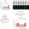
Fig. 1
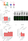
Fig. 1. Gamma-aminobutyric acid (GABA) non-cell-autonomously inhibits the mitochondrial unfolded protein response (UPRmt).
a, b Quantification (a) and DIC/fluorescent micrographs (b) of UPRmt reporter (hsp-6p::gfp) expression in L4 larvae of wild-type and mutant animals affecting neurotransmitter synthesis and transport. c Schematic of GABA synthesis (UNC-25/GAD), transport (UNC-47/VGAT), and release. d Schematic of GABA expressing neurons in C. elegans. e Quantification of UPRmt reporter (hsp-6p::gfp) expression in L4 larvae of wild-type, unc-47(n2409), unc-25(e156), unc-25(e156); unc-25p::unc-25 DNA, unc-25(e156); unc-30p::unc-25 DNA and unc-25(e156); unc-47ptruncated::unc-25 DNA animals. n = 30 animals. The hash symbol (#) refers to independent transgenic rescue lines. P values assessed by one-way analysis of variance (ANOVA) with Tukey’s post hoc test. Error bars indicate SEM. Scale bar, 250 μm. Source data are provided as a Source Data file.
Fig. 2
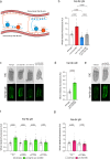
Fig. 2. Metabotropic GABAB receptors coordinate non-cell-autonomous UPRmt.
a Schematic of ionotropic GABAA (UNC-49 (inhibitory) and EXP-1 (excitatory)) and metabotropic (GBB-1 and GBB-2) GABAB receptors in C. elegans. b, c Quantification (b) and DIC/fluorescent micrographs (c) of UPRmt reporter (hsp-6p::gfp) expression in L4 larvae of wild-type, gbb-1(tm1406), gbb-2(tm1165), gbb-2(tm1165); gbb-1(tm1406), unc-49(e407) and exp-1(ok1131) animals. d Quantification of UPRmt reporter (hsp-6p::gfp) expression in L4 larvae of wild-type, gbb-1(tm1406), gbb-2(tm1165), gbb-2(tm1165); gbb-1(tm1406), unc-25(e156), unc-25(e156); gbb-1(tm1406), unc-25(e156); gbb-2(tm1165), and unc-25(e156); gbb-2(tm1165); gbb-1(tm1406) animals. e Quantification of UPRmt reporter (hsp-6p::gfp) expression in L4 larvae of wild-type, gbb-1(tm1406), gbb-1(tm1406); rgef-1p::gbb-1 cDNA and gbb-1(tm1406); acr-2p(s)::gbb-1 cDNA animals. n = 30 animals. P values assessed by one-way analysis of variance (ANOVA) with Tukey’s post hoc test. Error bars indicate SEM. Scale bar, 250 µm. Source data are provided as a Source Data file.
Fig. 3
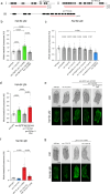
Fig. 3. Acetylcholine promotes non-cell-autonomous UPRmt through the intestinal ACR-11/nicotinic α7 receptor.
a Schematic of the C. elegans motor circuit. Orange = inhibitory GABAergic motor neurons; blue = excitatory cholinergic motor neurons; Pink = body wall muscle. b, c Quantification (b) and DIC/fluorescent micrographs (c) of UPRmt reporter (hsp-6p::gfp) expression in L4 larvae of wild-type, unc-25(e156), unc-17(e113), and unc-25(e156); unc-17(e113) animals. d, e Quantification (d) and DIC/fluorescent micrographs (e) of UPRmt reporter (hsp-6p::gfp) expression in L4 larvae of wild-type and ace-2(g72); ace-1(p1000) animals. f Quantification of UPRmt reporter (hsp-6p::gfp) expression in L4 larvae of wild-type and ace-2(g72); ace-1(p1000) animals grown on empty vector (EV), acr-6, acr-7 or acr-11 RNAi from the mother’s L4 stage. g Quantification of UPRmt reporter (hsp-6p::gfp) expression in L4 larvae of wild-type and unc-25(e156) animals grown on empty vector (EV) or acr-11 RNAi from the mother’s L4 stage. n = 30 animals. P values assessed by one-way analysis of variance (ANOVA) Tukey’s post hoc test (b, f, g) and two-way unpaired t test with Welch’s correction (d). Error bars indicate SEM. Scale bars, 250 µm. Source data are provided as a Source Data file.
Fig. 4
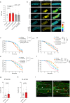
Fig. 4. The ACR-11/nicotinic α7 receptor cell autonomously mediates the UPRmt.
a (i) Structure of the acr-11 locus. rp191 = 3038 bp deletion in wild-type animals; rp192 = 3046 bp deletion in ace-2(g72); ace-1(p1000) animals. (ii) Structure of the acr-6 locus. rp209 = 3209 bp deletion in wild-type animals; rp210 = 3158 bp deletion in acr-11(rp191) animals. (iii) Structure of the acr-7 locus. rp212 = 1556 bp deletion. Black boxes, coding regions; grey boxes, untranslated regions; red lines, CRISPR/Cas9-generated deletion alleles. b Quantification of UPRmt reporter (hsp-6p::gfp) expression in L4 larvae of wild-type, acr-11(rp191), ace-2(g72); ace-1(p1000), and ace-2(g72) acr-11(rp192); ace-1(p1000) animals. c Quantification of UPRmt reporter (hsp-6p::gfp) expression in L4 larvae of wild-type, acr-6(rp209), acr-7(rp212), acr-11(rp191), acr-6(rp209); acr-7(rp212), acr-6(rp210) acr-11(rp191), acr-11(rp191); acr-7(rp212) and acr-6(rp210) acr-11(rp191); acr-7(rp212) animals. d, e Quantification (d) and DIC/fluorescent micrographs (e) of UPRmt reporter (hsp-6p::gfp) expression in L4 larvae of wild-type, ace-2(g72); ace-1(p1000), acr-11(rp191), ace-2(g72) acr-11(rp192); ace-1(p1000) and ace-2(g72) acr-11(rp192); ace-1(p1000); ges-1p::acr-11 DNA animals. f, g Quantification (f) and DIC/fluorescent micrographs (g) of UPRmt reporter (hsp-6p::gfp) expression in L4 larvae of wild-type, unc-17(e113), ges-1p::acr-11 DNA and unc-17(e113); ges-1p::acr-11 cDNA animals. n = 30 animals. P values assessed by one-way analysis of variance (ANOVA) with Tukey’s post hoc test. Error bars indicate SEM. Scale bars, 250 µm. Source data are provided as a Source Data file.
Fig. 5
Fig. 5. GABA/Ach regulation of ACR-11 influences mitochondrial stress resistance.
a, b Quantification (a) and micrographs (b) of calcium imaging by FRET microscopy of the two anterior intestinal cells of wild-type, unc-25(e156), ace-2(g72); ace-1(p1000), acr-11(rp191) and ace-2(g72) acr-11(rp192); ace-1(p1000) animals expressing nhx-2p::D3cpv, a ratiometric indicator. n = 38, 36, 36, 35, 36 animals (left to right). Scale bar, 50 µm. Note that induced expression of the ratiometric reporter in animals with excess ACh (unc-25 and ace2; ace-1 mutants) in intestinal cells Int1-3 was consistent in all animals and not due to mosaicism. c Survival analysis of wild-type, unc-25(e156), gbb-1(tm1406) and gbb-1(tm1406); rgef-1p::gbb-1 cDNA animals exposed to 200 mM paraquat from the L4 larval stage. n = 67, 74, 70, 67 animals (top to bottom). d Survival analysis of wild-type, ace-2(g72); ace-1(p1000), acr-11(rp191) and ace-2(g72) acr-11(rp192); ace-1(p1000) animals exposed to 200 mM paraquat from the L4 larval stage. n = 73, 72, 65, 67 animals (top to bottom). e Survival analysis of wild-type, unc-25(e156), unc-17(e113) and unc-25(e156); unc-17(e113) animals exposed to 200 mM paraquat from the L4 larval stage. n = 73, 74, 72, 71 animals (top to bottom). f Survival analysis of wild-type, unc-17(e113), ges-1p::acr-11 cDNA and unc-17(e113); ges-1p::acr-11 cDNA animals exposed to 200 mM paraquat from the L4 larval stage. n = 74, 74, 64, 75 animals (top to bottom). gi Quantification of (g) autophagosome puncta (AP), (h) autolysosome puncta (AL), and micrographs (i) of GFP and mCherry fluorescence in the two anterior intestinal cells of wild-type and acr-11(rp191) L4 larvae expressing LGG-1p::mCherry::GFP::LGG-1. White arrows = green puncta/autophagosomes, yellow arrows = red puncta/autolysosomes. n = 62 (wild type) and 59 (acr-11(rp191) animals). Scale bar, 5 µm. P values assessed by one-way analysis of variance (ANOVA) with Tukey’s post hoc test (a), two-way analysis of variance (ANOVA) with Tukey’s post hoc test (cf), and two-way unpaired t test with Welch’s correction (g, h). Error bars indicate SEM. Source data are provided as a Source Data file.

Similar articles

References

    1. Durieux, J., Wolff, S. & Dillin, A. The cell-non-autonomous nature of electron transport chain-mediated longevity. Cell144, 79–91 (2011). 10.1016/j.cell.2010.12.016 - DOI - PMC - PubMed
    1. Berendzen, K. M. et al. Neuroendocrine coordination of mitochondrial stress signaling and proteostasis. Cell166, 1553–1563.e1510 (2016). 10.1016/j.cell.2016.08.042 - DOI - PMC - PubMed
    1. Shao, L.-W., Niu, R. & Liu, Y. Neuropeptide signals cell non-autonomous mitochondrial unfolded protein response. Cell Res.26, 1182–1196 (2016). 10.1038/cr.2016.118 - DOI - PMC - PubMed
    1. Zhang, Q. et al. The mitochondrial unfolded protein response is mediated cell-non-autonomously by retromer-dependent Wnt signaling. Cell174, 870–883.e817 (2018). 10.1016/j.cell.2018.06.029 - DOI - PMC - PubMed
    1. Lan, J. et al. Translational regulation of non-autonomous mitochondrial stress response promotes longevity. Cell Rep.28, 1050–1062.e1056 (2019). 10.1016/j.celrep.2019.06.078 - DOI - PMC - PubMed

MeSH terms

Substances

LinkOut - more resources