Review
. 2024 Jun 27;25(13):7041.
doi: 10.3390/ijms25137041.

Extracellular Vesicles as Mediators of Neuroinflammation in Intercellular and Inter-Organ Crosstalk

Affiliations
Review

Extracellular Vesicles as Mediators of Neuroinflammation in Intercellular and Inter-Organ Crosstalk

Andrea Cabrera-Pastor. Int J Mol Sci. .

Abstract

Neuroinflammation, crucial in neurological disorders like Alzheimer's disease, multiple sclerosis, and hepatic encephalopathy, involves complex immune responses. Extracellular vesicles (EVs) play a pivotal role in intercellular and inter-organ communication, influencing disease progression. EVs serve as key mediators in the immune system, containing molecules capable of activating molecular pathways that exacerbate neuroinflammatory processes in neurological disorders. However, EVs from mesenchymal stem cells show promise in reducing neuroinflammation and cognitive deficits. EVs can cross CNS barriers, and peripheral immune signals can influence brain function via EV-mediated communication, impacting barrier function and neuroinflammatory responses. Understanding EV interactions within the brain and other organs could unveil novel therapeutic targets for neurological disorders.

Keywords: CNS barrier; exosomes; extracellular vesicles; glial cells; inter-organ crosstalk; neuroinflammation; neurological disorders; neuron.


Conflict of interest statement

The authors declare no conflict of interest.

Figures

Figure 1
Figure 1
Biogenesis of Extracellular Vesicles (EVs). The biogenesis of exosomes involves a tightly regulated process that begins with the formation of early endosomes. These early endosomes mature into multivesicular bodies (MVBs), which contain intraluminal vesicles harboring specific cargo molecules. Some of these MVBs directly fuse with lysosomes and degrade, some are transported to the Golgi for recovery, and some fuse with the cell membrane to release small vesicles outside of the cell and form exosomes. Many molecules play an important role in exosome biogenesis and abscission. First, the endosomal sorting complex required for transport (ESCRT) and other proteins, such as tumor susceptibility gene 101 protein (TSG101) and ALG-2 interacting protein X (ALIX), are involved in cargo sorting into exosomes. In addition, other ESCRT-independent mechanisms, including lipid rafts and tetraspanins CD63 and CD81, are conducive to exosome biogenesis. Finally, the Rab-GTPase family contributes to the intracellular trafficking and fusion of MVBs with the cell membrane to release exosomes. Exosomes are small EVs, typically ranging from 30 to 150 nanometers in diameter. In contrast, microvesicles are formed by the outward budding and shedding of the plasma membrane, resulting in the direct release of vesicles into the extracellular environment. Microvesicles are larger EVs, generally ranging from 100 to 1000 nanometers in size. The cargo of EVs includes a diverse array of bioactive molecules, such as nucleic acids (mRNA, miRNA, DNA, lncRNA), proteins, and lipids. The different surface proteins are transmembrane proteins such as tetraspanins (such as CD9, CD63, CD81), antigen-presenting molecules (MHC I and II), adhesion molecules (such as integrins, P-selectin), and other signaling receptors (such as TNFR, FasL, TfR); proteins in the EV lumen, such as heat shock proteins (HSPs), cytoskeletal proteins (such as actin, tubulin, vimentin), ESCRT components (such as Alix, TSG-101), membrane transport and fusion proteins (such as GTPases, Annexin, Flotillin, Clathrin), growth factors and cytokines (such as TNF-α, TGF-β, TRAIL), and metabolic enzymes (such as GAPDH, PKM2, PGK1, PDIA3). EVs also comprise multiple lipids, such as cholesterol, ceramides, sphingomyelin, phosphatidylinostol (PI), phosphatidylserine (PS), phosphatidylcholine (PC), phosphatidylethanolamine (PE), and gangliosides (GM). Importantly, the composition of EV cargo is influenced by the originating cell type and its physiological state. TNF-α = tumor necrosis alpha, TGF-β = transforming growth factor beta, TRAIL = TNF-related apoptosis-inducing ligand, messenger RNA (mRNA), microRNA (miRNA), lncRNA = long non-coding RNAs, GAPDH = Glyceraldehyde 3-phosphate dehydrogenase, PKM2 = Pyruvate kinase isozyme M2, PGK1 = Phosphoglycerate Kinase 1, PDIA3 = Protein disulfide-isomerase A3, TNFR = tumor necrosis factor receptor, FasL = Fas ligand, and TfR = Transferrin receptor.
Figure 2
Figure 2
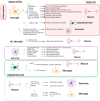
EV-mediated intercellular crosstalk among glial-neuron cells. EVs released by glial cells (astrocytes, oligodendrocytes, and microglia) or neurons have several target cells within the brain and not only orchestrate inflammatory reactions but also provide neurotrophic support and contribute to the maintenance of homeostasis. EVs from glial cells modulate synaptic activity, neuronal survival, neurogenesis, and myelination process, and, in an inflammatory environment, propagate the activation of inflammatory signaling pathways. Neuron-derived EVs also contribute to the homeostasis of astrocytes and microglia but, in neuroinflammatory conditions, they contribute to the activation of both. CAM/ECM: cell adhesion/extracellular matrix.
Figure 3
Figure 3
Scheme of EVs in inter-organ crosstalk. EVs disseminate inflammatory signals between organs. In brain–heart crosstalk, increased plasma astrocyte-derived EVs have been shown in post-ischemic stroke. The content of the EVs participates in the disruption of endothelial function and activation of coagulation factors. Moreover, several miRNAs have been found to be linked to both heart and brain pathophysiology. EVs from adipose tissue are involved in processes such as axonal growth, tract connectivity, oligodendrogenesis, and remyelination following subcortical ischemic stroke. Other emerging evidence suggests that EVs derived from mesenchymal stem cells, such as ADSCs (adipose-derived stem cells), possess anti-inflammatory properties and mitigate neuroinflammation in various pathological conditions. Plasma EVs from animal models with minimal hepatic encephalopathy (MHE) are able to induce altered neurotransmission. Age-related thyroid deficiency can enhance the transport of Apolipoprotein E4-containing EVs from the liver to the brain, contributing to Alzheimer’s disease-related dementia and neuronal dysfunction. In the gut–brain axis, EVs found in the intestinal microenvironment originate from both microorganisms, such as bacteria, and intestinal cells, and are involved in transmitting signals (LPS, DNA, RNA, miRNAs, etc.) to the brain through the vagus nerve or the bloodstream. These gut-derived EVs can induce neuroinflammation, modulate neuronal function, and increase the permeability of the blood–brain barrier (BBB).

Similar articles

Cited by

References

    1. Heneka M.T., Kummer M.P., Latz E. Innate immune activation in neurodegenerative disease. Nat. Rev. Immunol. 2014;14:463–477. doi: 10.1038/nri3705. - DOI - PubMed
    1. Cabrera-Pastor A., Llansola M., Montoliu C., Malaguarnera M., Balzano T., Taoro-Gonzalez L., García-García R., Mangas-Losada A., Izquierdo-Altarejos P., Arenas Y.M., et al. Peripheral inflammation induces neuroinflammation that alters neurotransmission and cognitive and motor function in hepatic encephalopathy: Underlying mechanisms and therapeutic implications. Acta Physiol. 2019;226:e13270. doi: 10.1111/apha.13270. - DOI - PubMed
    1. Prinz M., Priller J. Microglia and brain macrophages in the molecular age: From origin to neuropsychiatric disease. Nat. Rev. Neurosci. 2014;15:300–312. doi: 10.1038/nrn3722. - DOI - PubMed
    1. Gao H.M., Hong J.S. Why neurodegenerative diseases are progressive: Uncontrolled inflammation drives disease progression. Trends Immunol. 2008;29:357–365. doi: 10.1016/j.it.2008.05.002. - DOI - PMC - PubMed
    1. Izquierdo-Altarejos P., Cabrera-Pastor A., Gonzalez-King H., Montoliu C., Felipo V. Extracellular Vesicles from Hyperammonemic Rats Induce Neuroinflammation and Motor Incoordination in Control Rats. Cells. 2020;9:572. doi: 10.3390/cells9030572. - DOI - PMC - PubMed

Grants and funding

This research was funded by Instituto de Salud Carlos III (ISCIII) through the project “PI23/00204” and co-funded by the European Union; and Conselleria de Educación/Innovación, Universidades, Ciencia y Sociedad Digital, subvenciones para la realización de proyectos de I+D+i desarrollados por grupos de investigación emergentes (CIGE/083).

LinkOut - more resources