Scleral changes in systemic lupus erythematosus patients using swept source optical coherence tomography
- PMID: 38022606
- PMCID: PMC10656698
- DOI: 10.3389/fimmu.2023.1278893
Scleral changes in systemic lupus erythematosus patients using swept source optical coherence tomography
Abstract
Purpose: This study aims to examine scleral thickness in patients with systemic lupus erythematosus (SLE) without clinically evident scleritis and episcleritis, utilizing swept-source optical coherence tomography (SS-OCT).
Methods: This cross-sectional single center study compared scleral thickness (Nasal scleral thickness 1mm, 2mm, 3mm, 6mm from scleral spur; Temporal scleral thickness 1mm, 2mm, 3mm, 6mm from scleral spur) in 73 SLE patients without clinically evident scleritis and episcleritis and 48 healthy volunteers with SS-OCT. Further, we investigated the correlation between scleral thickness in SLE patients and various parameters including laboratory markers, disease duration, disease activity, and organ involvement.
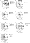
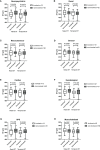
Results: Across all measured sites (nasal scleral thickness at distances of 1mm, 2mm, 3mm, and 6mm from the scleral spur, and temporal scleral thickness at the same distances), the scleral thickness in the SLE group was significantly greater than that in the control group (all p-values <0.001). SLE patients with a disease duration of 5 years or less exhibited a higher scleral thickness compared to those with a more prolonged disease duration. Patients with a higher erythrocyte sedimentation rate (ESR) had a thinner temporal scleral thickness. However, no significant associations were identified between scleral thickness and disease activity, organ involvement, or other laboratory markers.
Conclusion: Scleral thickness measured by SS-OCT was higher in SLE patients than healthy controls. Changes in scleral thickness in SLE patients are related to disease duration and ESR. SS-OCT can detect asymptomatic structural changes in SLE patients and may be a useful tool in the evaluation of early scleral abnormality.
Keywords: SS-OCT; disease duration; preclinical change; scleral thickness; systemic lupus erythematosus.
Copyright © 2023 Chen, Meng, Sun and Chen.
Conflict of interest statement
The authors declare that the research was conducted in the absence of any commercial or financial relationships that could be construed as a potential conflict of interest.
Figures


Similar articles
-
Measurements of scleral thickness and corneal optic densitometry in patients with systemic lupus erythematosus.Medicine (Baltimore). 2020 Jul 31;99(31):e21467. doi: 10.1097/MD.0000000000021467. Medicine (Baltimore). 2020. PMID: 32756168 Free PMC article.
-
Quantitative evaluation of retinal and choroidal vascularity in systemic lupus erythematosus by SS-OCT/OCTA.Graefes Arch Clin Exp Ophthalmol. 2023 Dec;261(12):3385-3393. doi: 10.1007/s00417-023-06155-5. Epub 2023 Jun 27. Graefes Arch Clin Exp Ophthalmol. 2023. PMID: 37367994
-
Morphological features in anterior scleral inflammation using swept-source optical coherence tomography with multiple B-scan averaging.Br J Ophthalmol. 2017 Apr;101(4):411-417. doi: 10.1136/bjophthalmol-2016-308561. Epub 2016 Jul 7. Br J Ophthalmol. 2017. PMID: 27388252
-
Preclinical ocular changes in systemic lupus erythematosus patients by optical coherence tomography.Rheumatology (Oxford). 2023 Jul 5;62(7):2475-2482. doi: 10.1093/rheumatology/keac626. Rheumatology (Oxford). 2023. PMID: 36331348
-
Role of anterior segment optical coherence tomography in scleral diseases: A review.Semin Ophthalmol. 2023 Apr;38(3):238-247. doi: 10.1080/08820538.2022.2112700. Epub 2022 Aug 22. Semin Ophthalmol. 2023. PMID: 35996334 Review.
Cited by
-
Anterior Segment Optical Coherence Tomography Angiography: A Review of Applications for the Cornea and Ocular Surface.Medicina (Kaunas). 2024 Sep 28;60(10):1597. doi: 10.3390/medicina60101597. Medicina (Kaunas). 2024. PMID: 39459384 Free PMC article. Review.
-
Retinal sublayer analysis in juvenile systemic lupus erythematosus without lupus retinopathy.Clin Rheumatol. 2024 Sep;43(9):2825-2831. doi: 10.1007/s10067-024-07064-6. Epub 2024 Jul 10. Clin Rheumatol. 2024. PMID: 38982013
References
MeSH terms
Substances
Grants and funding
LinkOut - more resources
Full Text Sources
Medical
Miscellaneous

