. 2023 Aug 29;42(8):112878.
doi: 10.1016/j.celrep.2023.112878. Epub 2023 Jul 25.

Structural homology screens reveal host-derived poxvirus protein families impacting inflammasome activity

Affiliations

Structural homology screens reveal host-derived poxvirus protein families impacting inflammasome activity

Ian N Boys et al. Cell Rep. .

Abstract

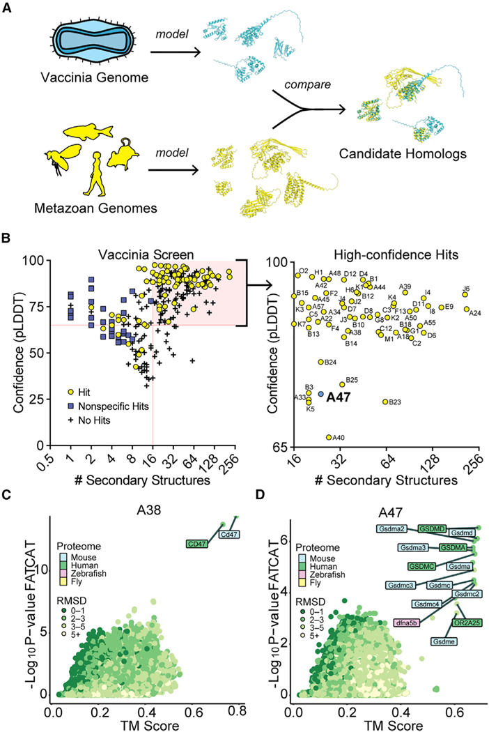
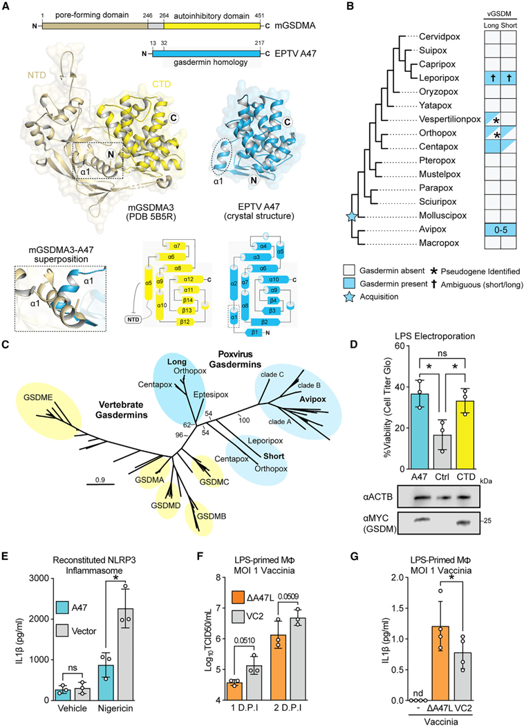
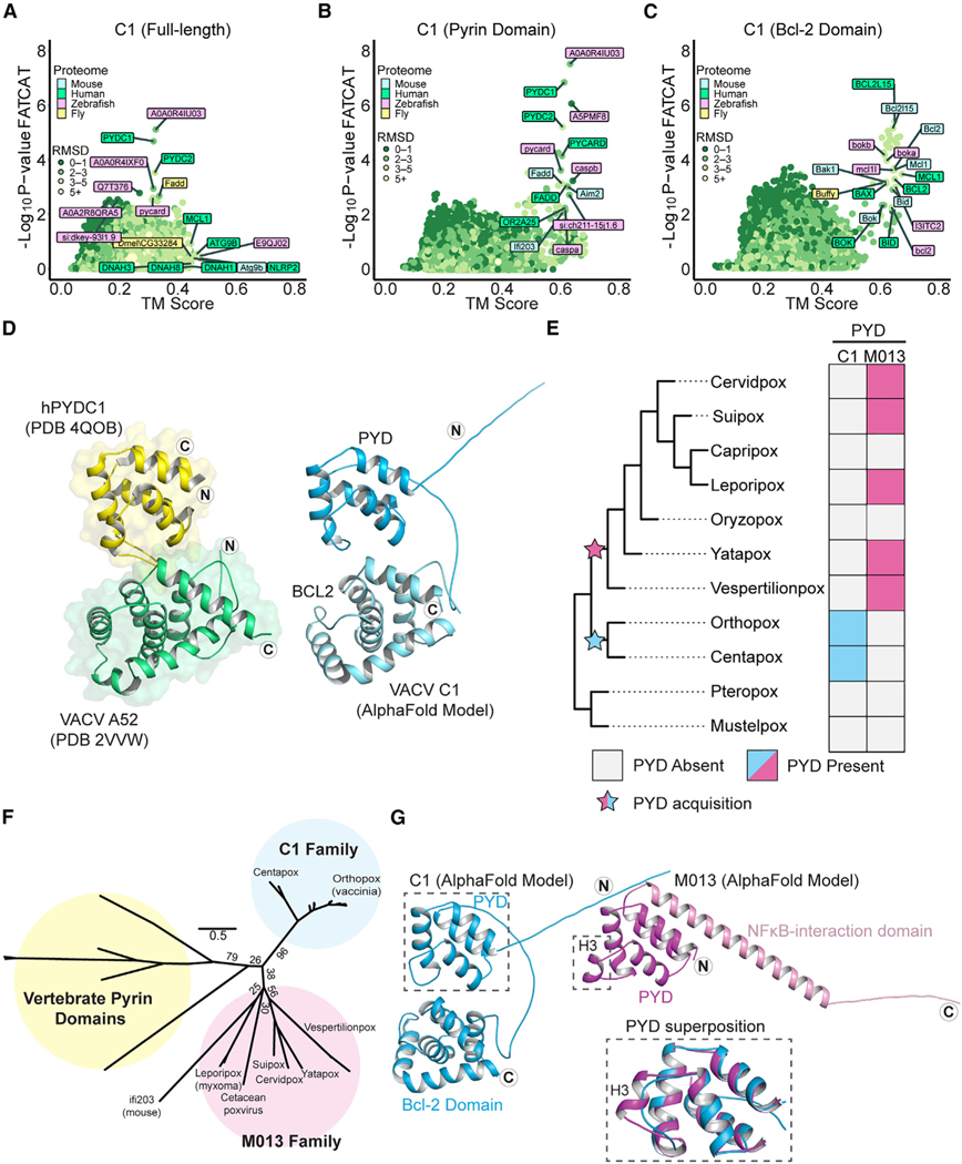
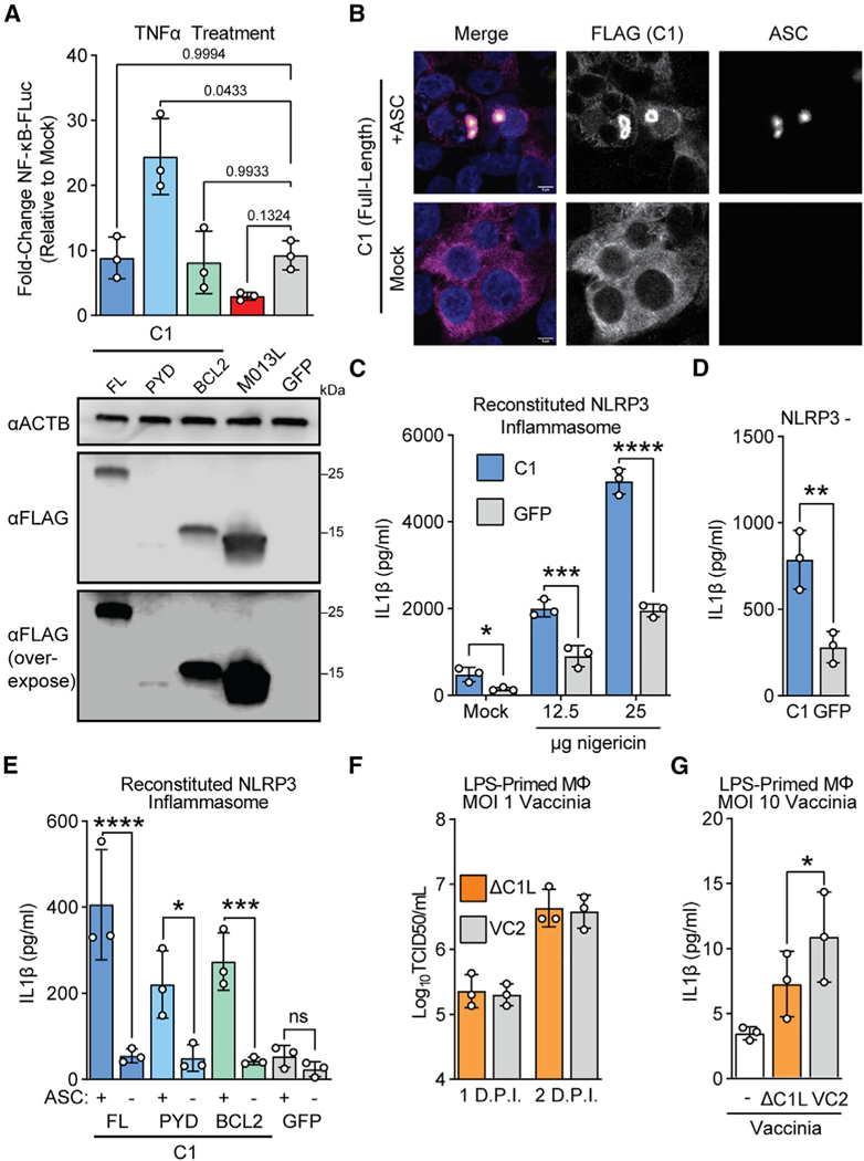
Viruses acquire host genes via horizontal transfer and can express them to manipulate host biology during infections. Some homologs retain sequence identity, but evolutionary divergence can obscure host origins. We use structural modeling to compare vaccinia virus proteins with metazoan proteomes. We identify vaccinia A47L as a homolog of gasdermins, the executioners of pyroptosis. An X-ray crystal structure of A47 confirms this homology, and cell-based assays reveal that A47 interferes with caspase function. We also identify vaccinia C1L as the product of a cryptic gene fusion event coupling a Bcl-2-related fold with a pyrin domain. C1 associates with components of the inflammasome, a cytosolic innate immune sensor involved in pyroptosis, yet paradoxically enhances inflammasome activity, suggesting differential modulation during infections. Our findings demonstrate the increasing power of structural homology screens to reveal proteins with unique combinations of domains that viruses capture from host genes and combine in unique ways.

Keywords: AlphaFold; CP: Immunology; CP: Microbiology; divergence; evolution; gasdermin; homology; inflammasome; poxvirus; pyroptosis; vaccinia; virus.


Conflict of interest statement

Declaration of interests The authors declare no competing interests.

Figures

Figure 1.
Figure 1.. Structure-based homology screens reveal candidate homologs of vaccinia proteins
(A) Homology screening pipeline. See STAR Methods for details. (B) Screen results. Right: high-confidence hits (those with model confidence [pLDDT] >65 and number of secondary structures >15 for which FATCAT and TM-align converged) are indicated. (C) FATCAT and TM-align results for A38, the poxvirus CD47 homolog. (D) FATCAT and TM-align results for A47.
Figure 2.
Figure 2.. A poxvirus homolog of gasdermins interferes with inflammatory caspases
(A) A crystal structure of Eptesipox virus (EPTV) gasdermin reveals homology to mammalian gasdermins. Top: domain organization of mouse GSDMA3 (mGSDMA3) and EPTV gasdermin indicating the pore-forming N-terminal domain (NTD) and the autoinhibitory C-terminal domain (CTD). Middle: crystal structures of full-length mGSDMA3 (PDB: 5B5R) and EPTV gasdermin (this study). The N and C termini are indicated with the circled letters N and C, and the dashed box and oval indicate the first α-helix of each structure. Bottom left: superposition of mGSDMA3 and EPTV gasdermin predicts a steric clash between the α1 helices of each protein. Bottom right: topology diagrams of the mGSDMA3 CTD and EPTV gasdermin indicate a conserved region of gasdermin homology. (B) Overview of probable gasdermin gain/loss events based on phylogenetic analyses in Figures 2C, S2H, and S2I. Gasdermins were identified by a combination of sequence and structure-based approaches; see STAR Methods for details. (C) Maximum likelihood tree of select vertebrate gasdermin regulatory domains and poxvirus gasdermins. Clusters of vertebrate gasdermins are labeled based on human gasdermins. Bootstrap values from 1,000 replicates are indicated for select branches. Scale: AA substitutions per site. (D) Top: HeLa cells expressing GSDMD-CTD, vaccina GSDM, or a vector control were electroporated with LPS to activate the noncanonical inflammasome. Viability as assessed by ATP levels relative to mock are indicated. n = 3 biological replicates. One-way ANOVA with Tukey’s multiple comparison test. Data are represented as mean ± SD. Bottom: alongside one replicate, levels of transfected gasdermin proteins in mock-electroporated wells were assessed by western blotting. (E) The murine NLRP3 inflammasome was reconstituted in 293T cells stably expressing A47L or a vector control. Following nigericin stimulation, IL-1β concentrations in supernatants were quantified by ELISA. n = 3 biological replicates. Two-way ANOVA with Šídák’s multiple comparisons test. Data are represented as mean ± SD. (F) Vaccinia replication (inoculum MOI of 1) in LPS-primed murine macrophages was measured by TCID50. n = 3 biological replicates. Two-way ANOVA with Fisher’s least significant difference (LSD) test. Data are represented as mean ± SD. ANOVA statistic for viral strain across time points: p = 0.0118. (G) LPS-primed murine macrophages were infected with wild-type (VC2) or A47-deficient vaccinia virus at an MOI of 1, and IL-1β was quantified by ELISA 1 day post-infection. n = 3 biological replicates. Paired t test. Data are represented as mean ± SD.
Figure 3.
Figure 3.. Vaccinia C1L encodes a unique pyrin-Bcl-2 fusion protein
(A) FATCAT and TM-align results for full-length C1 protein. Top results for each individual search tool are labeled. (B) FATCAT and TM-align results for the N-terminal (1–107) residues of C1 protein. Only a subset of hits are labeled. (C) FATCAT and TM-align results for the C-terminal (108–224) residues of C1 protein. Only a subset of hits are labeled. (D) C1 protein is a Pyrin-Bcl-2 fusion. Left: crystal structures of human PYDC1 (PDB: 4QOB) and vaccinia virus A52 (PDB: 2VVW). Right: AlphaFold model of vaccinia C1 protein. The N and C termini are indicated with the circled letters N and C. (E) Reconstructed gain/loss tree highlighting C1 and M013 pyrin-domain-containing proteins. (F) Maximum likelihood phylogenetic tree of vertebrate pyrin domains from Figure 4B alongside the M013 and C1 families. Bootstrap values from 1,000 replicates are indicated for select branches. Scale: AA substitutions per site. (G) AlphaFold models of vaccinia virus C1 protein and myxoma virus M013 protein. The N and C termini are indicated with circled letters N and C. Inset: superposition of the C1 and M013 pyrin domains showing the predicted absence of helix 3 in C1 protein.
Figure 4.
Figure 4.. C1 protein promotes ASC-dependent inflammasome activation
(A) Top: 293T cells were co-transfected with the indicated plasmids, a plasmid encoding firefly luciferase driven by an NF-κB promoter, and a constitutive Renilla luciferase plasmid as a transfection control. 24 h post-transfection, cells were treated with 1 ng/mL TNF-α, and promoter activity was assessed by luciferase assay 6 h post-transfection. n = 3 biological replicates. Repeated measures (RM) one-way ANOVA with Dunnett’s correction. Data are represented as mean ± SD. Bottom: in parallel, cells were transfected with equal amounts of the indicated plasmids, and protein expression was determined by western blotting. (B) 293T cells were co-transfected with a plasmid encoding FLAG-C1 and either ASC-GFP or an empty vector. 24 h post-transfection, cells were fixed and stained for FLAG (C1) and subsequently imaged. Representative image from n = 3 independent experiments. Scale bar: 5 μm. See also Figures S4B and S4C. (C) 293T cells were co-transfected with plasmids encoding the NLRP3 inflammasome components NLRP3, ASC, NEK7, caspase-1, and pro-IL-1β, in addition to either C1 or a GFP control. Following nigericin stimulation, IL-1β concentrations in supernatants were quantified by ELISA. n = 3 biological replicates. Two-way ANOVA with Šídák’s multiple comparisons test. Data are represented as mean ± SD. (D) 293T cells were co-transfected as in (C) but with NLRP3 replaced by an empty vector. Cells were not stimulated with nigericin. IL-1β concentrations in supernatants were quantified by ELISA. n = 3 biological replicates. Paired t test. Data are represented as mean ± SD. (E) 293T cells were co-transfected as in (C) but with ASC replaced by an empty vector where indicated. Cells were not stimulated with nigericin. IL-1β concentrations in supernatants were quantified by ELISA. n = 3 biological replicates. Two-way ANOVA with Šídák’s multiple comparisons test. Data are represented as mean ± SD. (F) Vaccinia replication (inoculum MOI of 1) in LPS-primed murine macrophages was measured by TCID50. Data are represented as mean ± SD. (G) LPS-primed murine macrophages were infected with wild-type (VC2) or C1L-deficient vaccinia virus at an MOI of 10, and IL-1β was quantified by ELISA 4 h post-infection. n = 3 biological replicates. Paired t test. Data are represented as mean ± SD.

Update of

Similar articles

Cited by

References

    1. Irwin NAT, Pittis AA, Richards TA, and Keeling PJ (2022). Systematic evaluation of horizontal gene transfer between eukaryotes and viruses. Nat. Microbiol 7, 327–336. 10.1038/s41564-021-01026-3. - DOI - PubMed
    1. Alcami A, and Koszinowski UH (2000). Viral mechanisms of immune evasion. Trends Microbiol. 8, 410–418. 10.1016/s0966-842x(00)01830-8. - DOI - PMC - PubMed
    1. Fixsen SM, Cone KR, Goldstein SA, Sasani TA, Quinlan AR, Rothenburg S, and Elde NC (2022). Poxviruses capture host genes by LINE-1 retrotransposition. Elife 11, e63332. 10.7554/eL-ife.63332. - DOI - PMC - PubMed
    1. Rahman MJ, Haller SL, Stoian AMM, Li J, Brennan G, and Rothenburg S. (2022). LINE-1 retrotransposons facilitate horizontal gene transfer into poxviruses. Elife 11, e63327. 10.7554/eLife.63327. - DOI - PMC - PubMed
    1. Salomon D, and Orth K. (2013). What pathogens have taught us about posttranslational modifications. Cell Host Microbe 14, 269–279. 10.1016/j.chom.2013.07.008. - DOI - PMC - PubMed

Publication types