. 2023 Jun 1;37(11-12):490-504.
doi: 10.1101/gad.350569.123. Epub 2023 Jun 26.

Major β cell-specific functions of NKX2.2 are mediated via the NK2-specific domain

Affiliations

Major β cell-specific functions of NKX2.2 are mediated via the NK2-specific domain

Vladimir Abarinov et al. Genes Dev. .

Abstract

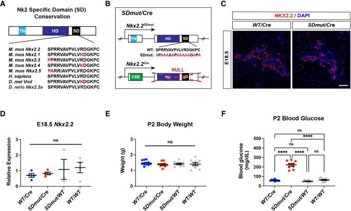
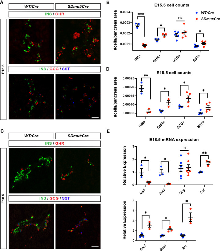
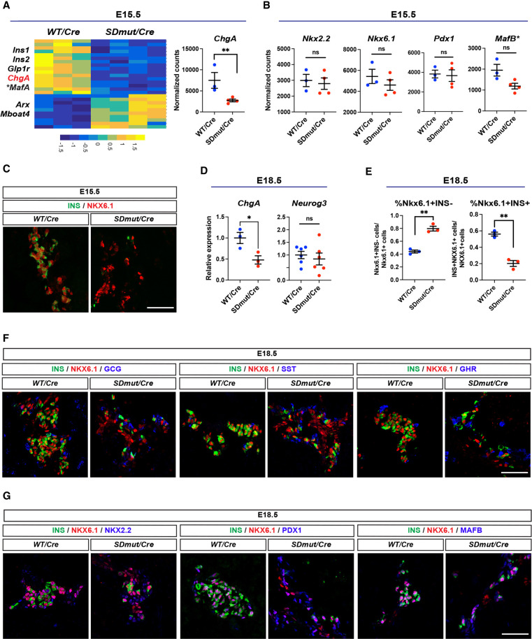
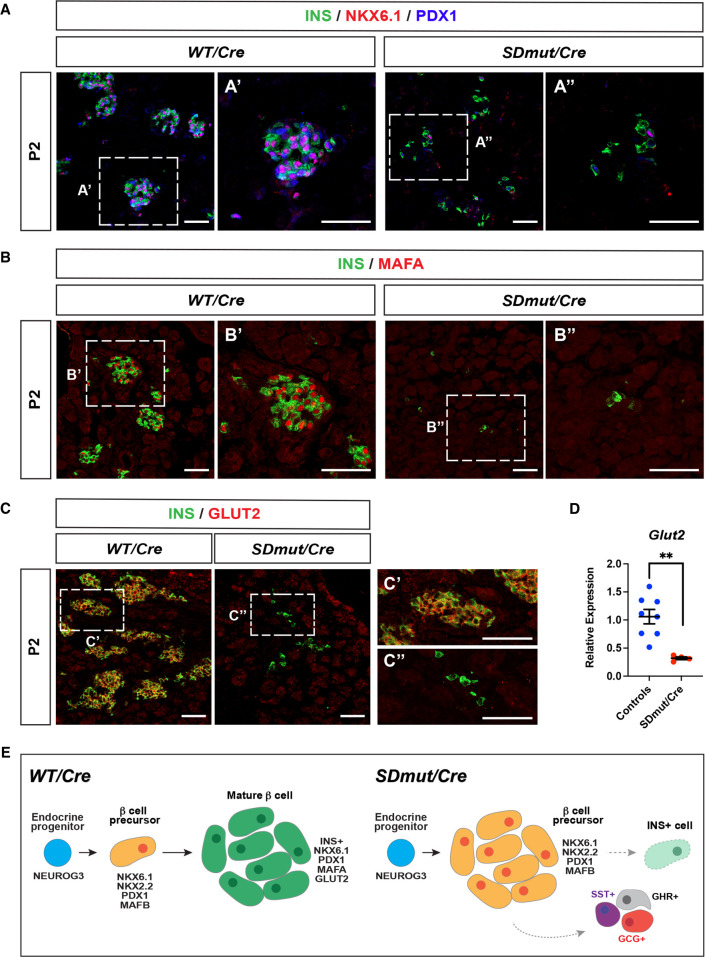
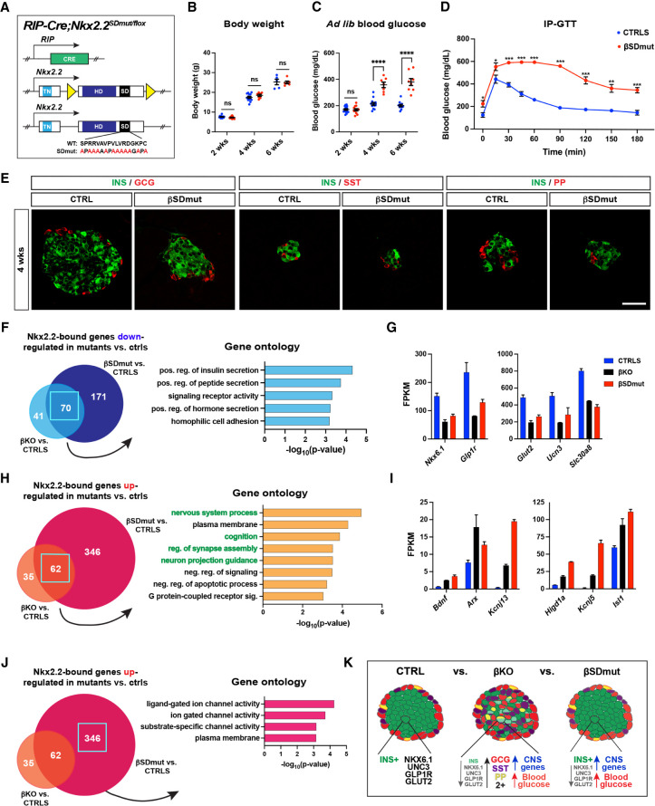
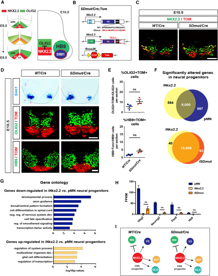
The consolidation of unambiguous cell fate commitment relies on the ability of transcription factors (TFs) to exert tissue-specific regulation of complex genetic networks. However, the mechanisms by which TFs establish such precise control over gene expression have remained elusive-especially in instances in which a single TF operates in two or more discrete cellular systems. In this study, we demonstrate that β cell-specific functions of NKX2.2 are driven by the highly conserved NK2-specific domain (SD). Mutation of the endogenous NKX2.2 SD prevents the developmental progression of β cell precursors into mature, insulin-expressing β cells, resulting in overt neonatal diabetes. Within the adult β cell, the SD stimulates β cell performance through the activation and repression of a subset of NKX2.2-regulated transcripts critical for β cell function. These irregularities in β cell gene expression may be mediated via SD-contingent interactions with components of chromatin remodelers and the nuclear pore complex. However, in stark contrast to these pancreatic phenotypes, the SD is entirely dispensable for the development of NKX2.2-dependent cell types within the CNS. Together, these results reveal a previously undetermined mechanism through which NKX2.2 directs disparate transcriptional programs in the pancreas versus neuroepithelium.

Keywords: NKX2.2; pancreatic islet; spinal cord; transcriptional regulation; β cells.


Figures

Figure 1.
Figure 1.
Nkx2.2SDmut/Cre animals develop overt neonatal diabetes. (A) Schematic highlighting amino acid conservation of the NK2-specific domain (SD). (B) Diagram of Nkx2.2 alleles used to generate Nkx2.2SDmut/Cre mice. (C) NKX2.2 protein expression is similar in Nkx2.2Cre/+ (WT/Cre) and Nkx2.2SDmut/Cre (SDmut/Cre) pancreata at E18.5. (D) E18.5 Nkx2.2 mRNA expression remains unchanged among wild-type, heterozygous, and Nkx2.2SDmut/Cre pancreata. (E,F) Body weight (E) and blood glucose levels (F) of P2 neonates. Data are presented as mean ± SEM. (ns) Not significant, (****) P < 0.0001. Scale bar, 50 µm.
Figure 2.
Figure 2.
Nkx2.2SDmut/Cre embryos show an expansion of other endocrine cell types at the expense of β cells beginning at E15.5. (AE) Characterization of insulin (INS+), glucagon (GCG+), ghrelin (GHR+), and somatostatin (SST+) cells in Nkx2.2SDmut/Cre (SDmut/Cre) and Nkx2.2Cre/+ (WT/Cre) embryos. (A,B) Immunostaining (A) and quantification (B) of endocrine cell types in E15.5 embryos. (CE) Immunostaining (C), quantification (D), and qRT-PCR analysis (E) at E18.8. Data are presented as mean ± SEM. (ns) Not significant, (*) P < 0.05, (**) P < 0.01, (***) P < 0.001. Scale bars represent 50 µm.
Figure 3.
Figure 3.
β Cells stall during differentiation in Nkx2.2SDmut/Cre embryos. (A, left) Heat map of significantly altered genes from RNA-seq analysis of E15.5 Nkx2.2Cre/+ (WT/Cre) and Nkx2.2SDmut/Cre (SDmut/Cre) pancreata. (Right) Normalized expression of ChgA, a pan-endocrine marker. (*) MafA adjusted P value = 0.057. (B) Normalized expression of key transcription factors (TFs) needed for β cell differentiation. (*) MafB adjusted P value = 0.79 or nonadjusted P value = 0.0040. (C) Immunostaining confirmed NKX6.1 expression in the E15.5 mutant pancreas. (D) qRT-PCR of ChgA and Neurog3 in E18.5 pancreata. (E) Percentage of NKX6.1+INS and NKX6.1+INS+ cells at E18.5. (F,G) NKX6.1+INS cells in Nkx2.2SDmut/Cre animals do not express GCG, SST, or GHR at E18.5 (F) but do express the β cell TFs NKX2.2, PDX1, and MAFB (G). Data are presented as mean ± SEM. (ns) Not significant, (*) P < 0.05, (**) P < 0.01. Scale bars, 50 µm.
Figure 4.
Figure 4.
Residual INS+ cells in Nkx2.2SDmut/Cre mice lack features characteristic of functionally mature β cells. (AC) The majority of the remaining INS+ cells in Nkx2.2SDmut/Cre (SDmut/Cre) animals do not exhibit NKX6.1 or PDX1 coexpression at P2 (shown in A). Similarly, MAFA (B) and GLUT2 (C) expression is also not observed. A, panel A′; B, panel B′; and C, panel C′ show higher magnification of Nkx2.2Cre/+ (WT/Cre) images, while A, panel A′′; B, panel B′′; and C, panel C′′ show higher magnification of Nkx2.2SDmut/Cre staining. (D) qRT-PCR of Glut2 in P2 pancreata. (E) Schematic summarizing changes in β cell development resulting from mutation of the SD. Data are presented as mean ± SEM. (**) P < 0.01. Scale bars, 50 µm.
Figure 5.
Figure 5.
RIP-Cre;Nkx2.2SDmut/flox (βSDmut) animals develop diabetes due to impaired β cell function. (A) Schematic of alleles used to generate βSDmut mice. (B,C) Body weight (B) and ad lib blood glucose levels (C) in male βSDmut animals. (D) βSDmut males show elevated fasting blood glucose levels and impaired glucose clearance at 4 wk compared with littermate RIP-Cre;Nkx2.2flox/+ control animals (CTRLS) during the intraperitoneal glucose tolerance test (IP-GTT). (E) INS, SST, and PP expression in 4-wk βSDmut animals. (FK) RNA-seq analysis of NKX2.2-bound genes that are significantly altered in βSDmut islets versus CTRLS compared with NKX2.2-bound genes that are significantly altered in RIP-Cre;Nkx2.2flox/flox (βKO) animals versus CTRLS. (F) Gene ontology (GO) analysis of the 70 genes bound by NKX2.2 and down-regulated in both βSDmut and βKO islets compared with CTRL mice. (G) FPKM values of selected genes from F. (H) GO analysis of the 62 genes bound by NKX2.2 and up-regulated in both βSDmut and βKO versus CTRL animals. (I) FPKM values of selected genes from H. (J) GO analysis of the 346 transcripts bound by NKX2.2 and only up-regulated in βSDmut animals. (K) Schematic comparing differences in islet morphology and function in βKO versus βSDmut mice. Data are presented as mean ± SEM. (ns) Not significant, (*) P < 0.05, (**) P < 0.01, (***) P < 0.001. Scale bars represent 50 µm.
Figure 6.
Figure 6.
NKX2.2-dependent CNS populations are not affected by mutation of the SD. (A) Schematic representing the onset of NKX2.2-mediated repression of motor neuron (MN) identity. (B) Diagram of NKX2.2 alleles used to generate Nkx2.2SDmut/Cre (SDmut/Cre) and Nkx2.2Cre/+ (WT/Cre) mice. The Rosa26-Tomato reporter allele (TOM) was included to enable lineage tracing. (C) NKX2.2 and TOM expression in E10.5 embryos. (D) In situ hybridization of Sim1, as well as OLIG2 and HB9 immunostaining, in Nkx2.2SDmut/Cre and Nkx2.2Cre/+ E10.5 spinal cords. (E) Quantification of the percentage of OLIG2+TOM+ and HB9+TOM+ cells from D. (F) RNA-seq analysis of mouse embryonic stem cells (ESCs) differentiated into control motor neuron progenitors (pMN) and pMN progenitors in which either wild-type (iNKX2.2) or SD mutant (iSDmut) NKX2.2 was ectopically induced. (G) Gene ontology (GO) analysis of transcripts differentially expressed in iNKX2.2 progenitors compared with pMN cells. (H) FPKM values of TFs necessary for MN development. (I) Schematic showing that all examined NKX2.2-dependent CNS populations (see Supplemental Fig. S8) are not affected by mutation of the SD. Data are presented as mean ± SEM. (ns) Not significant. Scale bars, 50 µm.

Similar articles

Cited by

References

    1. Apelqvist A, Li H, Sommer L, Beatus P, Anderson DJ, Honjo T, Hrabe De Angelis M, Lendahl U, Edlund H. 1999. Notch signalling controls pancreatic cell differentiation. Nature 400: 877–881. 10.1038/23716 - DOI - PubMed
    1. Arnes L, Leclerc K, Friel JM, Hipkens SB, Magnuson MA, Sussel L. 2012. Generation of Nkx2.2:lacZ mice using recombination-mediated cassette exchange technology. Genesis 50: 612–624. 10.1002/dvg.22037 - DOI - PMC - PubMed
    1. Artner I, Le Lay J, Hang Y, Elghazi L, Schisler JC, Henderson E, Sosa-Pineda B, Stein R. 2006. Mafb: an activator of the glucagon gene expressed in developing islet α- and β-cells. Diabetes 55: 297–304. 10.2337/diabetes.55.02.06.db05-0946 - DOI - PubMed
    1. Artner I, Blanchi B, Raum JC, Guo M, Kaneko T, Cordes S, Sieweke M, Stein R. 2007. Mafb is required for islet β cell maturation. Proc Natl Acad Sci 104: 3853–3858. 10.1073/pnas.0700013104 - DOI - PMC - PubMed
    1. Babu DA, Deering TG, Mirmira RG. 2007. A feat of metabolic proportions: Pdx1 orchestrates islet development and function in the maintenance of glucose homeostasis. Mol Genet Metab 92: 43–55. 10.1016/j.ymgme.2007.06.008 - DOI - PMC - PubMed

Publication types

MeSH terms

LinkOut - more resources