. 2023 May;12(13):e2201794.
doi: 10.1002/adhm.202201794. Epub 2023 Feb 24.

Design of Nanohydrogels for Targeted Intracellular Drug Transport to the Trans-Golgi Network

Affiliations

Design of Nanohydrogels for Targeted Intracellular Drug Transport to the Trans-Golgi Network

Thorsten Keller et al. Adv Healthc Mater. 2023 May.

Abstract

Nanohydrogels combine advantages of hydrogels and nanoparticles. In particular, they represent promising drug delivery systems. Nanogel synthesis by oxidative condensation of polyglycidol prepolymers, that are modified with thiol groups, results in crosslinking by disulfide bonds. Hereby, biomolecules like the antidiabetic peptide RS1-reg, derived from the regulatory protein RS1 of the Na+ -D-glucose cotransporter SGLT1, can be covalently bound by cysteine residues to the nanogel in a hydrophilic, stabilizing environment. After oral uptake, the acid-stable nanogels protect their loading during gastric passage from proteolytic degradation. Under alkaline conditions in small intestine the nanohydrogels become mucoadhesive, pass the intestinal mucosa and are taken up into small intestinal enterocytes by endocytosis. Using Caco-2 cells as a model for small intestinal enterocytes, by confocal laser scanning microscopy and structured illumination microscopy, the colocalization of fluorescent-labeled RS1-reg with markers of endosomes, lysosomes, and trans-Golgi-network after uptake with polyglycidol-based nanogels formed by precipitation polymerization is demonstrated. This indicates that RS1-reg follows the endosomal pathway. In the following, the design of bespoken nanohydrogels for specific targeting of RS1-reg to its site of action at the trans-Golgi network is described that might also represent a way of targeted transport for other drugs to their targets at the Golgi apparatus.

Keywords: drug delivery; nanohydrogels; regulation of the Na+-D-glucose cotransporter SGLT1 in intestine; regulatory protein RS1; targeted transport.


Conflict of interest statement

The authors declare no conflict of interest.

Figures

Figure 1
Figure 1
A) SDS‐PAGE showing purification of recombinant hRS1‐reg(S20E) and mRS1‐reg(S19E) expressed in E. coli. After protein expression for 3 h at 30°C, bacteria were lysed and centrifuged for 1 h at 100 000 × g. The supernatant (bacterial lysate) was incubated for 1 h with Ni2+‐NTA agarose, the suspension was poured into an empty gravity flow column and the flow‐through was removed. The resin was washed and purified RS1‐reg peptides were eluted with 500 × 10−3 m imidazole (eluate). The samples were subjected to SDS‐PAGE analysis. The gel was stained with Coomassie Brilliant Blue. Per lane, 10 µg (lysates and flow‐through) or 2 µg of protein (purified RS1‐reg) was applied. B) Scheme of nanohydrogel preparation by oxidative coupling of thiol‐functionalized polyglycidol prepolymers via crosslinking by formation of disulfide bridges. A cargo (for example the RS1‐reg peptide) can be coupled to the matrix via cysteine residues. Under the reducing conditions in the cytosol after cellular uptake, the nanogels are degraded under drug release (for further chemical details of the components see Figure S5, Supporting Information). C) Confocal laser scanning microscopy analysis, demonstrating uptake of ATTO488‐labeled RS1‐reg peptide (green fluorescence) after preincubation with polyglycidol‐based nanohydrogel, formed in inverse miniemulsion, into Caco‐2 cells (1) (staining of nuclei with Hoechst 33 342, staining of plasma membranes with Claret Far Red, Bar = 15 µm) and into small intestine of mouse (2), pig (3) and human (4) (Red Bars = 200 µm).
Figure 2
Figure 2
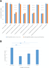
A) Downregulation of SGLT1‐mediated AMG uptake expressed in Caco‐2 cells or mouse small intestinal segments after preincubation with polyglycidol‐based nanohydrogels containing hRS1‐reg(S20E) (for experiments with Caco‐2 cells) or mRS1‐reg(S19E) (for measurements with mouse small intestinal segments), formed in inverse miniemulsion or by inverse nanoprecipitation either with or without TAT peptide. Independent of the way of preparation of nanogels containing RS1‐reg, AMG uptake was reduced to the same extent, by about 50 percent, in Caco‐2 cells and segments of mouse small intestine. No inhibition of AMG uptake was observed in approaches with empty nanogels or with free RS1reg peptides. Presence or absence of the cell‐penetrating TAT peptide did not change the results. B) Dose‐dependent inhibition of AMG uptake into Caco‐2 cells by a dilution series of hRS1‐reg(S20E) containing nanohydrogels out of nanoprecipitation without TAT peptide. Mean values ± S.E. of 9 wells with confluent Caco‐2 cells or rather mouse small intestinal segments from three independent experiments are shown, respectively. *P < 0.05; **P < 0.01; ***P < 0.001 for significance of difference from control tested by ANOVA with post hoc Tukey comparison.
Figure 3
Figure 3
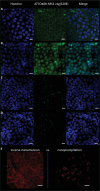
Confocal laser scanning microscopy (CLSM) analysis of the localization of ATTO488‐hRS1‐reg(S20E) in fixed Caco‐2 cells after uptake via nanohydrogels formed in A,B) inverse miniemulsion or C,D) by inverse nanoprecipitation either with B,D) or without A,C) cell‐penetrating TAT peptide. While in case of nanogel out of inverse miniemulsion without A) TAT an even distribution of the green fluorescent peptide in the cytosol can be detected, suggesting successful endosomal escape, the presence of B) TAT directs the majority of RS1‐reg signal with it into the cell nuclei. Uptake via nanoprecipitation nanogels in presence or absence of C,D) TAT results in a selective accumulation of ATTO488‐RS1‐reg in defined vesicular cytosolic compartments, suggesting impaired endosomal escape. A–D) The left column displays the staining of nuclei with Hoechst 33 342, in the middle column the signal of ATTO488‐labeled hRS1‐reg(S20E) can be seen and the right column shows merged signals. In E) a direct comparison of the localization of Alexa647‐labeled hRS1‐reg following uptake via nanohydrogels out of inverse miniemulsion (even distribution in the cytosol) versus nanoprecipitation (selective enrichment in cytosolic vesicles) without TAT is depicted, indicating that different ways of preparing nanohydrogels out of the same chemical compounds result in different intracellular localization of the cargo RS1‐reg. Bars = 15 µm.
Figure 4
Figure 4
Colocalization analysis in fixed, permeabilized, nonembedded Caco‐2 cells via structured illumination microscopy (SIM) of the distribution of Alexa488‐labeled hRS1‐reg(S20E) after uptake with nanogels formed by inverse nanoprecipitation. Colocalization was tested with marker proteins for endosomes (EEA1, row A), trans‐Golgi network (TGN46, row B), endoplasmatic reticulum (Calnexin, row C), lysosomes (Lamp‐1, row D) and with the enzyme ODC1 (row E). A partial colocalization was detected with EEA1, TGN46, Lamp‐1 and ODC1, respectively, but no colocalization with calnexin. In the left column the signal of the respective marker protein is visible, in the neighboring column the Alexa488‐RS1‐reg signal is displayed, followed by the staining of cell nuclei with Hoechst 34580 in the next column and merged signals in the right column. The results suggest impaired endosomal escape of RS1‐reg, which follows the endosomal way from early to late endosomes, where partial transport to the trans‐Golgi networks occurs via vesicle exchange. Residual RS1reg is finally degraded after fusion with lysosomes in the resulting endolysosomes. Bars = 15 µm.
Figure 5
Figure 5
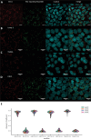
Colocalization analysis in fixed, permeabilized Caco‐2 cells via structured illumination microscopy (SIM) of the distribution of Alexa488‐labeled hRS1‐reg(S20E) after uptake with nanogels formed by inverse nanoprecipitation. Caco‐2 cells were embedded to allow for a precise channel alignment, resulting in a more precise localization of the RS1‐reg peptide and the marker proteins for different cellular structures. Marker proteins of endosomes (EEA1, row A), lysosomes (Lamp‐1, row B), trans‐Golgi network (TGN46, row C) and the enzyme ODC1 (row D) were tested for colocalization with Alexa488‐RS1‐reg. A partial colocalization was detected in all approaches. The left column shows the signal of the respective marker protein, in the neighboring column the Alexa488‐RS1‐reg signal can be seen, followed by the staining of cell nuclei with Hoechst 34580 in the next column and merged signals in the right column. The results indicate no endosomal escape of RS1‐reg. The peptide appears to pursue the endosomal route, by passing from early to late endosomes, where it can be translocated in part to the TGN by vesicle exchange. Remaining hRS1‐reg(S20E) is finally cleaved proteolytically in secondary lysosomes. Bars = 15 µm. E) Violin‐Plot of quantitative colocalization analysis employing all pictures of 4 stacks regarding the green signals of Alexa488‐RS1‐reg (Ch2) and red fluorescence of marker proteins of endosomes (EEA1), lysosomes (Lamp‐1), trans‐Golgi network (TGN46) and the enzyme ODC1 (Ch1, respectively). Pearson's coefficients were calculated using a custom‐written ImageJ macro (see Supporting Information). Pearson coefficients > 0.7 indicate a high positive linear correlation between the respective characteristics under consideration. As a control the colocalization between the signals of the marker proteins (Ch1) and nuclei (Ch3) was investigated, resulting in Pearson coefficients of approximately 0, indicating no dependence of the two characteristics on each other, as expected.
Figure 6
Figure 6
Model scheme for the uptake of the cargo RS1‐reg via nanohydrogels out of inverse miniemulsion (right side) or inverse nanoprecipitation (left side). Following cellular uptake with nanogels formed in inverse emulsion, that are softer probably due to a lower crosslinking degree, endosomal escape of RS1‐reg is possible, leading to an even distribution in the cytosol. Thus the antidiabetic peptide also reaches the trans‐Golgi network, where it can exert its biological function of inhibiting glucose uptake by blockage of the release of SGLT1‐containing vesicles. On the other hand after uptake via nanoprecipitation nanohydrogels the endosomal escape of RS1‐reg is impaired, because of the higher stability probably resulting from a higher crosslinking degree. The peptide follows the endosomal pathway from early to late endosomes, where it is partially transported to its site of action at the TGN by vesicle exchange. Residual RS1‐reg is finally degraded after fusion with lysosomes in the resulting endolysosomes.

Similar articles

References

    1. Peppas N. A., Bures P., Leobandung W., Ichikawa H., Eur. J. Pharm. Biopharm. 2000, 50, 27. - PubMed
    1. Kamath K. R., Park K., Adv. Drug Delivery Rev. 1993, 11, 59.
    1. Kim S. W., Bae Y. H., Okano T., Pharm Res 1992, 9, 283. - PubMed
    1. Schwall C. T., Banerjee I. A., Materials 2009, 2, 577.
    1. Cuggino J. C., Blanco E. R. O., Gugliotta L. M., Igarzabal C. I. A., Calderón M., J. Controlled Release 2019, 307, 221. - PubMed

Publication types

MeSH terms

Substances

LinkOut - more resources