. 2022 Oct 10:13:1002588.
doi: 10.3389/fphar.2022.1002588. eCollection 2022.

A comprehensive analysis of the prognostic value and immune infiltration of low expression DBT in clear cell renal cell carcinoma

Affiliations

A comprehensive analysis of the prognostic value and immune infiltration of low expression DBT in clear cell renal cell carcinoma

Wenjie Xie et al. Front Pharmacol. .

Abstract

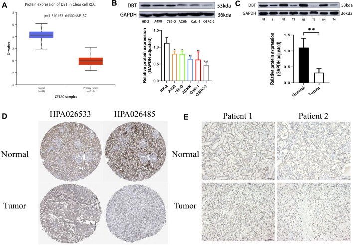
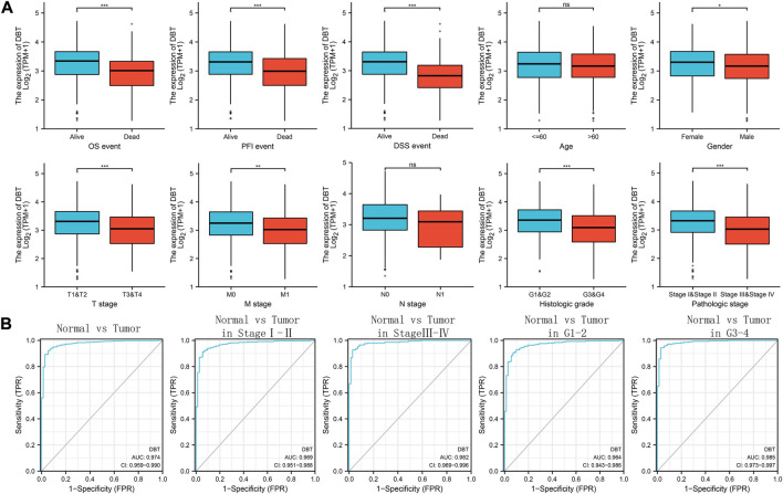
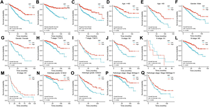
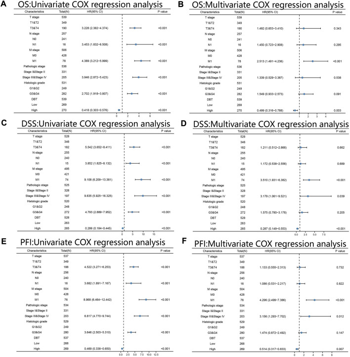
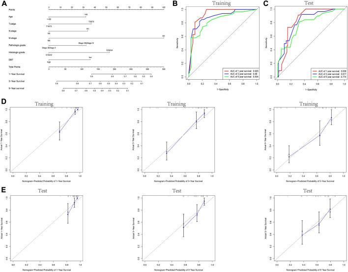
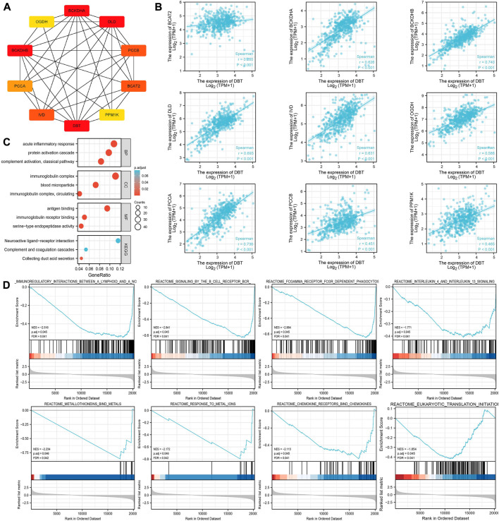
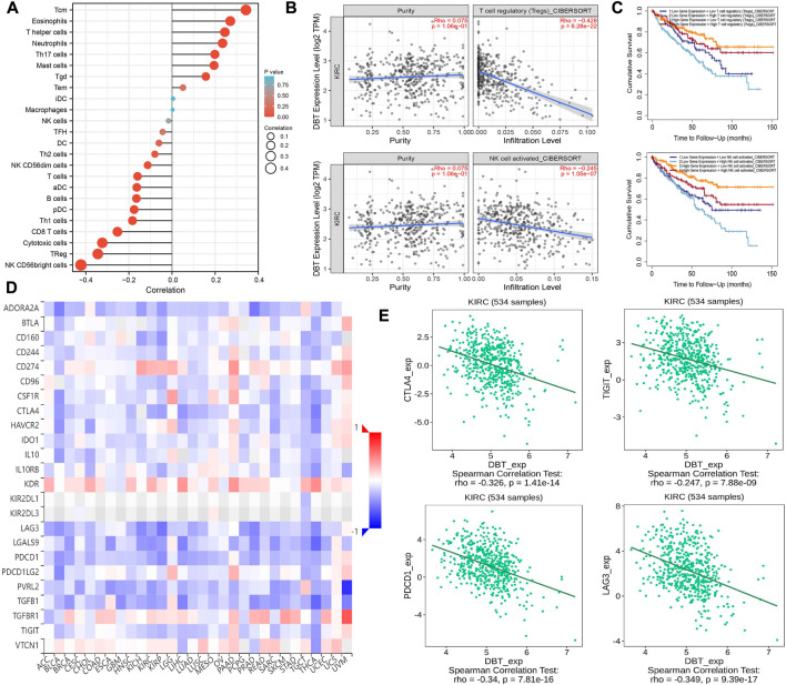
Background: Although DBT is strongly associated with human tumorigenesis and progression through a variety of pathways, the role of DBT in clear cell renal cell carcinoma (ccRCC) has not been well established. Materials and methods: The Cancer Genome Atlas (TCGA)-Kidney renal clear cell carcinoma (KIRC) databset provides RNA sequencing data and clinicopathological information on ccRCC. The Gene Expression Omnibus (GEO) database was used to validate the DBT expression levels, and qPCR was used to examine the DBT expression in renal cancer cell lines and ccRCC tissue samples from our centre. In parallel, DBT protein expression was explored in the Human Protein Atlas (HPA) database, and western blotting and immunohistochemistry of renal cancer cell lines and ccRCC tissues validated the results. Additionally, the diagnostic and prognostic value of DBT was comprehensively evaluated by receiver operating characteristic (ROC) curves, univariate and multivariate Cox regression analyses, and Kaplan‒Meier survival analysis. The protein‒protein interaction (PPI) network based on the STRING website, Gene Ontology (GO) analysis, Kyoto Gene and Genome Encyclopedia (KEGG) analysis and gene set enrichment analysis (GSEA) further provided a landscape of the molecular mechanisms of DBT in ccRCC. Finally, the TIMER 2.0, GEPIA and TISIDB websites were used to understand the relationship between DBT and immune characteristics. Results: The mRNA expression and protein expression of DBT were significantly downregulated in ccRCC tissues relative to normal tissues, which was associated with poor clinical outcomes. DBT has an encouraging discriminatory power for ccRCC and is an independent prognostic factor for ccRCC patients. Mechanistically, DBT is mainly involved in the regulation of immune-related signalling pathways in ccRCC; it is associated with a variety of immune infiltrating cells and immune checkpoints. Conclusion: DBT is a tumour suppressor gene in ccRCC and could be used as a new biomarker for diagnostic and prognostic purposes, and it is associated with immune infiltration in ccRCC.

Keywords: DBT; bioInformatic; clear cell renal cell carcinoma; immune infiltration; prognostic biomarker.


Conflict of interest statement

The authors declare that the research was conducted in the absence of any commercial or financial relationships that could be construed as a potential conflict of interest.

Figures

FIGURE 1
FIGURE 1
Low expression of DBT at the RNA level in ccRCC. (A) DBT showed low expression in the TCGA database and GSE40435, GSE5300, GSE53757, and GSE105261. (B) DBT was verified in renal cancer cell lines (A498, 786-O, ACHN, Caki-1, and OSRC-2) and renal tubular epithelial cells (HK-2) at the RNA expression level to have a low expression status. (C) DBT expression was lower in 14 renal clear cell carcinoma tissues than in the corresponding normal tissues. *p < 0.05, **p < 0.01, ***p < 0.001, “ns”: not statistically significant.
FIGURE 2
FIGURE 2
DBT protein expression is low in ccRCC. (A) The histogram obtained from the analysis of data from the Clinical Proteomic Tumor Analysis Consortium (CPTAC) on the UALCAN website shows significantly lower expression of the DBT protein in ccRCC tissues versus normal tissues (p = 1.31015316430268E-57). (B) The results of western blotting of A498, 786-O, ACHN, Caki-1, and OSRC-2 cell lysates showed lower protein expression of DBT in ccRCC cells compared to HK-2 cells, and the histogram quantifying the results also indicated a statistically significant difference. (C) Similarly, DBT protein expression was lower in renal clear cell carcinoma tissues than in matched pairs of paraneoplastic normal tissues, and the histogram quantification confirmed this finding. (D) Immunohistochemical results from the HPA database showed that DBT expression in ccRCC was mainly in the nucleus, whereas in normal renal tissue, it was in normal tubular epithelial cells. (E) This result was also validated by the immunohistochemical results of 2 pairs of ccRCC tissues from our study centre. *p < 0.05, **p < 0.01, ***pp < 0.001, “ns”: not statistically significant,“N”:Normal,“T”:Tumor.
FIGURE 3
FIGURE 3
Relationship between DBT expression and clinical features and its diagnostic value. (A) Low expression of DBT was significantly related to fatal events in terms of OS, PFI, and DSS, while it was also observed in men and those with higher T stage, M stage, histologic grade and pathologic stage, but there was no significant difference in age or N stage. (B) For the diagnostic ability of DBT for ccRCC tumours, the AUC value of DBT for distinguishing ccRCC tissues and normal tissues was 0.974, while the AUC values for distinguishing between different subtypes of ccRCC in pathological stages I-II and III-IV and histological grades 1–2 and 3–4 were 0.974, 0.969, 0.982, 0.964, and 0.985, respectively. *p < 0.05, **p < 0.01, ***p < 0.001, “ns”: Not statistically significant.
FIGURE 4
FIGURE 4
Relationship between DBT expression and the survival of ccRCC patients. In terms of outcomes such as OS (A), DSS (B), and PFI (C), low expression of DBT in ccRCC patients indicated a better prognosis in groups based on age (D, E), male sex (F), female sex (G), T1-T2 stage (H), N0 stage (J), M0 stage (L), M1 stage (M), histologic G1-G2 stage (N), histologic G3-G4 stage (O), and pathologic stages I-II (P). However, there were no significant differences in the prognosis of DBT expression groups for patients with T3-T4 stage (I), N1 stage (K), and pathologic stages III-IV (Q).
FIGURE 5
FIGURE 5
Univariate and multivariate Cox regression analyses. Univariate and multivariate Cox regression analyses of ccRCC based on the expression levels of DBT, T stage, N stage, M stage, pathologic stage, and histologic grade for OS (A,B), DSS (C,D) and PFI (E,F). All results consistently showed that DBT alone could be an independent factor in determining the prognosis of ccRCC patients. OS: overall survival; DSS: disease-specific survival; PFI: progression-free interval.
FIGURE 6
FIGURE 6
Construction and validation of the nomogram. (A) Prognostic nomogram constructed by combining data on clinically relevant factors (e.g., age, T stage, N stage, M stage, pathologic grade and histologic grade) and DBT expression. The TCGA-KIRC dataset was randomly divided into the training and test groups, and the ROC curves for 1, 3, and 5 years were plotted for the training and test groups based on the constructed prognostic judgement model. The AUC values predicting 1-year, 3-year, and 5-year survival were 0.925, 0.89, and 0.834 in the training group (B) and 0.839, 0.817, and 0.774 in the test group (C), respectively. (D,E) Calibration plots to assess the nomogram demonstrate its predictive power for 1-year, 3-year, and 5-year survival rates in the training and test groups of the TCGA-KIRC dataset.
FIGURE 7
FIGURE 7
Exploration of the potential functions of DBT in ccRCC. (A) The STRING website was used to analyse the DBT-related protein‒protein interaction network, which showed the associations between DBT and BCAT2, BCKDHA, BCKDHB, DLD, IVD, OGDH, PCCA, PCCB, and PPM1K. (B) Spearman analysis further demonstrated the associations between these genes, and significant positive associations between DBT and all of them were observed based on the results. (C) Gene Ontology (GO) analysis of biological processe (BP), cellular component (CC) and molecular function (MF), and Kyoto Encyclopedia of Genes and Genomes (KEGG) analysis were performed to obtain a comprehensive view of the potential functions of DBT in ccRCC. (D) Enrichment plots of DBT expression in terms of immunoregulatory interactions between a lymphoid and a nonlymphoid cell (NES = −2.516, p. adj = 0.045, FDR = 0.041), signalling by the B-cell receptor (NES = −2.841, p. adj = 0.045, FDR = 0.041), FCGAMMA receptor FCGR-dependent phagocytosis (NES = −2.884, p. adj = 0.045, FDR = 0.041), interleukin-4 and interleukin-13 signalling (NES = −1.771, p. adj = 0.045, FDR = 0.041), metallothioneins bind metals (NES = −2.234, p. adj = 0.046, FDR = 0.042), response to metal ions (NES = −2.172, p. adj = 0.046, FDR = 0.042), chemokine receptors bind chemokines (NES = -2.113, p. adj = 0.045, FDR = 0.041), and eukaryotic translation initiation (NES = −1.854, p. adj = 0.045, FDR = 0.041).
FIGURE 8
FIGURE 8
Exploration of the immune characteristics of DBT in ccRCC. (A) The correlations between TILs and DBT expression in different immune cells were calculated based on the “GSVA” package, and most of the immune cells were found to have some relationship with DBT expression. (B) DBT expression was found to be negatively correlated with the immune infiltration levels of regulatory T cells (Treg) and activated NK cells through the TIMER2.0 website based on the “CIBERSORT” algorithm. (C) Similarly, DBT gene expression was analysed in relation to Treg and activated NK-cell immune infiltration levels for the survival of ccRCC patients. (D) The relevance of immunosuppression across cancers. (E) There were significant negative correlations between DBT expression and immune checkpoint genes (CTLA4, TIGIT, LAG3, and PDCD1) in ccRCC.

Similar articles

Cited by

References

    1. Ahn S. H., Yang H. Y., Tran G. B., Kwon J., Son K. Y., Kim S., et al. (2015). Interaction of peroxiredoxin V with dihydrolipoamide branched chain transacylase E2 (DBT) in mouse kidney under hypoxia. Proteome Sci. 13, 4. 10.1186/s12953-014-0061-2 - DOI - PMC - PubMed
    1. Ardolino M., Zingoni A., Cerboni C., Cecere F., Soriani A., Iannitto M. L., et al. (2011). DNAM-1 ligand expression on Ag-stimulated T lymphocytes is mediated by ROS-dependent activation of DNA-damage response: Relevance for NK-T cell interaction. Blood 117 (18), 4778–4786. 10.1182/blood-2010-08-300954 - DOI - PubMed
    1. Atkins M. B., Tannir N. M. (2018). Current and emerging therapies for first-line treatment of metastatic clear cell renal cell carcinoma. Cancer Treat. Rev. 70, 127–137. 10.1016/j.ctrv.2018.07.009 - DOI - PubMed
    1. Braun D. A., Bakouny Z., Hirsch L., Flippot R., Van Allen E. M., Wu C. J., et al. (2021). Beyond conventional immune-checkpoint inhibition - novel immunotherapies for renal cell carcinoma. Nat. Rev. Clin. Oncol. 18 (4), 199–214. 10.1038/s41571-020-00455-z - DOI - PMC - PubMed
    1. Cerboni C., Zingoni A., Cippitelli M., Piccoli M., Frati L., Santoni A. (2007). Antigen-activated human T lymphocytes express cell-surface NKG2D ligands via an ATM/ATR-dependent mechanism and become susceptible to autologous NK- cell lysis. Blood 110 (2), 606–615. 10.1182/blood-2006-10-052720 - DOI - PubMed

LinkOut - more resources