. 2022 Oct 12;13(1):6005.
doi: 10.1038/s41467-022-33434-2.

The coilin N-terminus mediates multivalent interactions between coilin and Nopp140 to form and maintain Cajal bodies

Affiliations

The coilin N-terminus mediates multivalent interactions between coilin and Nopp140 to form and maintain Cajal bodies

Edward Courchaine et al. Nat Commun. .

Abstract

Cajal bodies (CBs) are ubiquitous nuclear membraneless organelles (MLOs) that concentrate and promote efficient biogenesis of snRNA-protein complexes involved in splicing (snRNPs). Depletion of the CB scaffolding protein coilin disperses snRNPs, making CBs a model system for studying the structure and function of MLOs. Although it is assumed that CBs form through condensation, the biomolecular interactions responsible remain elusive. Here, we discover the unexpected capacity of coilin's N-terminal domain (NTD) to form extensive fibrils in the cytoplasm and discrete nuclear puncta in vivo. Single amino acid mutational analysis reveals distinct molecular interactions between coilin NTD proteins to form fibrils and additional NTD interactions with the nuclear Nopp140 protein to form puncta. We provide evidence that Nopp140 has condensation capacity and is required for CB assembly. From these observations, we propose a model in which coilin NTD-NTD mediated assemblies make multivalent contacts with Nopp140 to achieve biomolecular condensation in the nucleus.


Conflict of interest statement

The authors declare no competing interests.

Figures

Fig. 1
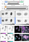
Fig. 1. The coilin NTD assembles into fibrils or puncta in vivo.
a Schematics of the coilin constructs used in this study. The number of amino acids (576 aa in full-length human coilin) included in the expressed protein is indicated below and to the left of each diagram. Coilin NTD is pink, the C-terminus is beige, the intrinsically disordered central region is shown in gray, the NLS shown in yellow is from SV40, and the GCN4 leucine zipper dimerization motif is in cyan. The NTD and NTDNLS constructs were tagged with either myc or HA on their C-termini (not shown) and immunostained. b Grayscale images of Coil−/− MEFs transfected with untagged coilinFL, coilinΔNTD (a deletion construct lacking the NTD) or coilinGCN4 in which the NTD was replaced by the dimerization motif of GCN4. Anti-coilin staining is shown. c Grayscale images NTD and NTDNLS transiently expressed in Coil−/− MEFs. d STED image of coilin NTD transfected Coil−/− MEFs. Scalebar = 2 µm. e Cytoplasmic NTD-myc fibrils (magenta) in HeLa cells do not stain with Thioflavin T (green). DNA visualized with Hoechst (blue). f Coil−/− MEFs transfected with NTD-myc (magenta) and counterstained with α-actin (cyan) to compare actin filaments with cytoplasmic coilin NTD fibrils. DNA visualized with Hoechst (gray). This experiment was repeated independently two times with similar results. All imaged with Leica SP8 laser scanning confocal microscope. Color bars are in analog-digital units to indicate differences in expression. Dotted lines in b, c indicate the nuclear limits. Scalebars (all except d) = 10 µm; insets, 2 µm.
Fig. 2
Fig. 2. Nopp140 is essential for Cajal body assembly.
a Western blots of total lysate from three biological replicates of HeLa cells depleted of Nopp140 or coilin using siRNA transfection targeting Nolc1 and coil mRNAs, respectively. Molecular weight markers indicated to the right of the blots. Representative images for cells undergoing transfection with a non-targeting siRNA (b), siCoil oligo pool (c), or siNolc1 oligo pool (d). Scale bars = 10 µm. SMN and Coilin signal imaged under identical conditions and displayed at the same levels. e Violin plots of Cajal body count per nucleus based on automated quantification of coilin immunostaining. N = 140 (non-targeting siRNA); 103 (siCoil); 142 (siNolc1). Violin plot area is normalized between samples, white dots represent the median, box denotes the interquartile range, and the whiskers note 1.5 the interquartile range. Source data are provided as a Source Data file.
Fig. 3
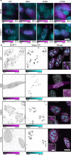
Fig. 3. Nopp140 forms inducible condensates and is required for nuclear NTD puncta, suggesting a mechanism for Cajal body condensation.
a Domain architectures of coilin and Nopp140 and predicted disorder score from RaptorX algorithm. b NIH-3T3 cells expressing CoilinΔNTD mCherry-Cry2 construct. Blue bars indicate 3-minute light activation of the Cry2 domain. c NIH-3T3 cells expressing Nopp140IDR mCherry-Cry2 construct. Red arrowheads indicate non-nucleolar bodies formed by blue light induction. d HeLa cells transfected with siRNA, non-targeting (top) and NOLC1 (bottom). Cells co-transfected with coilin NTDNLS (magenta) and counterstained for coilin (cyan). DNA visualized with Hoechst (gray). “No” denotes the nucleolus, white arrow denotes a representative Cajal body. Imaged with Leica SP8 laser scanning confocal microscope. Grayscale or color bars are given in analog-digital units. Scale bars = 10 µm; insets, 2 µm; Inset zoom, 2 µm.
Fig. 4
Fig. 4. Point mutations in the coilin NTD have distinct effects on self-association and Nopp140 interactions.
a Sequence alignment between mouse and human coilin NTDs with conservation scores from a Clustal Omega multiple alignment of vertebrate coilin sequences. Residues highlighted in red with high conservation scores were selected for alanine mutagenesis. b Wild-type or mutated coilinFL was transfected into Coil−/− MEFs and visually examined for CB formation (coilin is red, DNA is blue). Scale bars = 10 μm. Images acquired with DeltaVision. c, d In vivo acceptor photobleaching FRET measurements yield apparent FRET efficiencies calculated for constructs co-expressed in HeLa cells. FRET measurements between (c) coilin-CFP as the donor and coilin-YFP as the acceptor and d between coilin-CFP (donor) and Nopp140-YFP (acceptor). The indicated alanine mutations were present in all coilin molecules tested. Data is represented as mean values ± S.E.M. N = 10 (except R8A, where n = 9) (c) or 12 (d) cells measured. Black dots indicate individual data points. e Co-immunoprecipitation performed from HeLa cell total lysate after transfection with wild-type and mutant coilin-GFP constructs. Coilin-GFP acts as the bait for either endogenous coilin or Nopp140 as prey and detected by Western blotting as indicated in bold. Molecular weight markers are indicated to the right of the blots. f RaptorX-predicted structure of human coilin NTD, a ubiquitin-like fold. Amino acids implicated in coilin–coilin and coilin–Nopp140 interaction are represented as sticks. Source data are provided as a Source Data file.
Fig. 5
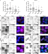
Fig. 5. Point mutations alter NTD capacity to form fibrils and puncta.
a Coil−/− MEFs transfected with constructs for cytoplasmic expression (NTD) or for nuclear expression (NTDNLS) of the coilin NTD bearing selected point mutations. The NTD was immunostained with anti-myc (magenta) and DNA visualized with DAPI (cyan). Color scale bars given in analog-digital units. Scale bar = 10 µm. Percentage to the lower right indicates the fraction of cells with the displayed phenotype where n = 300 cells. Images acquired with DeltaVision. b Coil−/− MEFs transfected with wild-type and mutant coilin nuclear expression constructs (NTDNLS). NTD was immunostained with anti-myc (magenta), counterstained with anti-Nopp140 (cyan), and DNA visualized with Hoechst (gray). The dashed boxes have been magnified in the inset panels. Imaged with Leica SP8 laser scanning confocal microscope. Color bars are in analog-digital units to indicate differences in expression. Scale bar = 10 µm; inset, 1 µm.
Fig. 6
Fig. 6. Coilin NTD mutants exert dominant negative effects on endogenous coilin and Cajal bodies.
Wild-type and mutant myc-tagged coilin NTD constructs expressed in the cytoplasm (NTD, a, b) and nucleus (NTDNLS, c, d) of HeLa cells. (a, c) Cells were immunostained for NTD-myc and endogenous coilin and Cajal bodies were quantified. Measures of center include the median (horizontal line) and mean (cross). The box is drawn from the 25th to the 75th percentile. The whiskers are drawn from the 5th percentile to the 95th percentiles. a n = 300 control, 93 WT, 277 R8A, 97 R36A, 51 D40A, and 302 D79A cells measured. c n = 300 control, 205 WT, 249 R8A, 48 R36A, 169 D40A, and 299 D79A cells measured. b, d Representative images show myc (magenta), coilin (green), and DAPI (blue). Grayscale bars given in analog-digital units. Scale bars = 10 µm. Images acquired with DeltaVision. Source data are provided as a Source Data file.
Fig. 7
Fig. 7. Working model for Cajal body assembly and maintenance via multivalent coilin NTD–NTD and NTD–Nopp140 interactions.
Schematic depicts coilin NTD fibrils in the cytoplasm, where Nopp140 is absent. In the nucleus, puncta may form by the remodeling of coilin NTD fibrils by Nopp140. Alternatively, biomolecular condensation by Nopp140 could limit coilin NTD fibril formation. Coilin NTD molecules are represented as magenta diamonds and the intrinsically disordered Nopp140 molecules are represented as cyan ribbons. Green, blue, and orange hooks represent NTD residues R8, D79, and R36, respectively.

Similar articles

Cited by

References

    1. Gall JG. Cajal bodies: The first 100 years. Annu. Rev. Cell Dev. Biol. 2000;16:273–300. doi: 10.1146/annurev.cellbio.16.1.273. - DOI - PubMed
    1. Machyna M, Neugebauer KM, Stanek D. Coilin: The first 25 years. RNA Biol. 2015;12:590–596. doi: 10.1080/15476286.2015.1034923. - DOI - PMC - PubMed
    1. Machyna M, et al. The coilin interactome identifies hundreds of small noncoding RNAs that traffic through Cajal bodies. Mol. cell. 2014;56:389–399. doi: 10.1016/j.molcel.2014.10.004. - DOI - PubMed
    1. Stanek D, Neugebauer KM. Detection of snRNP assembly intermediates in Cajal bodies by fluorescence resonance energy transfer. J. Cell Biol. 2004;166:1015–1025. doi: 10.1083/jcb.200405160. - DOI - PMC - PubMed
    1. Stanek D. Cajal bodies and snRNPs—friends with benefits. RNA Biol. 2017;14:671–679. doi: 10.1080/15476286.2016.1231359. - DOI - PMC - PubMed

Publication types