. 2022 Sep 29;185(20):3807-3822.e12.
doi: 10.1016/j.cell.2022.09.015.

A pan-cancer mycobiome analysis reveals fungal involvement in gastrointestinal and lung tumors

Affiliations

A pan-cancer mycobiome analysis reveals fungal involvement in gastrointestinal and lung tumors

Anders B Dohlman et al. Cell. .

Abstract

Fungal microorganisms (mycobiota) comprise a small but immunoreactive component of the human microbiome, yet little is known about their role in human cancers. Pan-cancer analysis of multiple body sites revealed tumor-associated mycobiomes at up to 1 fungal cell per 104 tumor cells. In lung cancer, Blastomyces was associated with tumor tissues. In stomach cancers, high rates of Candida were linked to the expression of pro-inflammatory immune pathways, while in colon cancers Candida was predictive of metastatic disease and attenuated cellular adhesions. Across multiple GI sites, several Candida species were enriched in tumor samples and tumor-associated Candida DNA was predictive of decreased survival. The presence of Candida in human GI tumors was confirmed by external ITS sequencing of tumor samples and by culture-dependent analysis in an independent cohort. These data implicate the mycobiota in the pathogenesis of GI cancers and suggest that tumor-associated fungal DNA may serve as diagnostic or prognostic biomarkers.

Keywords: Blastomyces; Candida; Malassezia; cancer; colon cancer; lung cancer; mycobiome; stomach cancer; trans-kingdom interactions; tumor-associated fungi.


Conflict of interest statement

Declaration of interests X.S. is the co-founder and CEO of Xilis, Inc. This study and its findings do not have any overlap or implications over Xilis’ commercial interests. A.B.D., X.L. and I.D.I. are inventors on a US provisional patent application, covering inventions described in this manuscript. The authors declare that they have no conflicts of interest with the contents of this article.

Figures
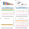
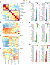
Figure 1.
Figure 1.. Fungal DNA is present in multiple cancer types not explained by contamination, See also Figure S1
(A) Geometric mean of reads per million (RPM) of fungal DNA detected in tumor and tumor-associated tissue samples from head-neck (HNSC), lung (LUSC), rectum (READ), colon (COAD), stomach (STAD), breast (BRCA), esophageal (ESCA) and brain (LGG) cancers. (B) Both bacterial and fungal reads were more abundant in the lower GI tract (COAD, READ) than the upper GI tract (HNSC, ESCA, STAD), and were more abundant in both GI groups compared to the brain (LGG) that was used here as a negative control. (C – D)Genome alignments to C. albicans (C) and S. cerevisiae (D) are largely absent in brain but present at high rates across other tumor types, especially upper GI. (E) Genome alignments to B. dermatidis are found at high rates in lung tumors, but not elsewhere. (F) The distribution of sequencing reads aligning to M. globosa displays similar depth across sequencing projects including brain. Reads are distributed randomly, a signature of biological contamination.
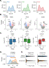
Figure 2.
Figure 2.. Primary tumor samples harbor disease-specific mycobiomes, See also Figure S2
(A) Principal coordinate analysis (PCoA) of normalized species abundances from head-neck (HNSC), esophageal (ESCA), stomach (STAD), colon (COAD), rectal (READ), lung (LUSC), breast (BRCA), and brain (LGG) reveal clustering by tumor type, after filtering contaminants and false-positive signals. (B) Clustered heatmap showing difference in relative fungal species abundances (RPM) between tissues from each TCGA cancer type, after filtering. Species are included if classified as tissue-associated in any of GI, lung, or breast samples, even if they were classified as contaminants in others. Heatmap values are z-scored by species abundance to highlight tissue-specific differences. (C) Boxplots showing distribution of relative abundances (RA) from the 10 or fewer most abundant species detected in each cancer type, after removing low-prevalence and contaminant species.
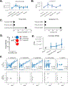
Figure 3.
Figure 3.. Trans-kingdom analysis reveals Candida- and Saccharomyces-associated GI cancer coabundance groups, See also Figure S3
(A) Clustered heatmap showing SparCC co-abundance among fungal species reveals species associated with C. albicans and S. cerevisiae (purple boxes). (B – D) Clustered heatmaps showing gene expression patterns in head-neck (HNSC; B), stomach (STAD; C), and colon (COAD; D) cancers. Heatmaps are clustered by row, while column clustering is determined by (A). Gray columns indicate species not detected in certain cancer types (E - G) SparCC co-abundance between Candida and Saccharomyces and bacterial genera found in matched tumor samples from TCMA, across head-neck (HNSC; E), stomach (STAD; F), and colon (COAD; G) cancers.
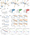
Figure 4.
Figure 4.. Candida is associated with late-stage and metastatic GI cancers, See also Figure S4
(A) Kernel density estimation (KDE) of Candida-to-Saccharomyces ratios in head-neck (HNSC), stomach (STAD), and colon (COAD) cancers. (B) Volcano plot showing genes differentially expressed in Candida-negative (blue) and Candida-high (red) tumor samples head-neck, stomach, and colon cancers. (C) Boxplots depicting Candida-to-Saccharomyces ratios in early-stage (I-III) and late-stage (IV) for head-neck (HNSC), stomach (STAD), and colon (COAD) cancers. (D) KDE analysis of Candida-to-Saccharomyces ratios in metastatic (orange) and non-metastatic (blue) tumor samples finds that Ca-type colon tumors are significantly more likely to be metastatic. (E) Violin-plots showing Bray-Curtis distances between fungal species compositions of patient-matched tumor and blood samples (blue) and unmatched tumor and blood samples (orange).
Figure 5.
Figure 5.. Live, transcriptionally active Candida species are associated with GI tumors, See also Figure S5
(A) Spatial distribution of Ascomycota abundance along the colorectal tract. Significance was calculated between adjacent tumor sites. (B) Targeted analysis showing spatial distribution of C. albicans abundance (RPKM). (C) Comparison of Candida abundance detected in TCGA WGS data (eRPKM; left) and matched original tissues by independent ITS sequencing (relative abundance; right). (D) Live C. albicans, C. lusitaniae, and C. tropicalis were isolated from the mucosa of adenocarcinomas from ascending colon of three individuals, Viable colony forming units (CFU) per mL of sample were determined by MALDI-TOF. (E) Abundance of RNA transcripts aligning to Candida in brain (gray) and sites across the lower GI tract (blue) from solid tissues in the HCMI cohort; no solid tumor samples were available from the ascending or transverse colon. (F) Correlation between fungal species abundances (log10-eRPKM) determined analysis of TCGA WGS and RNA-seq data in GI samples (blue) and brain samples (gray).
Figure 6.
Figure 6.. Candida species are present in GI cancers and high abundance is associated with early-stage stomach cancer
(A – B) Targeted analysis measuring abundance (RPKM) of C. albicans and C. tropicalis (A) and S. cerevisiae (B) across TCGA cancer types.* (C – D)Abundance of C. albicans, C. tropicalis (C), and S. cerevisiae (D) are elevated in stage 1 stomach cancer tumors and stage 4 colon cancer tumors. Significance was calculated between stage 1 tumors and each subsequent stage.* * The direction of the inequality symbol indicates which sample group is greater, while the number of symbols indicates the degree of statistical significance, determined by a two-sided Wilcoxon rank-sum statistic (1: p < 0.05, 2: p < 0.01, 3: p < 0.001).
Figure 7.
Figure 7.. Cancer-associated fungal mycobiota and clinical outcomes highlight predictive value of Candida, See also Figure S6
(A) Heat-tree depicting differential abundance of genera between tumor (blue) and matched adjacent normal tissue (yellow) in head-neck (HNSC), stomach (STAD), and colon (COAD) cancers. (B) Volcano plot showing differential abundance of genera between tumor (blue) and matched adjacent normal tissue (yellow) in stomach cancer. (C) Genera identified as important for distinguishing head-neck, stomach, and colon tumors from other tumor types, based on the Gini coefficient from RF classifiers. Site specific contaminants (#) were set to 0 prior to running the analysis and therefore may be predictive due to their absence. (D) Targeted analysis of Candida spp. shows that C. albicans and C. tropicalis increases in abundance from the proximal to distal stomach, while S. cerevisiae abundance remains relatively stable. (E) Survival analysis comparing outcomes for cancer patients with high rates of tumor-associated C. albicans, C. tropicalis, and S. cerevisiae, compared to patients whose head-neck, stomach, or colon tumors were negative for these species. (F) Across GI cancer types, patients with high levels of tumor-associated Candida experience decreased survival compared to Candida-negative patients. (G) GSEA reveals that genes related to cytosolic DNA sensing, Toll-like receptor, and Nod-like receptor signaling are up-regulated in stomach cancers with higher rates of Candida spp.

Comment in

Similar articles

Cited by

References

    1. Abraham A, Pedregosa F, Eickenberg M, Gervais P, Mueller A, Kossaifi J, Gramfort A, Thirion B, and Varoquaux G (2014). Machine learning for neuroimaging with scikit-learn. Front Neuroinform 8, 14. - PMC - PubMed
    1. Aggor FEY, Break TJ, Trevejo-Nunez G, Whibley N, Coleman BM, Bailey RD, Kaplan DH, Naglik JR, Shan W, Shetty AC, et al. (2020). Oral epithelial IL-22/STAT3 signaling licenses IL-17-mediated immunity to oral mucosal candidiasis. Sci Immunol 5. - PMC - PubMed
    1. Alam A, Levanduski E, Denz P, Villavicencio HS, Bhatta M, Alhorebi L, Zhang Y, Gomez EC, Morreale B, Senchanthisai S, et al. (2022). Fungal mycobiome drives IL-33 secretion and type 2 immunity in pancreatic cancer. Cancer Cell 40, 153–167 e111. - PMC - PubMed
    1. Ballou ER, Avelar GM, Childers DS, Mackie J, Bain JM, Wagener J, Kastora SL, Panea MD, Hardison SE, Walker LA, et al. (2016). Lactate signalling regulates fungal beta-glucan masking and immune evasion. Nat Microbiol 2, 16238. - PMC - PubMed
    1. Benedict K, Roy M, Chiller T, and Davis JP (2012). Epidemiologic and Ecologic Features of Blastomycosis: A Review. Current Fungal Infection Reports 6, 327–335.

Publication types

LinkOut - more resources