Review
. 2022 Oct 3;221(10):e202205135.
doi: 10.1083/jcb.202205135. Epub 2022 Sep 15.

ER as master regulator of membrane trafficking and organelle function

Affiliations
Review

ER as master regulator of membrane trafficking and organelle function

Eva Maria Wenzel et al. J Cell Biol. .

Abstract

The endoplasmic reticulum (ER), which occupies a large portion of the cytoplasm, is the cell's main site for the biosynthesis of lipids and carbohydrate conjugates, and it is essential for folding, assembly, and biosynthetic transport of secreted proteins and integral membrane proteins. The discovery of abundant membrane contact sites (MCSs) between the ER and other membrane compartments has revealed that, in addition to its biosynthetic and secretory functions, the ER plays key roles in the regulation of organelle dynamics and functions. In this review, we will discuss how the ER regulates endosomes, lysosomes, autophagosomes, mitochondria, peroxisomes, and the Golgi apparatus via MCSs. Such regulation occurs via lipid and Ca2+ transfer and also via control of in trans dephosphorylation reactions and organelle motility, positioning, fusion, and fission. The diverse controls of other organelles via MCSs manifest the ER as master regulator of organelle biology.


Figures

Figure 1.
Figure 1.
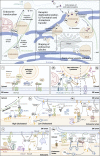
ER-mediated control of endosome dynamics. Overview of cell biological functions of ER-endosome contact sites and the involved molecules. The molecular composition of ER–endosome contact sites. OSBP, ORPs, and VAPs function as dimers or multimers. For simplicity, this is not displayed in the figure. VAP family members (see text box) are depicted as “VAP.” (1) Perinuclear vesicle tethering: The E2 ubiquitin-conjugating enzyme UBE2J1 activates the E3 ubiquitin ligase RNF26, which then ubiquitinates SQSTM1/p62. Ubiquitinated SQSTM1/p62 in turn binds to organelle-specific adaptor proteins, such as T6BP/TAX1BP1, on TGN vesicles and EPS15B or TOLLIP on endosomes. The release of the tethered vesicles is mediated by the deubiquitinase USP15. (2) Endosome translocation. (2a) The BORC complex recruits the small GTPase ARL8B to endosomes, which in turn recruits and activates the Kinesin-1 adaptor protein SKIP/PLEKHM2, resulting in plus-end directed movement of endosomes and lysosomes. Upon ER stress, IRE1 inhibits BORC-dependent anterograde endosome translocation. (2b) The ER-resident protein Protrudin contacts endosomes by binding to RAB7 and PtdIns3P. At these contact sites, Protrudin mediates the hand-over of Kinesin-1 to the endosomal adaptor protein FYCO1, allowing plus-end translocation of endosomes along microtubules. The activity of Protrudin can be regulated by CPT1C, which promotes anterograde endosome transport under nutrient-rich conditions and blocks it under cellular stress conditions. PDZD8 interacts with Protrudin and RAB7, also mediating ER-endosome contact. In addition, PDZD8 might mediate contact with mitochondria. (2c) Endosomes containing high levels of cholesterol move along microtubules in the minus-end direction by dynein/dynactin motor proteins, which connect to the endosome through RILP, RAB7, and ORP1L. Under low concentrations of cholesterol, ORP1L makes contact with VAP in the ER, which leads to the dissociation of dynein/dynactin and the HOPS complex. ER-endosome contact enables ORP1L to transfer cholesterol from the ER to endosomes. Sufficient levels of endosomal cholesterol are a prerequisite for ILV formation (see also legend to 4b). (3) Shaping of endosomal tubules: The formation of recycling tubules requires transient accumulation of PtdIns4P on endosomes to allow WASH-dependent actin nucleation and retromer function. OSBP interacts with PtdIns4P on endosomes via its PH domain and tethers endosomes to the ER via interaction with VAP. PtdIns4P is then dephosphorylated by the ER-resident lipid phosphatase SAC1, securing a transient PtdIns4P pool on endosomes. WASH is linked to the retromer by its subunit FAM21, which marks the site of tubule scission. The PtdIns3P-binding retromer subunit SNX2 is also able to interact with the ER through VAP. The ER protein TMCC1 and Coronin 1C on endosomes are required for contact site formation and fission of WASH-containing endosome tubules. It is not known whether Coronin 1C and TMCC1 interact directly, or if there are additional proteins required to generate these membrane contact sites. (4) Receptor dephosphorylation, ILV formation, and cholesterol transfer. (4a) EGFR-induced phosphorylation of Annexin A1 induces the formation of Annexin A1/S100A11-mediated ER-endosome contact sites, aided by the local increase in Ca2+ through the endosomal Ca2+ channel TPC1. PTP1B in the ER dephosphorylates EGFRs and ESCRT-0, facilitating the sorting of EGFRs into forming ILVs. (4b) In addition, Annexin A1/S100A11-mediated ER-endosome contact sites facilitate cholesterol transfer from ER to forming ILVs by ORP1L (see also legend to 2c). (4c) STARD3 and its paralog STARD3NL (not shown) mediate cholesterol transfer from ER to EGFR-negative endosomes. ORP5 facilitates cholesterol transport from endosomal membranes to the ER. The cholesterol is provided by NPC2 and NPC1, which interacts with ORP5, forming an ER–endosome contact. Direct shuttling of sterols using the ORD domain of ORP5 remains to be confirmed (Santos et al., 2020).
Figure 2.
Figure 2.
Biogenesis of the phagophore membrane via ER contacts. Autophagy is initiated by sequestration of cytoplasmic material by a double-membrane phagophore, whose seed is thought to be composed of ATG9-containing vesicles originating from the Golgi. The phagophore elongates and closes to form an autophagosome, and the sequestered material is degraded once the autophagosome fuses with a lysosome. Phagophore elongation is promoted by a flux of lipids from the ER to the phagophore membrane via the lipid channel transporter ATG2, which tethers subdomains of the ER to growing phagophores by interaction with the ER-localized lipid scramblases TMEM41B and VMP1, and the lipid scramblase ATG9 in the phagophore membrane (additional contacts between the membranes are likely). TMEM41B-VMP1 and ATG9 serve to maintain transbilayer lipid balance in the ER and phagophore membrane, respectively.
Figure 3.
Figure 3.
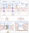
Control of mitochondrial functions via contacts with ER. The figure shows an overview of some of the best-studied functional contacts between the ER and mitochondrial membranes. (a) Calcium transport for homeostasis or apoptosis. In healthy cells, Ca2+ flows from the lumen of ER via the IP3R and through the VDAC1 channel in the outer mitochondria membrane (OMM). GRP75 binds both channels to stabilize the synapse. Inside the mitochondria, ions pass the inner mitochondria membrane (IMM) via MCU1 where Ca2+ is needed for the Krebs cycle. Several protein–protein interactions are required to strengthen the contact site. Examples of such contacts are the ER proteins MFN2 and VAP-B which can interact with mitochondria-resident proteins MFN1/2 and PTPIP51, respectively. During apoptosis, a membrane complex consisting of BAP31, procaspase-8, CDIP1, and FIS1 tethers mitochondria and ER together in addition to the complex required for calcium transport. BAP31 from the ER bind both CDIP1 and procaspase-8, the latter is activated by interacting via its DED domain to bind a vDED domain on BAP31. FIS1 on the mitochondria interacts with BAP31 to bridge the two organelles. These apoptotic cues lead to increased Ca2+ levels in the mitochondria matrix and open the PTP. This disrupts the proton gradient and eventually leads to swelling and rupture of the mitochondria membrane, allowing cytochrome c to leak into the cytosol. APAF1 binds cytochrome c and assembles the apoptosome to execute apoptosis. (b) Mitochondria fission and fusion. ER marks the position for mitochondria fission or fusion by wrapping tubules around the mitochondria. Spire1C nucleates actin filaments and binds INF2 on the ER. INF2 stimulates the mitochondrial Ca2+ uptake and polymerizes actin filaments to further connect ER and mitochondria, allowing the IMM to divide first. DRP1 self assembles into a spiral guided by MFF and FIS1, and with the help of actin filaments constricts to separate the OMM. The final separation of the mitochondria can be aided by lysosomes or trans-Golgi network vesicles containing PtdIns4P at the ER–mitochondria contact site. Fusion is engaged by homodimerization between MFN1 or MFN2 in the OMM through their GTPase domain. Similarly, the GTPase domain on OPA1 interacts to fuse the inner membranes. Miro can bind motor proteins on both microtubules and actin filaments, possibly to strengthen the ER–mitochondria contact by reducing mitochondria movements.
Figure 4.
Figure 4.
Contact sites with ER control peroxisome and Golgi functions. (a) The peroxisomal proteins ACBD4 and ACBD5 interact with the VAP proteins in the ER via their FFAT motifs, anchoring peroxisomes to the ER to facilitate lipid transfer. ER anchored E-Syts contact peroxisomal PtdIns(4,5)P2 to allow cholesterol transport from lysosomes via peroxisomes to the ER. The ER protein BAP31 can potentially interact with FIS1 on peroxisomes, presumably required for peroxisome fission similar to mitochondria. (b) PtdIns4P, the signature phosphoinositide of the Golgi. A PtdIns4P gradient is maintained by phosphorylation of PtdIns by PI4KIIIβ in the TGN and dephosphorylation of PtdIns4P in the ER by the phosphatase SAC1. CERT recognizes PtdIns4P in the Golgi and is tethered to the ER by binding to VAP, where it uses its START domain to transfer ceramide from the ER to the trans-Golgi network. OSBP and OSBP-related proteins (ORPs) interact with PtdIns4P in the Golgi and VAP in the ER. Here, the transfer of PtdIns4P from the Golgi to the ER along the PtdIns4P gradient fuels the counter-transfer of cholesterol or PS to the Golgi. NIR2 binds and transfers PtdIns from the ER to the Golgi, thereby replenishing the PtdIns pool. FAPP1 promotes the activity of SAC1 to dephosphorylate PtdIns4P in trans in narrow membrane contact sites. CPT1C inhibits SAC1 activity to maintain normal levels of PtdIns4P in the Golgi under basal conditions.

Similar articles

Cited by

References

    1. Abrisch, R.G., Gumbin S.C., Wisniewski B.T., Lackner L.L., and Voeltz G.K.. 2020. Fission and fusion machineries converge at ER contact sites to regulate mitochondrial morphology. J. Cell Biol. 219:e201911122. 10.1083/jcb.201911122 - DOI - PMC - PubMed
    1. Alanko, J., Hamidi H., and Ivaska J.. 2016. Signaling from endosomes. In Encyclopedia of Cell Biology. Bradshaw R.A., and Stahl P.D., editors. Academic Press, Waltham. 211–224
    1. Allison, R., Lumb J.H., Fassier C., Connell J.W., Ten Martin D., Seaman M.N., Hazan J., and Reid E.. 2013. An ESCRT–spastin interaction promotes fission of recycling tubules from the endosome. J. Cell Biol. 202:527–543. 10.1083/jcb.201211045 - DOI - PMC - PubMed
    1. Alpy, F., Rousseau A., Schwab Y., Legueux F., Stoll I., Wendling C., Spiegelhalter C., Kessler P., Mathelin C., Rio M.C., et al. . 2013. STARD3 or STARD3NL and VAP form a novel molecular tether between late endosomes and the ER. J. Cell Sci. 126:5500–5512. 10.1242/jcs.139295 - DOI - PubMed
    1. Amarilio, R., Ramachandran S., Sabanay H., and Lev S.. 2005. Differential regulation of endoplasmic reticulum structure through VAP-Nir protein interaction. J. Biol. Chem. 280:5934–5944. 10.1074/jbc.M409566200 - DOI - PubMed

Publication types