. 2022 Aug 1;20(8):e3001714.
doi: 10.1371/journal.pbio.3001714. eCollection 2022 Aug.

Structure of the human galanin receptor 2 bound to galanin and Gq reveals the basis of ligand specificity and how binding affects the G-protein interface

Affiliations

Structure of the human galanin receptor 2 bound to galanin and Gq reveals the basis of ligand specificity and how binding affects the G-protein interface

Yunseok Heo et al. PLoS Biol. .

Abstract

Galanin is a neuropeptide expressed in the central and peripheral nervous systems, where it regulates various processes including neuroendocrine release, cognition, and nerve regeneration. Three G-protein coupled receptors (GPCRs) for galanin have been discovered, which is the focus of efforts to treat diseases including Alzheimer's disease, anxiety, and addiction. To understand the basis of the ligand preferences of the receptors and to assist structure-based drug design, we used cryo-electron microscopy (cryo-EM) to solve the molecular structure of GALR2 bound to galanin and a cognate heterotrimeric G-protein, providing a molecular view of the neuropeptide binding site. Mutant proteins were assayed to help reveal the basis of ligand specificity, and structural comparison between the activated GALR2 and inactive hβ2AR was used to relate galanin binding to the movements of transmembrane (TM) helices and the G-protein interface.


Conflict of interest statement

The authors have declared that no competing interests exist.

Figures

Fig 1
Fig 1. Cryo-EM structure of GALR2 complex with galanin.
(A) The molecular structure of GALR2 complex is shown both as cryo-EM map and ribbon diagram. GALR2, mGαqiN, Gβ1, Gγ2, and scFv16 are colored in blue, red, yellow-orange, marine, and lemon, respectively. The bound galanin peptide is colored in light orange. (B) The C-terminal helix of mGαqiN is inserted in the pocket formed by TM5–7 of GALR2 forming hydrophobic and hydrophilic interactions. Arg32 of mGαqiN forms a salt bridge with Glu135ICL2 of GALR2. (C) The galanin is bound to the pocket formed among the 7 TMs of GALR2 at the extracellular side (left panel). GALR2 and galanin are shown as an electrostatic surface representation (right panel). cryo-EM, cryo-electron microscopy; TM, transmembrane.
Fig 2
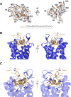
Fig 2. Galanin recognition of GALR2.
(A) The cryo-EM map near the galanin ligand in 2 different orientations. The sequence of galanin is written below, and the C-terminal 14 amino acids, which are not visible in the map, are colored in gray. (B) The ligand binding pocket of GALR2 with galanin is shown in 2 different orientations. GALR2 is shown as a ribbon diagram, and galanin as a ribbon diagram and stick model. (C) Detailed interactions between galanin and GALR2. The hydrophobic interactions between Leu10P and Phe264ECL3 and Trp2P and Leu266ECL3 are indicated with yellow dotted lines. cryo-EM, cryo-electron microscopy.
Fig 3
Fig 3. Gq-signaling activity of GALR2 mutants.
Galanin-induced Gq-signaling activity of WT GALR2 (titrated plasmid volume) and mutant GALR2 was assessed by the NanoBiT Gq-PLCβ assay. Symbols and error bars indicate mean and SEM, respectively, of 3 independent experiments with each performed in duplicate. The dashed lines, the dotted lines, and long dashed dotted lines represent response curves of WT, mock transfection, and surface expression–matched WT, respectively. Surface expression levels of WT and the mutants both of which contained the N-terminal FLAG-epitope tag were assessed by the flow cytometry using a FLAG-epitope tag antibody (S4A Fig). Note that, in many data points, error bars are smaller than the size of symbols and thus are not visible. The data underlying this figure can be found in S1 Data. WT, wild type.
Fig 4
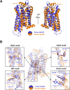
Fig 4. Comparison of the conserved motifs between active GALR2 and inactive hβ2AR.
(A) The overall structures of activated GALR2 and inactive hβ2AR in 2 different orientations. GALR2, hβ2AR, and galanin are shown as a ribbon diagram. Active GALR2, inactive hβ2AR, and galanin are colored in blue, orange, and light orange, respectively. The movements of TM1, TM6, and TM7 are indicated by red arrows. PDB code of the inactive structure of hβ2AR is 2RH1. (B) The conserved motifs are shown as a stick model. The movements of the residues are indicated by black arrows, and the movements of TM are indicated by red arrows. TM, transmembrane.

Similar articles

References

    1. Habert-Ortoli E, Amiranoff B, Loquet I, Laburthe M, Mayaux JF. Molecular cloning of a functional human galanin receptor. Proc Natl Acad Sci U S A. 1994. Oct 11;91(21):9780–9783. doi: 10.1073/pnas.91.21.9780 - DOI - PMC - PubMed
    1. Šípková J, Kramáriková I, Hynie S, Klenerová V. The galanin and galanin receptor subtypes, its regulatory role in the biological and pathological functions. Physiol Res. 2017. Nov 24;66(5):729–740. doi: 10.33549/physiolres.933576 - DOI - PubMed
    1. Webling KEB, Runesson J, Bartfai T, Langel U. Galanin Receptors and Ligands. Front Endocrinol (Lausanne). 2012. Dec 7;3:146. doi: 10.3389/fendo.2012.00146 - DOI - PMC - PubMed
    1. Land T, Langel U, Löw M, Berthold M, Undén A, Bartfai T. Linear and cyclic N-terminal galanin fragments and analogs as ligands at the hypothalamic galanin receptor. Int J Pept Protein Res. 1991. Sep;38(3):267–272. doi: 10.1111/j.1399-3011.1991.tb01438.x - DOI - PubMed
    1. Wang S, He C, Hashemi T, Bayne M. Cloning and expressional characterization of a novel galanin receptor. Identification of different pharmacophores within galanin for the three galanin receptor subtypes. J Biol Chem. 1997. Dec 19;272(51):31949–31952. doi: 10.1074/jbc.272.51.31949 - DOI - PubMed

Grants and funding

This work was supported by Japan Agency for Medical Research and Development, AMED under grant numbers JP21fk0310103 (S.-Y.P.), JP20ae0101047 (K.M.), and by JSPS/MEXT KAKENHI grant (JP19H05779, 21H02449 to S.-Y.P.), (JP20H05873 (K.M.)). This work was also supported by a grant (NRF-2020M3A9G7103934 to W.L.) from National Research Foundation (NRF) of Korea. A.I. was funded by KAKENHI 21H04791 and 21H05113 from the Japan Society for the Promotion of Science (JSPS); the LEAP JP20gm0010004 and the BINDS JP20am0101095 from the Japan Agency for Medical Research and Development (AMED). K.M. was funded by the BINDS JP20am0101093 from AMED. A.I. was funded by FOREST Program JPMJFR215T and JST Moonshot Research and Development Program JPMJMS2023 from the Japan Science and Technology Agency (JST); Daiichi Sankyo Foundation of Life Science; Takeda Science Foundation; The Uehara Memorial Foundation. The funders had no role in study design, data collection and analysis, decision to publish, or preparation of the manuscript.

LinkOut - more resources