Review
. 2022 Jul 15;14(7):1476.
doi: 10.3390/pharmaceutics14071476.

IgG Fusion Proteins for Brain Delivery of Biologics via Blood-Brain Barrier Receptor-Mediated Transport

Affiliations
Review

IgG Fusion Proteins for Brain Delivery of Biologics via Blood-Brain Barrier Receptor-Mediated Transport

Ruben J Boado. Pharmaceutics. .

Abstract

The treatment of neurological disorders with large-molecule biotherapeutics requires that the therapeutic drug be transported across the blood-brain barrier (BBB). However, recombinant biotherapeutics, such as neurotrophins, enzymes, decoy receptors, and monoclonal antibodies (MAb), do not cross the BBB. These biotherapeutics can be re-engineered as brain-penetrating bifunctional IgG fusion proteins. These recombinant proteins comprise two domains, the transport domain and the therapeutic domain, respectively. The transport domain is an MAb that acts as a molecular Trojan horse by targeting a BBB-specific endogenous receptor that induces receptor-mediated transcytosis into the brain, such as the human insulin receptor (HIR) or the transferrin receptor (TfR). The therapeutic domain of the IgG fusion protein exerts its pharmacological effect in the brain once across the BBB. A generation of bifunctional IgG fusion proteins has been engineered using genetically engineered MAbs directed to either the BBB HIR or TfR as the transport domain. These IgG fusion proteins were validated in animal models of lysosomal storage disorders; acute brain conditions, such as stroke; or chronic neurodegeneration, such as Parkinson's disease and Alzheimer's disease. Human phase I-III clinical trials were also completed for Hurler MPSI and Hunter MPSII using brain-penetrating IgG-iduronidase and -iduronate-2-sulfatase fusion protein, respectively.

Keywords: Alzheimer’s disease; Parkinson’s disease; blood–brain barrier; decoy receptors; fusion proteins; insulin receptor; lysosomal storage disorders; monoclonal antibody; neurotrophic factors; protein-based therapy; transferrin receptor.


Conflict of interest statement

The author is the co-inventor of patents on the delivery of biological drugs to the brain.

Figures

Figure 1
Figure 1
Receptor-mediated transport of IgG fusion proteins across the BBB. Biologicals (red circle) do not cross the BBB and stay in circulation following IV administration, as in the case of enzymes, MAbs, decoy receptors, and/or neurotrophic factors. These potential therapeutic agents for the CNS can be re-engineered as fusion proteins with an MAb targeting a BBB receptor that induces receptor-mediated transcytosis (R1), such as the human BBB insulin receptor (HIR) or the transferrin receptor (TfR). The transport domain of the IgG fusion protein targets the BBB R1 endogenous receptor to gain access to the brain. The transport MAb binds to an exofacial epitope of the receptor without interfering with the normal transport of its endogenous ligand (green rectangle) to gain access to the brain. Depending on the therapeutic domain of the IgG fusion protein, it can (i) bind to its ligand in the brain interstitial compartment, as in the case of bispecific MAbs or decoy receptors; (ii) target a brain cell membrane receptor (R2), such as neurotrophic factors; or (iii) be endocytosed via the same targeted R1 receptor in brain cells as lysosomal enzymes to produce physiological and/or neuropharmacological effect.
Figure 2
Figure 2
Genetic engineering of IgG fusion proteins. The therapeutic domain of the IgG bifunctional fusion protein can be fused to the C-terminus of either the heavy or light chain of the transport monoclonal antibody (MAb), in this case targeting the BBB human insulin receptor (HIR). The indication for these IgG fusion proteins is: HIRMAb-PPT1, Batten disease type 1; HIRMAb-GLB1, GM1-gangliosidosis; HIRMAb-HEXA, Tay–Sachs disease; and HIRMAb-ASM, Niemann–Pick disease types A and B. From reference [28].
Figure 3
Figure 3
Autoradiography through eight parallel sagittal sections of the cerebral hemisphere of the rhesus monkey obtained 2 h after the IV administration of either the [125I]-HIRMAb-IDUA fusion protein (bottom) or [125I]-IDUA (top). The section on the left-hand side is the most lateral part of brain, and the section on the right-hand side is the most medial part of brain. The cerebellum is visible in the more medial sections of the brain. The BBB-penetrating HIRMAb-IDUA gained access to the brain, producing a global distribution throughout this organ. On the contrary, IDUA does not cross the BBB, showing just background activity in the primate brain. From [62] with permission.
Figure 4
Figure 4
Reversal of lysosomal storage in brain of adult MPSI mice with IV injections of mouse TfRMAb-IDUA fusion protein. Six-month-old MPSI mice were treated with 1 mg/kg BW TfRMAb-IDUA IV twice weekly for 8 weeks. Electron microscopy showed a marked reduction in lysosomal inclusion bodies in animals treated with brain-penetrating IDUA fusion protein (B) compared with saline (A), resulting in a 73% reduction in brain lysosomal inclusion bodies (right top). The administration of the TfRMAb-IDUA produced a marked reduction in glycosaminoglycans (GAG) in the peripheral organs (right bottom) that was comparable to that reported for the recombinant IDUA. The data (means ± SE) are terminal organ assays at the end of the 8-week study of MPSI mice treated with either saline or 1 mg/kg BW of the TfRMAb-IDUA fusion protein. From [35] with permission.
Figure 5
Figure 5
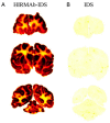
Film autoradiogram (20 µm sections) of rhesus monkey brain removed 2 h after IV injection of the HIRMAb-IDS fusion protein (A) or IDS (B). Scans were produced after labeling of the HIRMAb-IDS fusion protein or IDS with [125I]-Bolton–Hunter reagent. The forebrain section is on the top, the midbrain section is in the middle, and the hindbrain section with cerebellum is on the bottom. From reference [68] with permission.
Figure 6
Figure 6
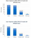
Reduction in brain heparan sulfate (HS) in the MPSIIIA mouse with systemic administration of a mouse TfRMAb-SGSH fusion protein. Two-week-old MPSIIIA mice (JAX) were treated three times per week for 6 weeks with IP 5 mg/kg of the TfRMAb-SGSH fusion protein or the isotype control (TfRMAb). The mice were euthanized 1 week after the last dose. HS was measured in brain and liver by LC-MS following enzymatic digestion into disaccharides using HS disaccharide standards. The 30-fold elevation in HS in the brain was reduced 70% by the chronic treatment with the IgG fusion protein (top). HS was also elevated in liver, and treatment with the mouse TfRMAb-SGSH reduced hepatic HS by 85% (bottom). Data are expressed as means ± SD (n = 8 mice/group). From [37] with permission.
Figure 7
Figure 7
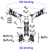
Schematic representation of a tetravalent bispecific MAb. In this construct, the transport domain of the fusion protein targets the BBB human insulin receptor (HIR), and the therapeutic domain is a single-chain anti-Aβ antibody monomer (ScFv) fused to the carboxyl terminus of the heavy chain of the HIRMAb. This tetravalent bispecific Mab maintains a high affinity for both Aβ and the BBB insulin receptor [29].
Figure 8
Figure 8
Pharmacokinetics and brain uptake of a tetravalent bispecific MAb fusion protein in the rhesus monkey. The structure of the tetravalent bispecific MAb fusion protein is shown in Figure 7. This fusion protein, designated [125I]-fusion antibody in this figure, comprises the transport domain, which targets the BBB human insulin receptor, and the therapeutic domain, which is a single-chain anti-Aβ antibody monomer (ScFv). (A) Plasma pharmacokinetics analysis showing no measurable clearance from the blood of the control [3H]-mouse Abeta MAb, whereas the [125I]-fusion antibody is rapidly cleared from blood due to uptake via the insulin receptor. (B) Brain VD for the [125I]-fusion antibody is >100 μL/g in both the brain homogenate and the post-vascular supernatant, which indicates the [125I]-fusion antibody is transported across the BBB. The VD for the [3H]-mouse Abeta MAb, 10 μL/g, is equal to the brain arterial blood volume, which indicates this antibody is not transported across the primate BBB in vivo. (C) Global distribution of fusion antibody to primate brain. Brain scans of adult rhesus monkey at 3 h after the intravenous administration of the [125I]-fusion antibody demonstrates the widespread distribution of the fusion antibody into the primate brain in vivo. The top scan is the most frontal part of brain, and the bottom scan is the most caudal part of brain and includes the cerebellum. From [29] with permission.
Figure 9
Figure 9
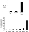
Selective targeting of a TNFR decoy receptor pharmaceutical to the primate brain as a receptor-specific IgG fusion protein. This fusion protein, HIRMAb-TNFR comprises a transport domain targeting the BBB human insulin receptor and the TNFR ECD as therapeutic domain. The brain uptake and peripheral biodistribution of the HIRMAb-TNFR were investigated in the rhesus monkey and compared with those of the control TNFR:Fc, etanercept, with [125I]-Bolton–Hunter reagent-labeled articles. The HIRMAb-TNFR fusion protein was transported across the BBB, producing a brain uptake of 3% ID. On the other hand, the non-brain-penetrating TNFR:Fc produced a brain uptake comparable to IgG1, which is confined to the blood compartment in the brain (top). The ratio for the organ permeability–surface area (PS) of the HIRMAb-TNFR relative to the organ PS for the TNFR:Fc in the rhesus monkey approximates 1 (bottom), as both molecules are transported into the peripheral organs. The PS ratio was >30 in the brain, as just the HIRMAb-TNFR is transported across the BBB and into the primate brain. From [30] with permission.
Figure 10
Figure 10
Neuroprotection of the mouse TfRMAb-GDNF fusion protein in a reversible middle cerebral artery occlusion (MCAO) stroke model. Brain coronal sections were obtained 24 h after MCAO and stained with 2,3,5-triphenyltetrazolium chloride (TTC), and representative brains are shown in the figure. Saline, 1 mg/kg BW mouse TfRMAb-GDNF fusion protein, and an equimolar dose of GDNF (0.17 mg/kg BW), were injected IV 45 min after MCAO. The mouse TfRMAb-GDNF fusion protein produced a 30% reduction in cortical stroke volume (right) compared with the control treated with saline (left), whereas GDNF alone had no effect on stroke volume (center). From [125] with permission.

Similar articles

Cited by

References

    1. Pardridge W.M., Boado R.J., Black K.L., Cancilla P.A. Blood-brain barrier and new approaches to brain drug delivery. West. J. Med. 1992;156:281–286. - PMC - PubMed
    1. Pardridge W.M. The blood-brain barrier: Bottleneck in brain drug development. NeuroRx. 2005;2:3–14. doi: 10.1602/neurorx.2.1.3. - DOI - PMC - PubMed
    1. Abbott N.J., Patabendige A.A.K., Dolman D.E.M., Yusof S.R., Begley D.J. Structure and function of the blood-brain barrier. Neurobiol. Dis. 2010;37:13–25. doi: 10.1016/j.nbd.2009.07.030. - DOI - PubMed
    1. Pardridge W.M. CSF, blood-brain barrier, and brain drug delivery. Expert Opin. Drug Deliv. 2016;13:963–975. doi: 10.1517/17425247.2016.1171315. - DOI - PubMed
    1. Boado R.J., Pardridge W.M. Molecular cloning of the bovine blood-brain barrier glucose transporter cDNA and demonstration of phylogenetic conservation of the 5′-untranslated region. Mol. Cell. Neurosci. 1990;1:224–232. doi: 10.1016/1044-7431(90)90005-O. - DOI - PubMed

Grants and funding

This review received no external funding.

LinkOut - more resources