. 2022 Aug 2;146(5):412-426.
doi: 10.1161/CIRCULATIONAHA.121.057276. Epub 2022 Jul 6.

Restoration of Cardiomyogenesis in Aged Mouse Hearts by Voluntary Exercise

Affiliations

Restoration of Cardiomyogenesis in Aged Mouse Hearts by Voluntary Exercise

Carolin Lerchenmüller et al. Circulation. .

Abstract

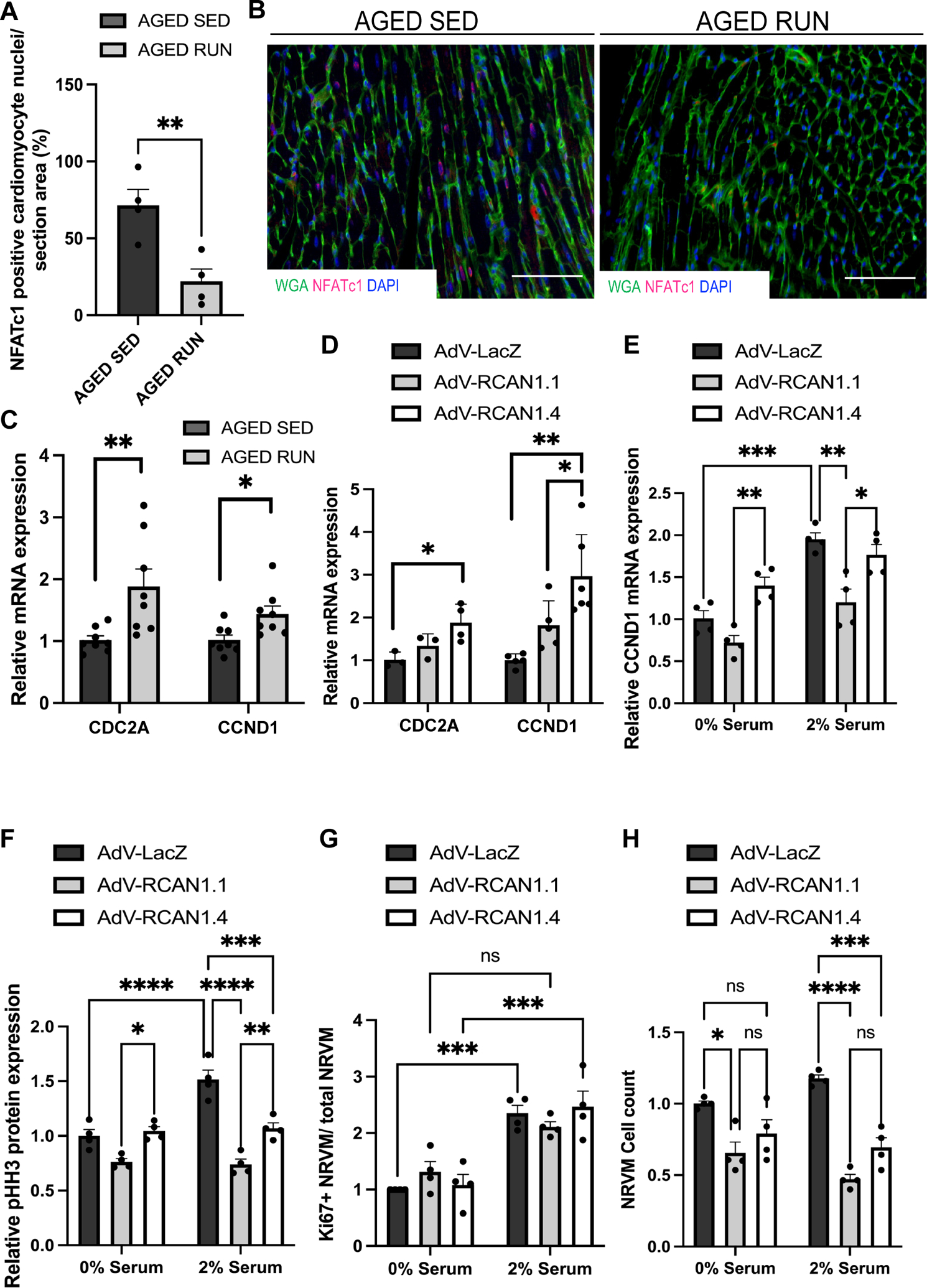
Background: The human heart has limited capacity to generate new cardiomyocytes and this capacity declines with age. Because loss of cardiomyocytes may contribute to heart failure, it is crucial to explore stimuli of endogenous cardiac regeneration to favorably shift the balance between loss of cardiomyocytes and the birth of new cardiomyocytes in the aged heart. We have previously shown that cardiomyogenesis can be activated by exercise in the young adult mouse heart. Whether exercise also induces cardiomyogenesis in aged hearts, however, is still unknown. Here, we aim to investigate the effect of exercise on the generation of new cardiomyocytes in the aged heart.

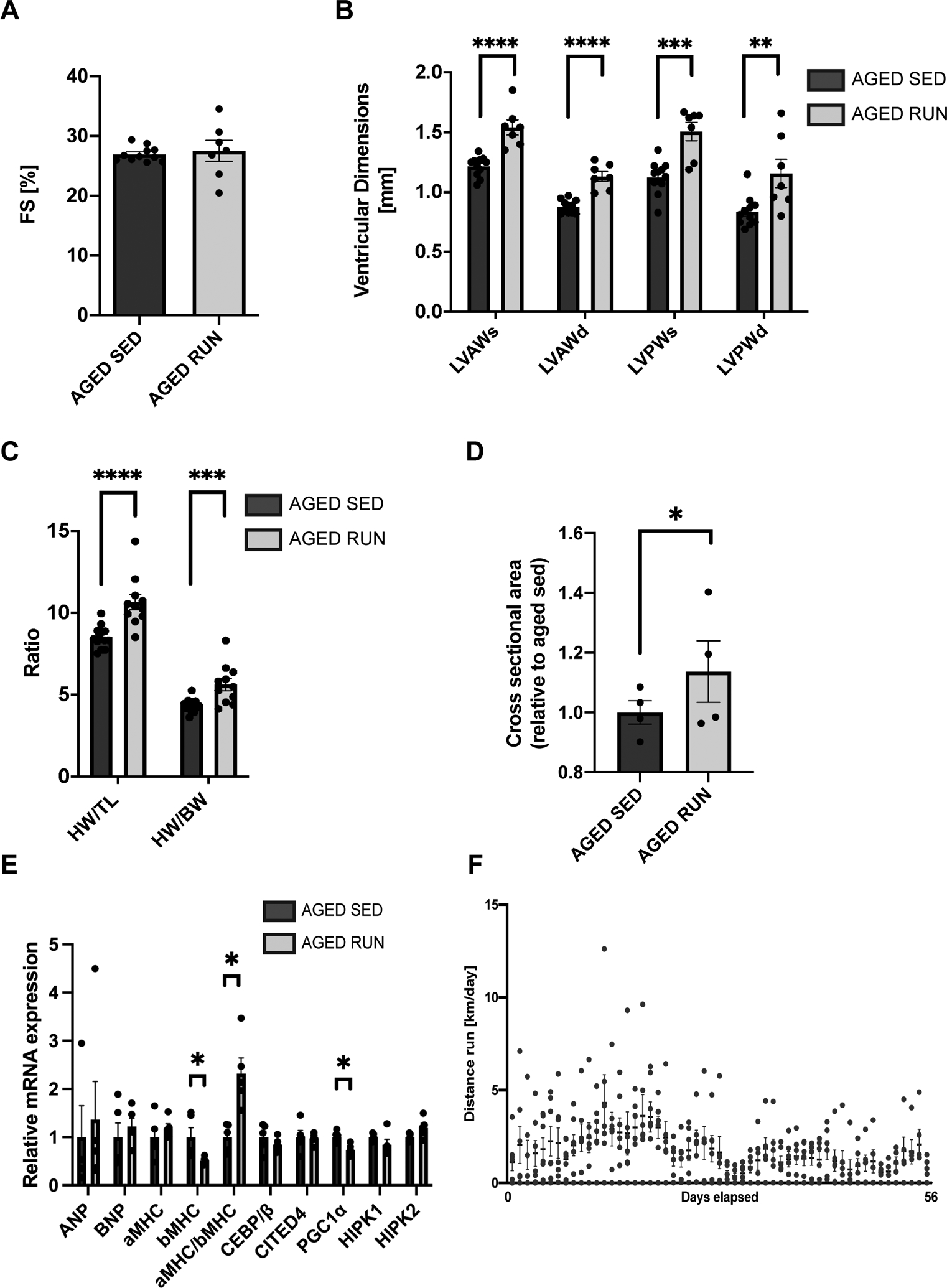
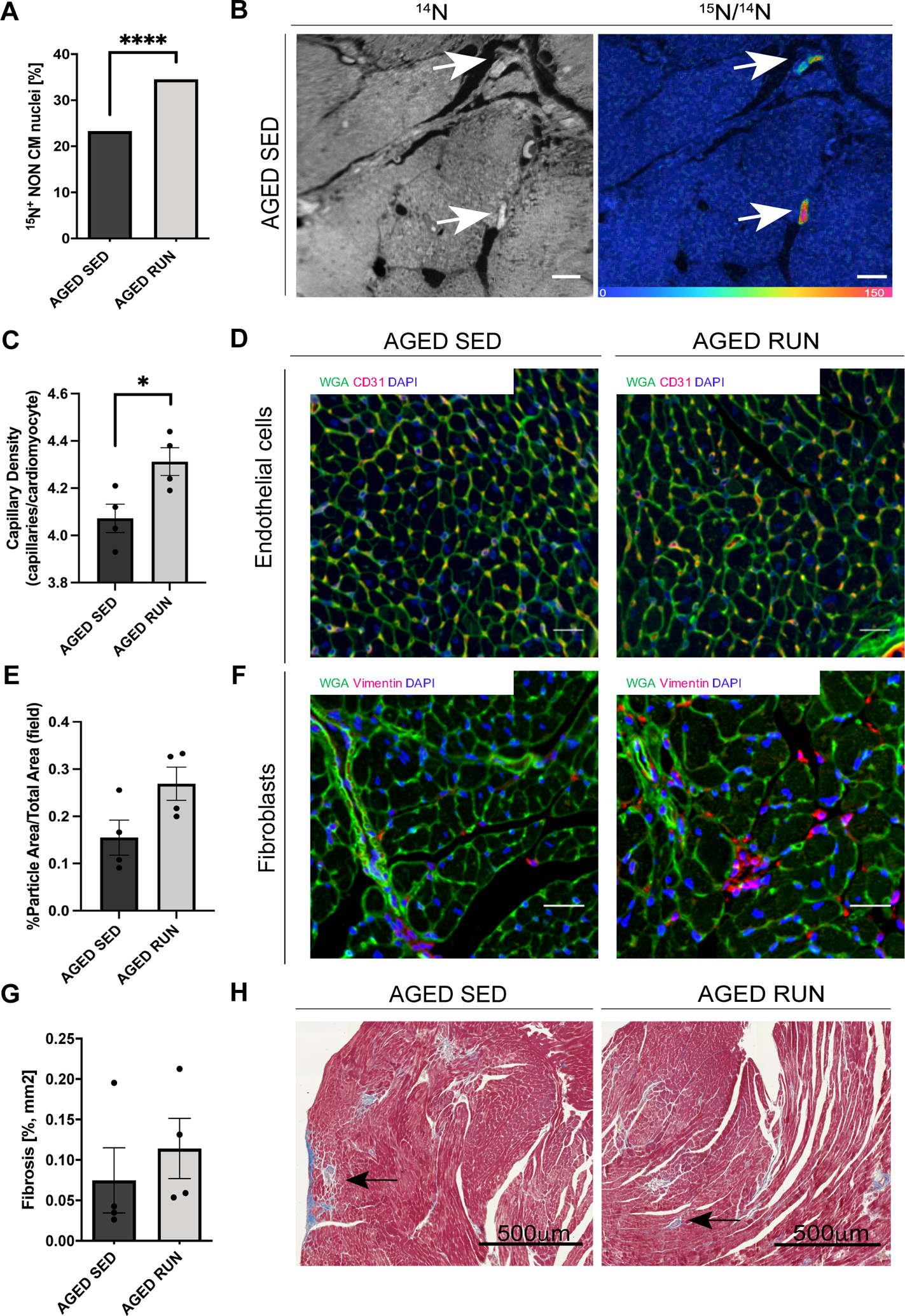
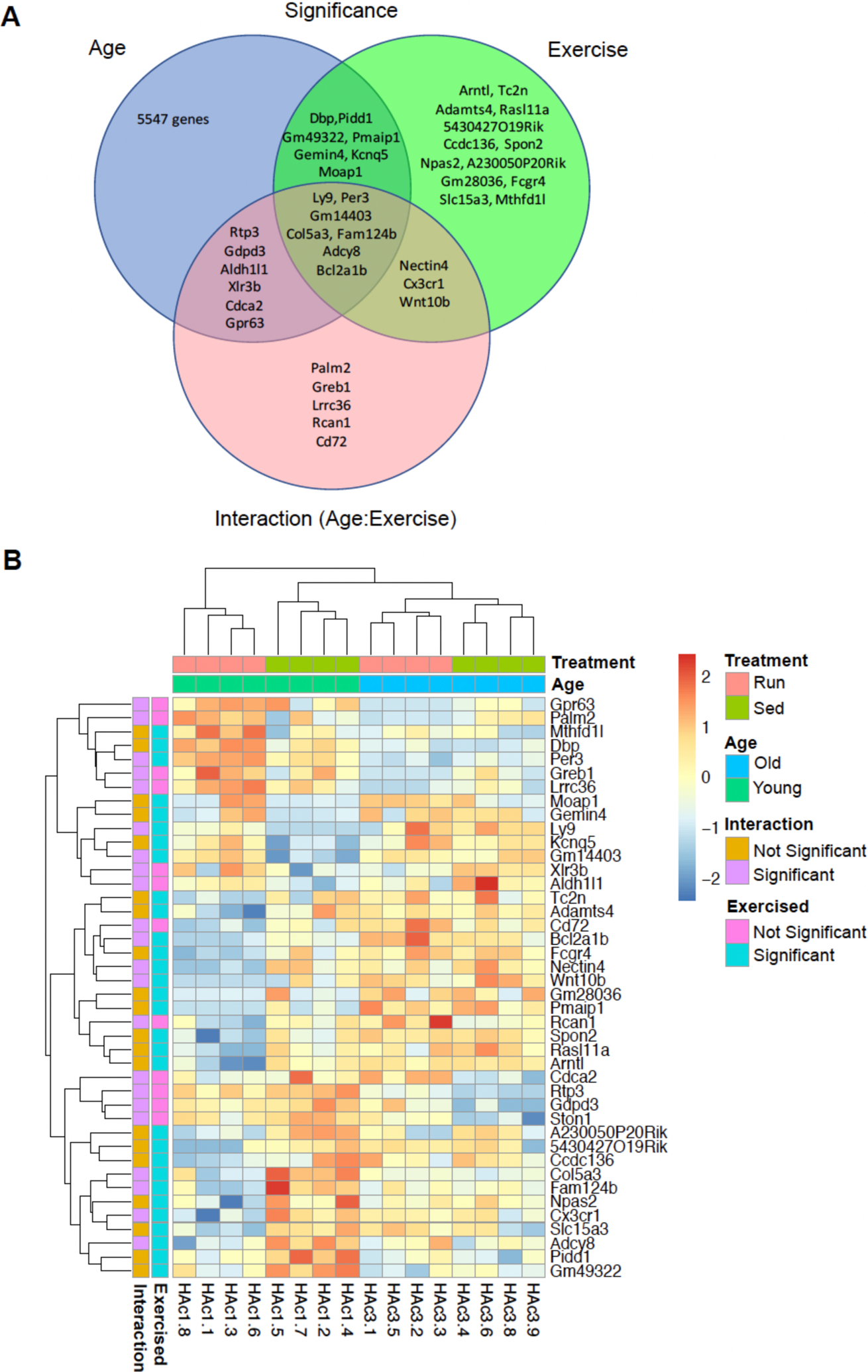
Methods: Aged (20-month-old) mice were subjected to an 8-week voluntary running protocol, and age-matched sedentary animals served as controls. Cardiomyogenesis in aged hearts was assessed on the basis of 15N-thymidine incorporation and multi-isotope imaging mass spectrometry. We analyzed 1793 cardiomyocytes from 5 aged sedentary mice and compared these with 2002 cardiomyocytes from 5 aged exercised mice, followed by advanced histology and imaging to account for ploidy and nucleation status of the cell. RNA sequencing and subsequent bioinformatic analyses were performed to investigate transcriptional changes induced by exercise specifically in aged hearts in comparison with young hearts.

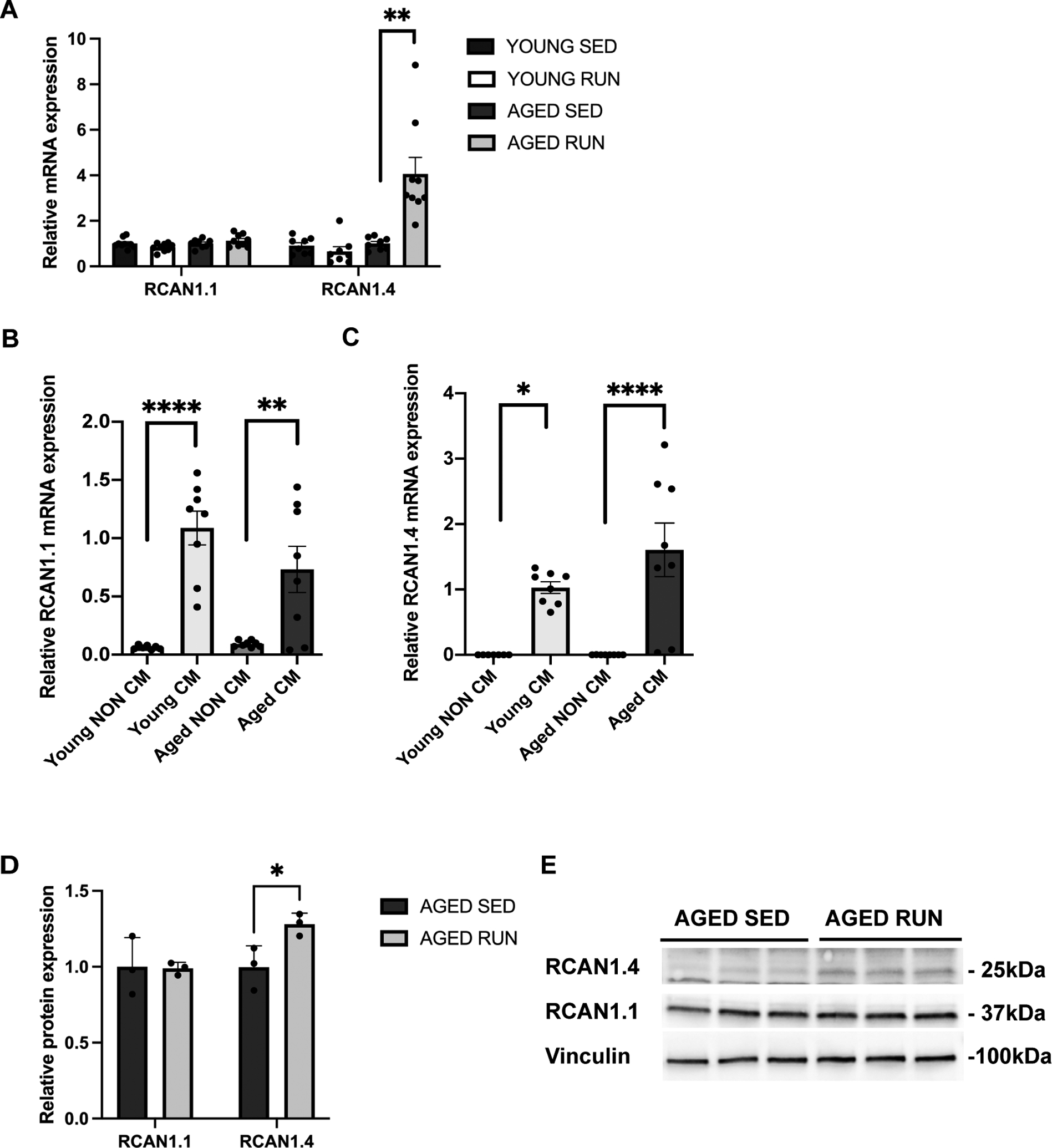
Results: Cardiomyogenesis was observed at a significantly higher frequency in exercised compared with sedentary aged hearts on the basis of the detection of mononucleated/diploid 15N-thymidine-labeled cardiomyocytes. No mononucleated/diploid 15N-thymidine-labeled cardiomyocyte was detected in sedentary aged mice. The annual rate of mononucleated/diploid 15N-thymidine-labeled cardiomyocytes in aged exercised mice was 2.3% per year. This compares with our previously reported annual rate of 7.5% in young exercised mice and 1.63% in young sedentary mice. Transcriptional profiling of young and aged exercised murine hearts and their sedentary controls revealed that exercise induces pathways related to circadian rhythm, irrespective of age. One known oscillating transcript, however, that was exclusively upregulated in aged exercised hearts, was isoform 1.4 of regulator of calcineurin, whose regulation and functional role were explored further.

Conclusions: Our data demonstrate that voluntary running in part restores cardiomyogenesis in aged mice and suggest that pathways associated with circadian rhythm may play a role in physiologically stimulated cardiomyogenesis.

Keywords: age factors; circadian rhythm; exercise; heart failure; muscle development.


Figures

Figure 1.
Figure 1.. Voluntary wheel running induces physiological cardiac adaptation in aged mice.
Echocardiographic analysis showed preserved fractional shortening (FS) (A), with (B) increased anterior and posterior wall thickness in systole and diastole (LVAWs/d, LVPWs/d) in exercised, aged animals when compared to aged, sedentary controls, (n= 7–11 mice/group, ****p<0.0001, ***p=0.0001, **p=0.0081, Student’s t-test). (C) Both heart weight to tibia length and heart weight to body weight ratios were increased in exercised, aged mice (n=11–15 mice/group, HW/TL ****p<0.0001, HW/BW ***p=0.0007 Student’s t-test). (D) Likewise, Cardiomyocyte Cross Sectional Area (CSA) analyzed from wheat-germ-agglutinin-stained transverse LV sections showed an increase in cardiomyocyte size with exercise (>100 cardiomyocytes were counted per group, n=4 mice/group, *p<0.05, Student’s t-test). (E) Anticipated switch of α/β myosin heavy chain gene (MHC) expression after 8 weeks of voluntary wheel running was also detected. PGC1-α was downregulated after 8 weeks (n=5 mice/group, *p<0.05, Student’s t-test). (F) Running activity (running distance per 24h) was measured for each mouse for 8 weeks. Aged mice ran on average 2.4 km per 24h (n=4 mice/group). All data are presented as Mean ± s.e.m.
Figure 2.
Figure 2.. Exercise stimulates an increase in mononuclear diploid 15N-thymidine-labeled cardiomyocytes in the aged mouse heart.
15N-thymidine was administered continuously for 8 weeks to aged mice (20 months old at the beginning of the experiment) undergoing voluntary wheel running or sedentary activity. (A) Representative images of the myocardium in sedentary aged mouse hearts, labeled with 15N-thymidine. Mass 14N image (left side) show histological details such as cell architecture, while the hue-saturation-intensity-image (mosaics, right side) demonstrate nuclear 15N-thymidine labeling. 15N-thymidine labeling was detected predominantly in non-cardiomyocyte cells in aged sedentary hearts. Scale bar = 10 μm. (B) Representative image of 15N-thymidine labeled cardiomyocyte (yellow asterisks), together with one non-labeled-cardiomyocyte (white asterisk), and one labeled non-cardiomyocyte (yellow arrow) in the myocardium of an aged runner. The scale ranges from blue, where the ratio is equivalent to natural ratio (0.37%, expressed as 0% above natural ratio (enrichment over natural ratio)), to red, where the ratio is 150% above natural ratio. 15N-thymidine has labeled the nucleus while the cytoplasm is at the natural abundance level. Scale bar = 10μm. (C) Data presented as comparison of the percentages (%) of 15N-labeled cardiomyocyte nuclei in exercised to sedentary aged hearts. Exercise increases cardiomyocyte cell cycle activity (sedentary: exercise = 0.06:1.20%; >1,700 cardiomyocytes from four mice per group were counted, ****p<0.0001, Fisher’s exact test). Out of 1.20% 15N-labeled cardiomyocyte nuclei, 0.35% were mononuclear and diploid. (D) Periodic acid Schiff staining and fluorescent in situ hybridization (Y-chromosome) were performed on serial adjacent sections in both directions from the 15N-thymidine-labeled cardiomyocyte to define the number of nuclei and ploidy status in each 15N-thymidine-labeled cardiomyocyte. The table shows absolute numbers and percentages (%) of 15N-thymidine labeled cardiomyocytes, as well as the absolute numbers and percentages (%) of mononucleate/diploid cells among all 15N-thymidine-labeled cardiomyocytes from each group (sedentary: exercised= 0.00:0.35%, n=4 mice per group, *p<0.05, Fisher’s exact test).
Figure 3.
Figure 3.. Angiogenic and fibrotic response in the aged, exercised heart.
(A) Exercise increases the 15N-thymidine-labeled non-cardiomyocyte fraction (aged sedentary:exercised = 23.3:34.5%) (>10,000 non-cardiomyocytes from four mice/group were counted, ****p<0.0001, Fisher’s exact test). (B) Representative images of 15N-thymdine-labeled non-cardiomyocyte nuclei in aged mouse hearts (mosaic, right side white arrow). Mass 14N images (left side) show histological details such as localization and structure, scale bar = 10μm. (C) Number of CD31 (PECAM1) positive capillaries per cardiomyocyte after voluntary wheel running (>100 cardiomyocytes counted, n=4 mice/group, *p<0.05, Student’s t-test) (left panel). (D) Representative images of endothelial CD31 (PECAM1) staining (red), in combination with wheat germ agglutinin (WGA, green) to determine the capillary density (capillaries/cardiomyocyte). Nuclei were counterstained with DAPI (blue), scale bar = 50μm (right panel). (E) Expression of Vimentin in aged exercised and sedentary mouse hearts, shown as % Vimentin positive area/ Total area, (n=4 mice/group, p=0.07, Student’s t-test) (left panel). (F) Representative images of Vimentin staining (red), in combination with wheat germ agglutinin (WGA, green). Nuclei were counterstained with DAPI (blue) (right panel). (G) Cardiac fibrosis was not significantly affected by exercise in aged mice, n=4 mice/group, Student’s t-test). (H) Representative Masson Trichrome stained images from aged sedentary and exercised mice, scale bar = 500μm. Collagen rich areas stained blue (black arrows). Data presented as Mean ± s.e.m.
Figure 4.
Figure 4.. Differentially regulated genes in exercised hearts.
Global gene expression in the heart was determined using RNAseq in young (8 weeks) and aged mice (20 months) sacrificed 8 weeks after voluntary wheel running or sedentary activity (n=4 per group). (A) Venn diagram comparing functionally enriched biological genes in young and aged mice exposed to either exercise or sedentary activity. Differentially expressed genes unique to age (blue), exercise (green), or the interaction thereof (exercise:age; red) are shown. (B) Heat map depicts hierarchically clustered differentially regulated genes in the myocardium of young and aged exercised mice, compared to their age-matched sedentary controls by either age, exercise, or the interaction thereof (n=8 mice per treatment, 4 young and 4 aged).
Figure 5.
Figure 5.. Exercise induces expression of RCAN1.4 in aged hearts.
(A) Validation of increased Rcan1.1 and Rcan1.4 gene expression by qPCR analysis in exercised, aged hearts in both the hearts that were analyzed by RNAseq and an independent exercised cohort (n=7–9 mice/group, *p<0.05, Student’s t-test). (B-C) Rcan1.1 and Rcan1.4 gene expression was quantified from isolated young (2 months old) and aged (20 months old) cardiomyocytes and noncardiomyocytes (n=8 mice/group, *p<0.05, **p<0.01, ****p<0.0001, one-way ANOVA). (D-E) RCAN1.1 (~37 kDa) and RCAN1.4 (~25 kDa) protein levels were determined in aged, sedentary, and exercised mouse hearts by immunoblotting. RCAN1.4 protein levels were increased in aged, exercised hearts (n=3 mice/group, *p<0.05, Student’s t-test). Vinculin (~100 kDa) was used as a protein loading control. All data are presented as Mean ± s.e.m.
Figure 6.
Figure 6.. Exercise reduces calcineurin activity in aged hearts and RCAN1.4 overexpression in aged cardiomyocytes increases cardiomyocyte cell cycle markers.
(A) The fraction of NFATc1 positive cardiomyocyte nuclei per section area (%) was quantified in aged mouse hearts. Exercise treatment reduced NFATc1 expression in cardiomyocyte nuclei, compared to sedentary control hearts (n=4 mice/group, **p<0.001, Student’s t-test). (B) Representative images from aged sedentary and exercised mouse hearts show NFATc1 (red) stained nuclei (DAPI, blue), in combination with wheat germ agglutinin (WGA, green), scale bar = 100μm. (C) Validation of cell cycle regulators Cdca2 and Ccnd1 mRNA expression in aged sedentary and exercised mouse hearts (n=8 mice/group, *p<0.05, **p<0.01, Student’s t-test). (D) Isolated murine aged cardiomyocytes (20 months) were infected with RCAN1.1 (AdV-RCAN1.1) and RCAN1.4 (AdV-RCAN1.4) and LacZ control (AdV-LacZ) adenoviruses, respectively, and Cdc2a and Ccnd1 mRNA expression was analyzed (n=3–6 replicates per treatment from a total of 6 hearts, *p<0.05, one-way ANOVA). (E) NRVM were infected with RCAN1.1 (AdV-RCAN1.1) and RCAN1.4 (AdV-RCAN1.4) and LacZ control (AdV-LacZ) adenoviruses, respectively, and Ccnd1 mRNA expression was analyzed after stimulation with indicated serum concentrations (n=4, *p<0.05, **p<0.01, ***p<0.001, two-way ANOVA) (F) NRVM were infected with RCAN1.1 (AdV-RCAN1.1) and RCAN1.4 (AdV-RCAN1.4) and LacZ control (AdV-LacZ) adenoviruses, respectively, and pHH3 protein expression was analyzed after stimulation with indicated serum concentrations (n=4, *p<0.05, **p<0.01, ***p<0.001****p<0.0001, two-way ANOVA). (G) NRVM were infected with RCAN1.1 (AdV-RCAN1.1) and RCAN1.4 (AdV-RCAN1.4) and LacZ control (AdV-LacZ) adenoviruses, respectively, and Ki67 positive cardiomyocytes (immunofluorescence staining) were counted and normalized to total cardiomyocyte cell count per slide after treatment with indicated serum concentrations (n=4, ***p<0.001, two-way ANOVA). (H) Cell count of NRVM after infection with RCAN1.1 (AdV-RCAN1.1) and RCAN1.4 (AdV-RCAN1.4) and LacZ control (AdV-LacZ) adenoviruses followed by indicated serum stimulation (n=4, *p<0.05, ***p<0.001, two-way ANOVA). All data are presented as Mean ± s.e.m.

Similar articles

Cited by

References

    1. Yazdanyar A, Newman AB. The burden of cardiovascular disease in the elderly: morbidity, mortality, and costs. Clinics in geriatric medicine. 2009;25:563–577, vii. doi: 10.1016/j.cger.2009.07.007 - DOI - PMC - PubMed
    1. United Nations Department of Economic and Social Affairs U. World Population Ageing. United Nations New York. 2020. Accessed Nov 30th, 2020.
    1. Go AS, Mozaffarian D, Roger VL, Benjamin EJ, Berry JD, Blaha MJ, Dai S, Ford ES, Fox CS, Franco S, et al. Heart disease and stroke statistics--2014 update: a report from the American Heart Association. Circulation. 2014;129:e28–e292. doi: 10.1161/01.cir.0000441139.02102.80 - DOI - PMC - PubMed
    1. Roh J, Rhee J, Chaudhari V, Rosenzweig A. The Role of Exercise in Cardiac Aging: From Physiology to Molecular Mechanisms. Circ Res. 2016;118:279–295. doi: 10.1161/CIRCRESAHA.115.305250 - DOI - PMC - PubMed
    1. Wencker D, Chandra M, Nguyen K, Miao W, Garantziotis S, Factor SM, Shirani J, Armstrong RC, Kitsis RN. A mechanistic role for cardiac myocyte apoptosis in heart failure. J Clin Invest. 2003;111:1497–1504. doi: 10.1172/JCI17664 - DOI - PMC - PubMed

Publication types