. 2022 Aug;41(33):4003-4017.
doi: 10.1038/s41388-022-02391-x. Epub 2022 Jul 8.

PFKFB3 regulates cancer stemness through the hippo pathway in small cell lung carcinoma

Affiliations

PFKFB3 regulates cancer stemness through the hippo pathway in small cell lung carcinoma

Prabhu Thirusangu et al. Oncogene. 2022 Aug.

Erratum in

Abstract

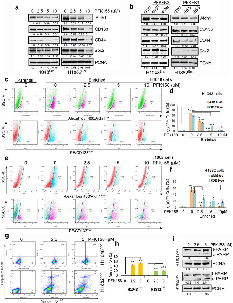
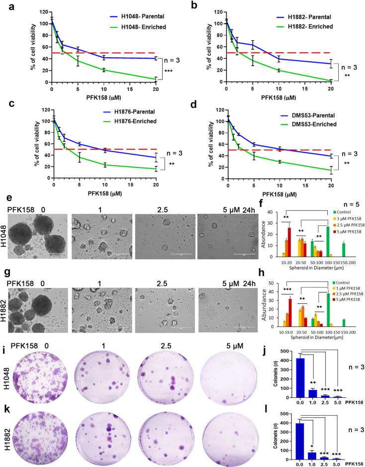
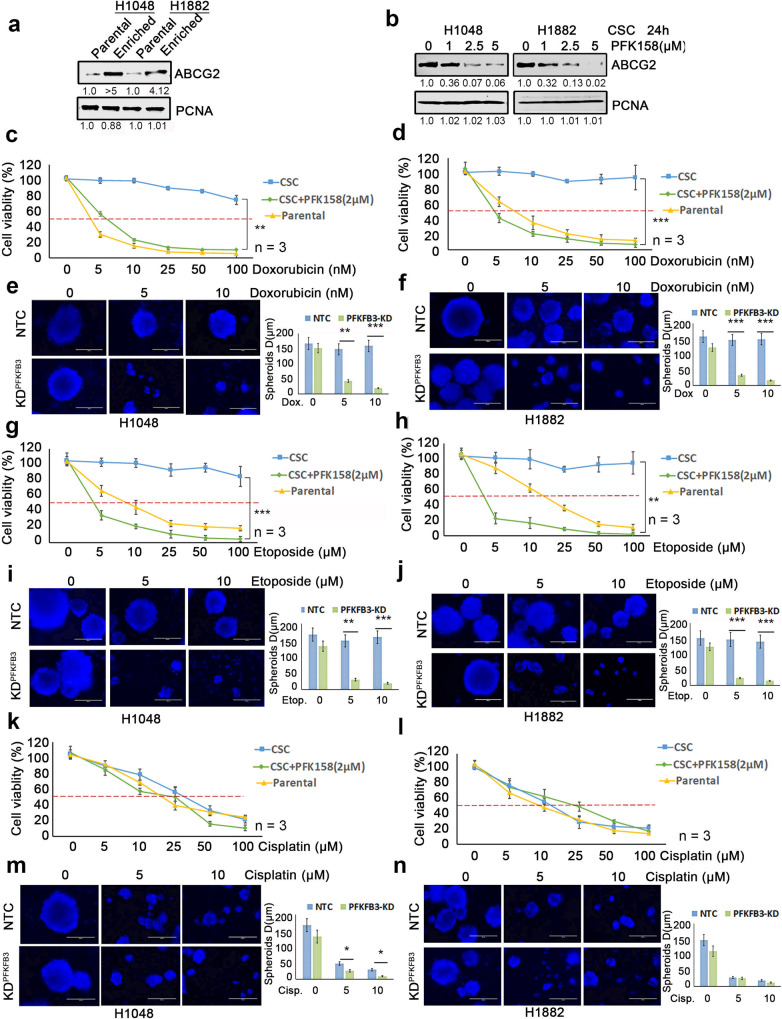
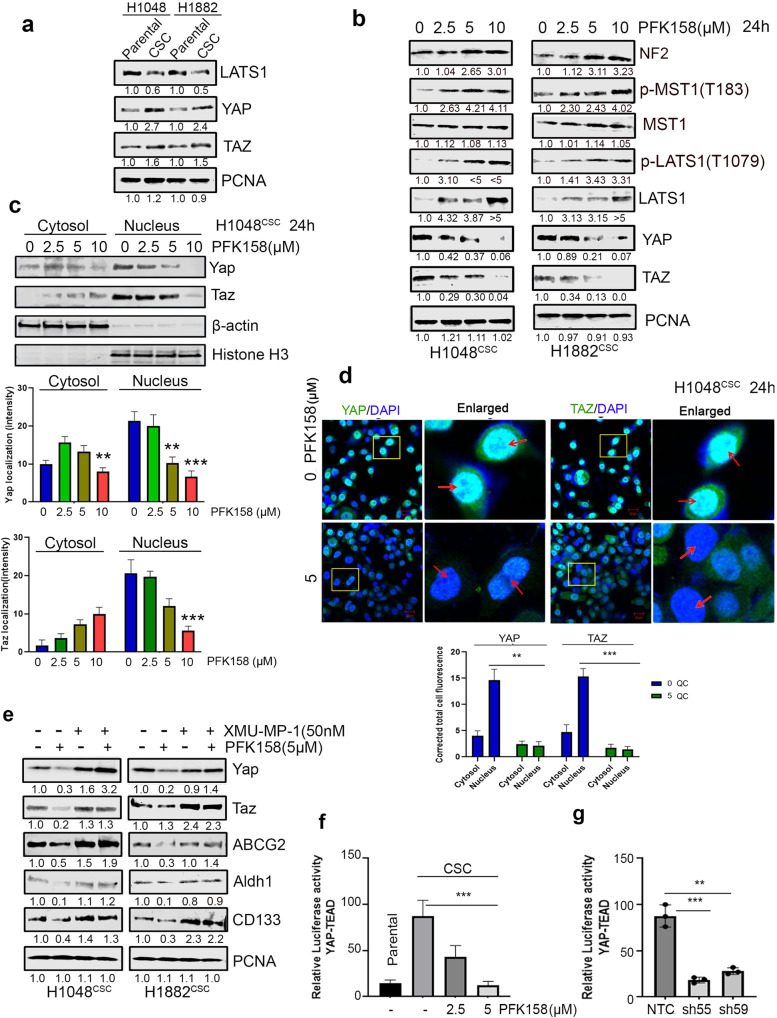
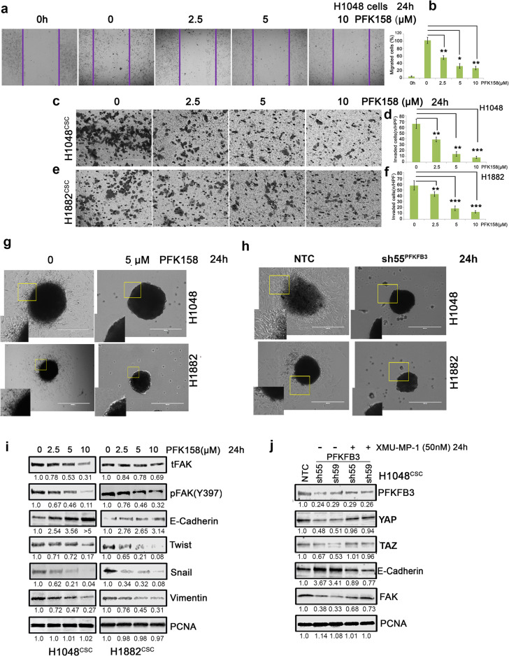
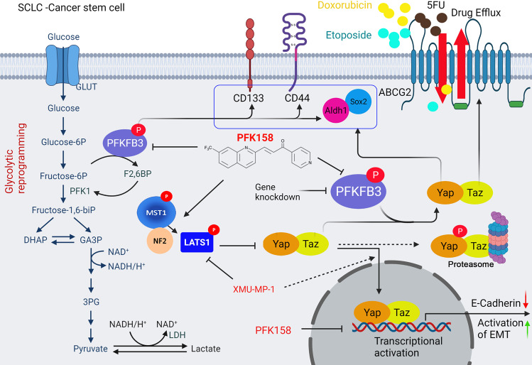
PFKFB3 (6-phosphofructo-2-kinase) is the rate-limiting enzyme of glycolysis and is overexpressed in several human cancers that are associated with poor prognosis. High PFKFB3 expression in cancer stem cells promotes glycolysis and survival in the tumor microenvironment. Inhibition of PFKFB3 by the glycolytic inhibitor PFK158 and by shRNA stable knockdown in small cell lung carcinoma (SCLC) cell lines inhibited glycolysis, proliferation, spheroid formation, and the expression of cancer stem cell markers CD133, Aldh1, CD44, Sox2, and ABCG2. These factors are also associated with chemotherapy resistance. We found that PFK158 treatment and PFKFB3 knockdown enhanced the ABCG2-interacting drugs doxorubicin, etoposide, and 5-fluorouracil in reducing cell viability under conditions of enriched cancer stem cells (CSC). Additionally, PFKFB3 inhibition attenuated the invasion/migration of SCLC cells by downregulating YAP/TAZ signaling while increasing pLATS1 via activation of pMST1 and NF2 and by reducing the mesenchymal protein expression. PFKFB3 knockdown and PFK158 treatment in a H1048 SCLC cancer stem cell-enriched mouse xenograft model showed significant reduction in tumor growth and weight with reduced expression of cancer stem cell markers, ABCG2, and YAP/TAZ. Our findings identify that PFKFB3 is a novel target to regulate cancer stem cells and its associated therapeutic resistance markers YAP/TAZ and ABCG2 in SCLC models.


Conflict of interest statement

DBO transitioned to postdoctoral training at AstraZeneca during this project. AstraZeneca was not involved in the funding, experimental design, and results of this manuscript. The authors declare no further competing interests.

Figures

Fig. 1
Fig. 1. Deficient PFKFB3 causes downregulation of cancer stem cell markers and cell death.
a Expression levels of CSC markers including Aldh1, CD133, CD44, and Sox2 in enriched 3D cultures of H1048 and H1882 cells by western blot analysis. b The effect of PFKFB3 depletion by shRNAs on CSC markers level in enriched 3D cultures of H1048 and H1882 cells. c Aldh1 (pink)/CD133 (blue) was very higher level in CSC enriched 3D culture compared to parental cells and exposure to PFK158 at indicated concentrations decreased detection of both Aldh1+ve/CD133+ve levels in H1048 spheroids. The pink (first panel) and blue (second panel) gating boxed represent unique Aldh1high /CD133high cells in H1048 spheroids. d PFK158 mediated decrease of CSC markers Aldh1+ve/CD133+ve cells. e Aldh1 (blue)/ CD133 (pink) expressing cells are more in 3D spheroids compared to parental 2D cultures and Dot plot for expression of cancer stemness markers Aldh1 and CD133 analyzed with FACS depicting concentration dependant decrease CSC markers upon PFK158 treatment. The blue (first panel) and pink (second panel) gating boxed represent unique Aldh1high /CD133high cells respectively, in H1882 spheroids. f Percentage of CSC Aldh1+ve/CD133+ve cells in H1882 spheroids. g Determination of the early and late apoptotic cells after exposure to PFK158 at specified concentrations by Annexin V-Pacific Blue and PI dual stain in H1048CSC and H1882CSC spheroids. h Bar graph depicts percentage of Annexin-V-positive/apoptotic cells. i immunoblot analysis of cleaved PARP level after PFK158 treatment in H1048CSC and H1882CSC cells. PCNA used as loading control. The experiment was repeated thrice (n = 3), and the graphs are represented as mean ± S.D. Significance measured comparing individual groups with control is represented as *p < 0.05, **p < 0.01, ***p < 0.001.
Fig. 2
Fig. 2. Cytotoxicity of PFK158 on SCLC cells and CSC enriched tumor spheroids.
Cell viability was measured following 24 h of PFK158 treatment at 0, 1, 2.5, 5, 10, 20 μM concentrations using MTT assay in both parental and CSC enriched tumor spheroids of (a) H1048, (b) H1882, (c) H1876 and (d) DMS53 cells. Values are expressed as the mean ± SD and experiments were conducted in triplicate (n = 3) and repeated independently three times. H1048 (e) and H1882 (g) Cells grown as spheroids were exposed to the indicated concentrations of PFK158 for 24 h and the effects on spheroids were measured (n = 5) and (f and h) Graphs depict the abundance of different size of spheroids upon PFK158 treatment in H1048 and H1882 cells, respectively. Scale bar: 200 μm, ×20 magnification. The effect of PFK158 on colony forming abilities of enriched (i) H1048 and (k) H1882 cells were assessed and (j and l) The number of colonies was counted and plotted (n = 3) in triplicates. Data are shown as mean ± SD and significance was determined comparing test samples to untreated control and expressed as *p < 0.05, **p < 0.01, ***p < 0.001.
Fig. 3
Fig. 3. Pharmacological PFKFB3 inhibition with PFK158 suppresses the glycolytic activity of SCLC cells in vitro.
a Expression levels of total (t)-PFKFB3 and phospho (p)-PFKFB3 (ser461) in parental (epithelial-2D culture) and enriched (spheroidal 3D culture) conditions of H1048 and H1882 cells by immunoblot (IB) analysis. b Expression of t-PFKFB3 and p-PFKFB3 (ser461) in H1048 and H1882 3D-spheroids were determined after PFK158 (0, 2.5, 5, 10 μM) treatment for 24 h by IB analysis. Densitometric analysis showing fold change was calculated using Image J software. c Glucose uptake was analyzed using 2-NBDG in CSC enriched spheroids of H1048, H1882 and H1876 after treatment with PFK158 at indicated concentrations (Scale bar: 100 μm, ×40 magnification) and (d) Pictograph shows percentage of glucose uptake. Intracellular LDH activity (e) and ATP generation (f) were measured in H1048, H1882 and H1876 cells with or without PFK158. g Genetic knockdown of PFKFB3 inhibits glucose uptake in spheroids of H1048 and H1882 (Scale bar, 100 μm) and (h) Bar graph shows percentage of glucose uptake. PCNA used as loading control. All experiments were repeated at least three times (n = 3). Data are shown as mean ± SD and significance was determined comparing test samples to untreated control and expressed as *p < 0.05, **p < 0.01, ***p < 0.001.
Fig. 4
Fig. 4. Loss of PFKFB3 induces synergistic activity with ABCG2 substrate chemo therapeutic agents to inhibit CSC mediated proliferation of SCLC cells.
a The expression levels of ABCG2 in parental (2D culture) and enriched CSC (spheroidal 3D culture) conditions of H1048 and H1882 cells by western blot analysis. b Inhibition of ABCG2 in H1048 and H1882 CSC (Aldh1high/CD133high) cells were determined after PFK158 (0, 1, 2.5, 5 μM) treatment for 24 h by IB analysis. Both 2D (parental) and 3D-hangdrop (CSC) H1048 (c) and H1882 (d) cells were incubated with Doxorubicin at specified range of concentrations in plus or minus PFK158 (2 µM) for 24 h and cell viability was measured by Trypan blue dye exclusion assay. Depletion of PFKFB3 by shRNA enhances the sensitivity of H1048 (e) and H1882 (f) CSC cells to Doxorubicin at different concentration and graph shows range of tumor spheres size in NTC versus PFKFB3 knockdown. Both 2D and 3D type H1048 (g) and H1882 (h) Cells were incubated with for Etoposide (0–100 µM) with or without PFK158 for 24 h and cell viability was measured. Knock down of PFKFB3 sensitize H1048 (i) and H1882 (j) CSC cells to Etoposide and reduces tumor-sphere sizes (respective bar graphs). Cisplatin (0–100 µM) induced cytotoxicity on Parental and CSC cells of H1048 (k) and H1882 (l) cells were measured in presence or absence of PFK158 for 24 h. Genetic inhibition of PFKFB3 enhances sensitivity of H1048 (m) and H1882 (n) CSC cells to Cisplatin with reduced tumor-sphere sizes (respective bar graphs). All experiments were performed in triplicate (n = 3) and Data are represented as mean ± S.D. Significance measured comparing individual groups with control is represented as *p < 0.05, **p < 0.01, ***p < 0.001. Magnification ×20, Scale 200 µm.
Fig. 5
Fig. 5. Impairment of PFKFB3 significantly reticences SCLC pluripotency targeting YAP/TAZ expression.
a Immunoblot analysis of level of LATS1 and YAP/TAZ in parental and CSC (Aldh1high/CD133high) of H1048 and H1882 cell lines. b Inhibition of YAP/TAZ via p-LATS1(T1079) activation through p-MST1(T183) and NF2 by PFK158 at specified concentrations was verified by western blot analysis. c Nuclear localization of YAP/TAZ in H1048CSC after exposure to PFK158 was assessed by cytosolic and nuclear fractionation studies and bar graphs shows the level of YAP/TAZ in cytosol vs nucleus. d Representative confocal images show PFK158 mediated nuclear translocation of YAP/TAZ in H1048CSC cells and bar graph shows the corrected total cell fluorescence for YAP/TAZ localization. e YAP/TAZ activator XMU-MP-1 reverses PFK158 mediated inhibition of ABCG2 and CSC markers Aldh1/CD133 in H1048CSC and H1882CSC cells. f H1048CSC co-transfected with pGL3b-YAP/TAZ -TEAD and pNL1.1.TK constructs and then treated with PFK158 (5 µM) for 24 h and activity reported as relative luminescence units (RLU). g H1048CSC NTC, PFKFB3 KD cells co-transfected with pGL3b-YAP/TAZ--TEAD and pNL1.1.TK constructs and luciferase activity reported. PCNA, β-actin or histone H3 are used as loading control. Data are represented as mean ± S.D. Significance measured comparing individual groups with control is represented as *p < 0.05, **p < 0.01, ***p < 0.001. Magnification x20, Scale 200 µm.
Fig. 6
Fig. 6. Pharmacological impairment of PFKFB3 inhibits migration/invasion of SCLC.
a The effect of PFK158 on H1048 motile ability was measured by scratch wound healing assay and (b) Pictogram shows PFK158 exhibited migration inhibition (Magnification x20 and Scale bar 200um). Transwell migration assay of (c) H1048 and (e) H1882 CSC (Aldh1high/CD133high) cells treated with PFK158 at 0 2.5, 5,10 µM for 24 h (Magnification x40 and Scale bar 100 um). (d and f) Bar graphs represent the mean account of cells invaded high per field. (g and h) Validation of tumor spheroid invasion distance migrated by H1048 and H1882 after inhibition of PFKFB3 with PFK158 or knockdown, using 3D tumor spheroid invasion assay. i Concentration dependant inhibition of epithelial to mesenchymal transition (EMT) marker expressions upon PFK158 treatment in H1048 and H1882 3D-spheroids were verified by immunoblot analysis. j YAP/TAZ activator XMU-MP-1 reverses PFKFB3KD mediated regression of EMT markers in H1048CSC cells. PCNA was used as loading control. Statistical significance calculated by comparing test group with untreated control and expressed as *p < 0.05, **p < 0.01, ***p < 0.001.
Fig. 7
Fig. 7. Antitumor role of PFKFB3 in PFK158 treatment or PFKFB3 knock down in SCLC xenograft model.
a Schematic diagram displaying the time course of tumor induction and treatment in mice. b Representation of tumor growth inhibition by PFK158 and PFKFB3 genetic down regulation c Physical morphology and tumor images of xenograft from each group are shown. d Effect of pharmacological and genetic down regulation of PFKFB3 on tumor growth in NSG mice (n = 10 per group) and final tumor weights from different groups are shown. e Western analysis of the effect of PFKFB3 deficiency on cancer stemness markers including Aldh1, CD133, CD44, and Sox2. f Western blot analysis was performed to assess phospho and total LATS1/MST1, YAP/TAZ, and ABCG2 expression in lysates from the randomly selected xenografts in each group. g Immunoblot analysis of proliferative and apoptotic markers expressions such as Ki67, cleaved caspase3 and PARP from tumors of each group. α-tubulin was used as loading control. Each bar represents the mean ± SD (n = 10), Significant value expressed as *p < 0.05, **p < 0.01, and ***p < 0.001 by comparing test groups with control group.
Fig. 8
Fig. 8. A working model that PFKFB3 inhibition plays an important role of deregulating cancer stemness, EMT and chemosensitivity in small cell lung cancer.
PFKFB3 inhibition led to glycolytic rewiring (impaired glycolysis), inhibited the cancer stemness and EMT by downregulating Aldh1, CD133, CD44 and Sox2, and ABCG2 through inactivation of YAP/TAZ pathway which consequently enhanced chemosensitivity and cell death.

Similar articles

Cited by

References

    1. Gong J, Salgia R. Managing patients with relapsed small-cell lung cancer. J Oncol Pract. 2018;14:359–66. doi: 10.1200/JOP.18.00204. - DOI - PMC - PubMed
    1. Qin A, Kalemkerian GP. Treatment options for relapsed small-cell lung cancer: what progress have we made? J Oncol Pract. 2018;14:369–70. doi: 10.1200/JOP.18.00278. - DOI - PubMed
    1. Altan M, Chiang AC. Management of small cell lung cancer: progress and updates. Cancer J. 2015;21:425–33. doi: 10.1097/PPO.0000000000000148. - DOI - PubMed
    1. Thirusangu P, Vigneshwaran V Lung cancer: pathophysiology and current advancements in therapeutics. In: Rayees S, Din I, Singh G, Malik FA (eds). Chronic Lung Diseases: Pathophysiology and Therapeutics. Springer Singapore: Singapore, 2020, pp 129-41.
    1. MacDonagh L, Gray SG, Breen E, Cuffe S, Finn SP, O’Byrne KJ, et al. Lung cancer stem cells: the root of resistance. Cancer Lett. 2016;372:147–56. doi: 10.1016/j.canlet.2016.01.012. - DOI - PubMed

Publication types