. 2022 Jun 28;119(26):e2115190119.
doi: 10.1073/pnas.2115190119. Epub 2022 Jun 23.

Unbalanced bidirectional radial stiffness gradients within the organ of Corti promoted by TRIOBP

Affiliations

Unbalanced bidirectional radial stiffness gradients within the organ of Corti promoted by TRIOBP

Hesam Babahosseini et al. Proc Natl Acad Sci U S A. .

Abstract

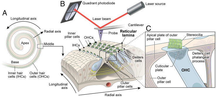
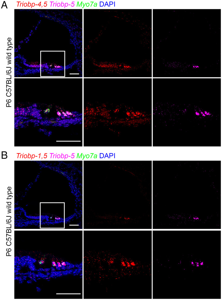
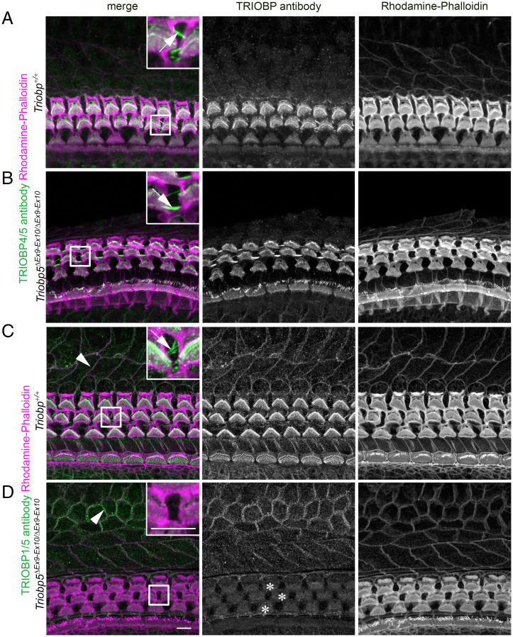
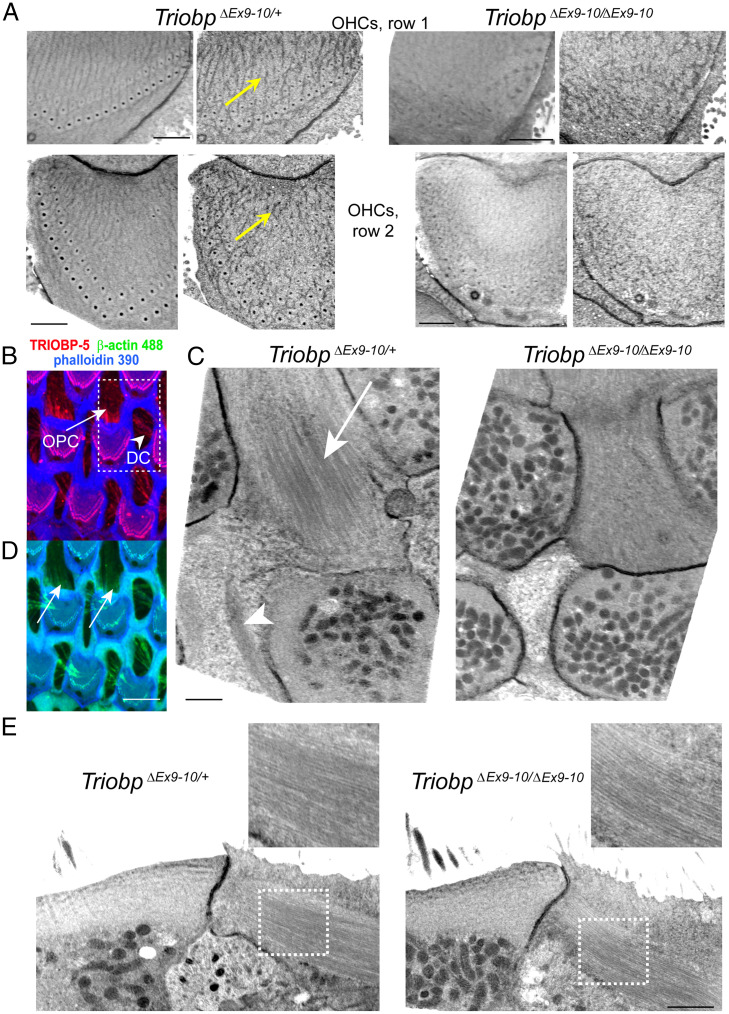
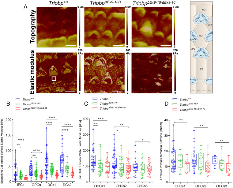
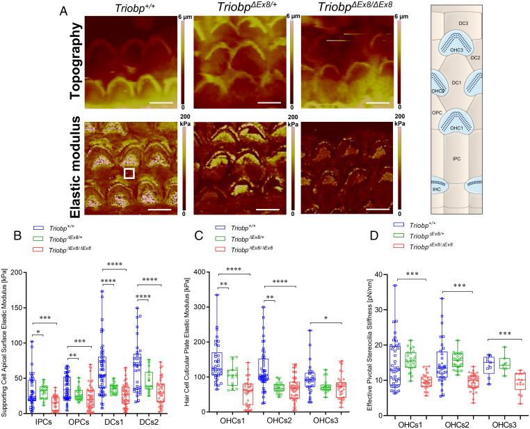
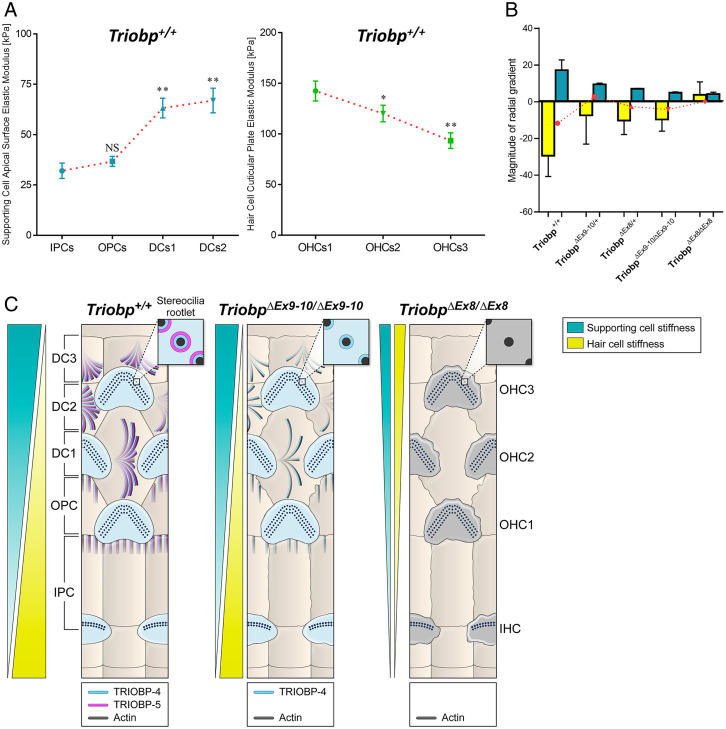
Hearing depends on intricate morphologies and mechanical properties of diverse inner ear cell types. The individual contributions of various inner ear cell types into mechanical properties of the organ of Corti and the mechanisms of their integration are yet largely unknown. Using sub-100-nm spatial resolution atomic force microscopy (AFM), we mapped the Young's modulus (stiffness) of the apical surface of the different cells of the freshly dissected P5-P6 cochlear epithelium from wild-type and mice lacking either Trio and F-actin binding protein (TRIOBP) isoforms 4 and 5 or isoform 5 only. Variants of TRIOBP are associated with deafness in human and in Triobp mutant mouse models. Remarkably, nanoscale AFM mapping revealed unrecognized bidirectional radial stiffness gradients of different magnitudes and opposite orientations between rows of wild-type supporting cells and sensory hair cells. Moreover, the observed bidirectional radial stiffness gradients are unbalanced, with sensory cells being stiffer overall compared to neighboring supporting cells. Deafness-associated TRIOBP deficiencies significantly disrupted the magnitude and orientation of these bidirectional radial stiffness gradients. In addition, serial sectioning with focused ion beam and backscatter scanning electron microscopy shows that a TRIOBP deficiency results in ultrastructural changes of supporting cell apical phalangeal microfilaments and bundled cortical F-actin of hair cell cuticular plates, correlating with messenger RNA and protein expression levels and AFM stiffness measurements that exposed a softening of the apical surface of the sensory epithelium in mutant mice. Altogether, this additional complexity in the mechanical properties of the sensory epithelium is hypothesized to be an essential contributor to frequency selectivity and sensitivity of mammalian hearing.

Keywords: actin cytoskeleton; atomic force microscopy; biophysics; hearing mechanics; mechanobiology.


Conflict of interest statement

The authors declare no competing interest.

Figures

Fig. 1.
Fig. 1.
Schematic of the organ of Corti sensory epithelium, hair cells, supporting cells cellular structures, and the experimental system for PFT-AFM measurements. (A) The schematic illustrates the coiled shape of the inner ear auditory sensory epithelium and the orientations of the longitudinal and radial axes. (B) The PFT-AFM experimental setup that was used to measure nanoscale stiffness of the organ of Corti reticular lamina of sensory epithelium segment including three rows of OHCs and the supporting cells (IPCs, OPCs, and three rows of DCs). (C) Close-up view of the longitudinal cross-section through the OHC and surrounding supporting cells showing the F-actin–based cuticular plate, stereocilia bundle of OHC, OPC apical plate, and DC’s phalangeal process.
Fig. 2.
Fig. 2.
In situ hybridization using RNAscope probe in P6 wild-type mouse cochlea. (A) Expression of Triobp-4 and Triobp-5 mRNAs (Triobp-4/5) (red, Probe-Mm-Triobp-O1), Triobp-5 only mRNA (magenta, Probe-Mm-Triobp-O2-C3), and Myo7a mRNA (green, Probe-Mm-Myo7a-C2). Triobp-4/5 mRNA (red) is expressed in IHCs and OHCs and supporting cells, whereas mRNA of Triobp-5 alone is expressed mainly in OHCs. (B) Expression of both Triobp-1 and Triobp-5 mRNA (Triobp-1/5) (red, Probe-Mm-Triobp-O3), Triobp-5 only mRNA (magenta, Probe-Mm-Triobp-O2-C3), and Myo7a mRNA (green, Probe-Mm-Myo7a-C2). Triobp-1/5 mRNA was detected mainly in hair cells. (Scale bars: 50 µm.)
Fig. 3.
Fig. 3.
Localization of TRIOBP-1 and TRIOBP-4 proteins are unaffected by the absence of TRIOBP-5 in TriobpΔEx9-10/ΔEx9-10 mouse organ of Corti. (A) P8 wild-type mouse organ of Corti stained with TRIOBP-4/5 antibody. (Inset) An enlarged image of the area outlined by the square shape depicting TRIOBP-4/5 immunoreactivity (green) present in stereocilia rootlets and the cuticular plate of OHCs (Inset) and in microfilaments of DCs (arrow in the Inset). (B) TRIOBP-4 (green) but not TRIOBP-5 is detected and localized to the stereocilia rootlets of OHCs (Inset) and microfilaments of DCs (arrow in the Inset) in P8 TRIOBP-5–deficient littermate mouse. (C) P8 wild-type mouse organ of Corti stained with anti-TARA antibody developed against the C-terminal 374 residues present in both TRIOBP-5 and TRIOBP-1. (Inset) Enlarged image of the area outlined by the square depicting TRIOBP-1/5 immunoreactivity (green) present in stereocilia rootlets and the cuticular plates of OHCs (Inset) and in microfilaments of DCs (arrow in the Inset). (D) Immunostaining of TriobpΔEx9-10/ΔEx9-10 mouse organ of Corti using TRIOBP-1/5 antibody reveals that stereocilia rootlet and DC signals observed in wild-type mouse (C) correspond to TRIOBP-5, while nonsensory cell junction signals of the external and internal sulcus cells represent TRIOBP-1 (arrowheads in C and D). TRIOBP-1 was detected also at the junctions of the hair cells and supporting cells of the organ of Corti and diffusely distributed in the cuticular plates of OHCs (asterisks). (Scale bars in D and D, Inset: 5 µm [applies to all panels and Insets].)
Fig. 4.
Fig. 4.
Ultrastructural effects of TRIOBP-5 deficiency. (A) Regular cortical actin “tangles” at the upper surfaces of the OHC cuticular plates in P6 TriobpΔEx9-10/+ mice (Left, yellow arrows) and their disruption in TriobpΔEx9-10/ΔEx9-10 mice (Right). Similar regular actin “tangles” at the top of the OHC cuticular plate were observed in wild-type Triobp+/+ mice (SI Appendix, Fig. S3B). Actin filaments were stabilized with tannic acid during fixation before staining with uranyl acetate in the freeze-substitution step (see Materials and Methods). Upper images illustrate first row OHCs while bottom images illustrate second row OHCs. Each cuticular plate is shown first as a median Z-projection of a 400-nm-thick FIB-SEM volume and then as a single representative FIB-SEM section. The data are representative of five TriobpΔEx9-10/+ OHCs and eight TriobpΔEx9-10/ΔEx9-10 OHCs. (B) Maximum intensity projection view of the Z-stack volume through the hair cell cuticular plates of wild-type Triobp+/+ mice showing TRIOBP-5 immunofluorescence (red) and phalloidin 390 labeling of F-actin (blue) in the OHCs cuticular plate and in the OPC and DC microfilaments. (C) Median Z-projections of a 400-nm-thick FIB-SEM volume at the level of the bottom of the OHC cuticular plates, which show microfilaments/microtubules in OPCs (arrow) and actin patches in DCs (arrowhead) in P6 TriobpΔEx9-10/+ mice (Left) and their disruption in TriobpΔEx9-10/ΔEx9-10 mice (Right). Similar regular microfilaments/microtubules structures in OPCs were observed in wild-type Triobp+/+ mice (SI Appendix, Fig. S3D). The data are representative of two TriobpΔEx9-10/+ and two TriobpΔEx9-10/ΔEx9-10 OPCs and two TriobpΔEx9-10/+ and three TriobpΔEx9-10/ΔEx9-10 DCs. (D) Fluorescent β-actin labeling (green) could be revealed in wild-type Triobp+/+ mice OPCs (arrows) and in DCs in a maximum intensity projection view shown in B. (E) Lateral views of microfilaments/microtubules shown in C. The Insets show a smaller density of these structures in TriobpΔEx9-10/ΔEx9-10 OPCs. (Scale bars: 1 µm in A, C, and E; 5 µm in D.)
Fig. 5.
Fig. 5.
TRIOBP-5 deficiency results in a significant decrease in supporting cell apical surfaces stiffness and a decrease to a lesser degree in hair cell apical surface and stereocilia bundle stiffness. (A) Topography and stiffness maps of the organ of Corti explants for wild-type Triobp+/+, heterozygous TriobpΔEx9-10/+, and homozygous TriobpΔEx9-10/ΔEx9-10 mice. The schematic illustrates the locations of the apical surfaces of supporting cells phalangeal process and the hair cells cuticular plate within the reticular lamina. (B) E values for apical processes of IPCs, OPCs, DCs row 1 (DCs1), and DCs row 2 (DCs2) of Triobp+/+, TriobpΔEx9-10/+, and TriobpΔEx9-10/ΔEx9-10, respectively. (C) E values for cuticular plates of three rows of OHCs including OHCs1, OHCs2, and OHCs3 of Triobp+/+, TriobpΔEx9-10/+, and TriobpΔEx9-10/ΔEx9-10, respectively. The E values of the reticular lamina were measured in regions of interest (white square box in Triobp+/+ Young’s modulus image) overlaying the apical surface of supporting and hair cells. (D) Effective pivotal stereocilia stiffness values within hair bundles of three rows of OHCs. Data are represented as mean (kilopascals or piconewtons per nanometer) ± SD. Significant differences between conditions by unpaired two-tailed Student’s t test with Welch’s correction indicated as ****P ˂ 0.0001, ***P ˂ 0.001, **P ˂ 0.01, and *P ˂ 0.05. (Scale bars: 5 µm.) SI Appendix, Table S1 includes detailed statistical analysis.
Fig. 6.
Fig. 6.
Deficiency of both TRIOBP-4 and TRIOBP-5 proteins results in significant decreases in stiffness of supporting and hair cell apical surfaces and stereocilia bundles. (A) Topography and stiffness maps of the organ of Corti explants for wild-type Triobp+/+, heterozygous TriobpΔEx8/+, and homozygous TriobpΔEx8/ΔEx8 mice. The schematic illustrates the locations of the apical surfaces of supporting cells phalangeal process and the hair cells cuticular plate within the reticular lamina. (B) E values for apical processes of IPCs, OPCs, DCs row 1 (DCs1), and DCs row 2 (DCs2) of Triobp+/+, TriobpΔEx8/+, and TriobpΔEx8/ΔEx8, respectively. (C) E values for cuticular plates of three rows of OHCs including OHCs1, OHCs2 and OHCs3 of Triobp+/+, TriobpΔEx8/+ and TriobpΔEx8/ΔEx8, respectively. The E values of the reticular lamina were measured in regions of interest (white square box in Triobp+/+ Young’s modulus image) overlaying the apical surface of supporting and hair cells. (D) Effective pivotal stereocilia stiffness values within hair bundles of three rows of OHCs. Note that Triobp+/+ wild-type control results are the same for Figs. 4 and 5 since both mice strains used in this study are on the same C57BL/6 background. Data are represented as mean (kilopascals or piconewtons per nanometer) ± SD. Significant differences between conditions by unpaired two-tailed Student’s t test with Welch’s correction indicated as ****P ˂ 0.0001, ***P ˂ 0.001, **P ˂ 0.01, and *P ˂ 0.05. (Scale bars: 5 µm.) SI Appendix, Table S2 includes detailed statistical analysis.
Fig. 7.
Fig. 7.
Novel bidirectional radial stiffness gradients within the organ of Corti are promoted by TRIOBP expression. (A) Graphs show changes in measured Young’s modulus of supporting cells (cyan) and OHCs (green) within the reticular lamina of P5-P6 wild-type mice. Data are represented as means ± SEM; NS indicates nonsignificant differences, P > 0.05 (by unpaired two-tailed Student’s t test with Welch’s correction); **P < 0.01 and *P < 0.05 indicates significant differences by unpaired two-tailed Student’s t test with Welch’s correction). SI Appendix, Tables S1 and S2 includes detailed statistical analysis. (B) Average slope magnitudes of radial gradients in Young’s modulus values against cell position within the reticular lamina (slope; E/radial position) of supporting and hair cells for Triobp+/+, heterozygous TriobpΔEx9-10/+, and homozygous TriobpΔEx9-10/ΔEx9-10 mice as well as heterozygous TriobpΔEx8/+ and homozygous TriobpΔEx8/ΔEx8 mice. Note that when TRIOBP-4 and TRIOBP-5 isoforms are both absent the reticular lamina radial stiffness gradients become unidirectional. The superimposed plot in red represents the comparison between supporting cells and hair cells for each condition indicating the residual stiffness between sensorial and nonsensorial cells. Data are represented as mean ± SEM. SI Appendix, Table S3 includes detailed statistical analysis. (C) Summary model describing opposing radial stiffness gradients in the reticular lamina of the organ of Corti. The illustration of TRIOBP-4 and TRIOBP-5 distribution in the stereocilia bundles, cuticular plate of hair cells and apical processes of supporting cells of wild-type (Triobp+/+), TriobpΔEx9-10/ΔEx9-10 homozygous deficient for TRIOBP-5, and TriobpΔEx8/ΔEx8 homozygous deficient for both TRIOBP-4 and TRIOBP-5 (TRIOBP-4/5) and additionally demonstrating radial stiffness gradients in the organ of Corti reticular lamina of each one.

Similar articles

Cited by

References

    1. Anniko M., Cytodifferentiation of cochlear hair cells. Am. J. Otolaryngol. 4, 375–388 (1983). - PubMed
    1. Robles L., Ruggero M. A., Mechanics of the mammalian cochlea. Physiol. Rev. 81, 1305–1352 (2001). - PMC - PubMed
    1. Kimura R. S., The ultrastructure of the organ of Corti. Int. Rev. Cytol. 42, 173–222 (1975). - PubMed
    1. Breglio A. M., et al. , Exosomes mediate sensory hair cell protection in the inner ear. J. Clin. Invest. 130, 2657–2672 (2020). - PMC - PubMed
    1. McGrath J., Roy P., Perrin B. J., Stereocilia morphogenesis and maintenance through regulation of actin stability. Semin. Cell Dev. Biol. 65, 88–95 (2017). - PMC - PubMed

Publication types