. 2022 Apr 13;17(4):e0266937.
doi: 10.1371/journal.pone.0266937. eCollection 2022.

Evolutionarily conserved properties of CLCA proteins 1, 3 and 4, as revealed by phylogenetic and biochemical studies in avian homologues

Affiliations

Evolutionarily conserved properties of CLCA proteins 1, 3 and 4, as revealed by phylogenetic and biochemical studies in avian homologues

Florian Bartenschlager et al. PLoS One. .

Abstract

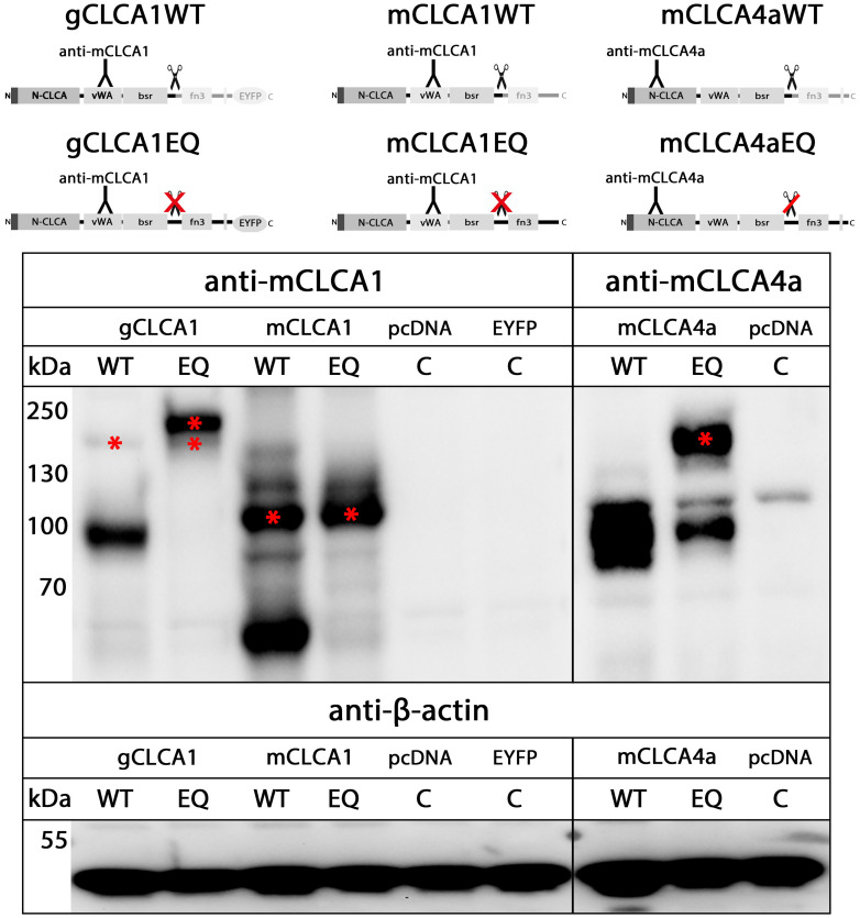
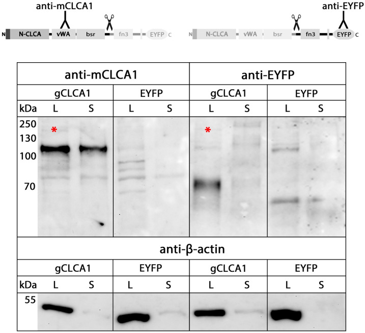
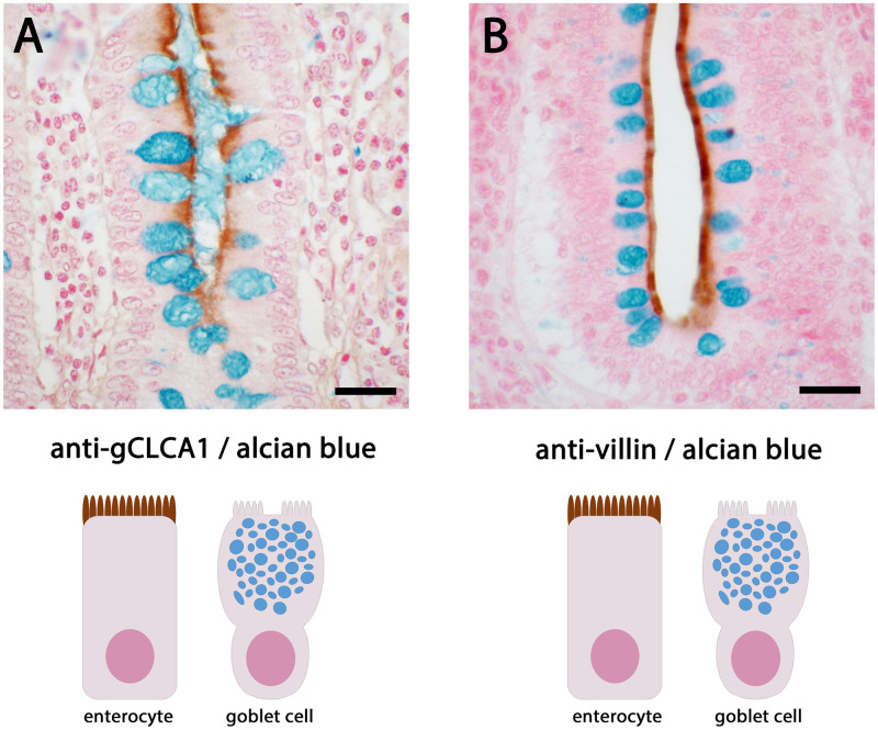
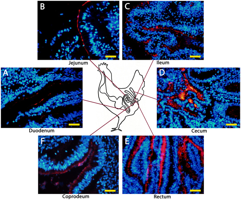
Species-specific diversities are particular features of mammalian chloride channel regulator, calcium activated (CLCA) genes. In contrast to four complex gene clusters in mammals, only two CLCA genes appear to exist in chickens. CLCA2 is conserved in both, while only the galline CLCA1 (gCLCA1) displays close genetic distance to mammalian clusters 1, 3 and 4. In this study, sequence analyses and biochemical characterizations revealed that gCLCA1 as a putative avian prototype shares common protein domains and processing features with all mammalian CLCA homologues. It has a transmembrane (TM) domain in the carboxy terminal region and its mRNA and protein were detected in the alimentary canal, where the protein was localized in the apical membrane of enterocytes, similar to CLCA4. Both mammals and birds seem to have at least one TM domain containing CLCA protein with complex glycosylation in the apical membrane of enterocytes. However, some characteristic features of mammalian CLCA1 and 3 including entire protein secretion and expression in cell types other than enterocytes seem to be dispensable for chicken. Phylogenetic analyses including twelve bird species revealed that avian CLCA1 and mammalian CLCA3 form clades separate from a major branch containing mammalian CLCA1 and 4. Overall, our data suggest that gCLCA1 and mammalian CLCA clusters 1, 3 and 4 stem from a common ancestor which underwent complex gene diversification in mammals but not in birds.


Conflict of interest statement

The authors have declared that no competing interests exist.

Figures

Fig 1
Fig 1. gCLCA1 gene locus and structure as compared with mammalian homologues.
(A) Comparisons of CLCA gene loci of the chicken and of the three mammalian representatives human, pig, and mouse. The galline CLCA locus consists of two CLCA genes, gCLCA1 and gCLCA2, and is shorter than that of mammals. Genes are depicted as boxes and noncoding, intergenic segments are represented as black lines. The chicken locus is scaled 2-fold larger for illustrative purposes. P = pseudogene, kb = kilobases. (B) Comparative gene structure of gCLCA1 and functional mammalian CLCA1, 3 and 4 genes. Like the mammalian CLCA genes of clusters 1, 3, and 4, gCLCA1 is encoded by 14 exons (vertical boxes). Due to short intronic segments (horizontal black lines), the gCLCA1 gene is shorter than its orthologues of the mammal cluster 1, 3, and 4.
Fig 2
Fig 2. The basic functional domains of mammalian CLCA proteins are conserved in gCLCA1.
Schematic depiction of the gCLCA1 protein and of CLCA proteins of clusters 1, 3, and 4 in three mammalian representatives (human, pig and mouse). A cleavable N-terminal signal peptide (dark brown box), an N-CLCA domain (N-CLCA, light brown box), a vWA-domain (vWA, light green box), a β-sheet rich domain (bsr, dark green box) and a fibronectin type III domain (fn3, light blue box) were predicted by in silico analyses and manual sequence alignments. The N-CLCA domain comprises an intact zinc-dependent metalloprotease motif (HExxH, vertical yellow bar). The vWA domain contains a MIDAS site (DxSxS, T, D, vertical yellow bar). Vertical dark red bars indicate cysteine residues, predominantly in the N-CLCA domain. The gCLCA1 protein is putatively cleaved after the amino acids QNR at aa position 705 (scissor). gCLCA1 has a putative C-terminal transmembrane domain (TM, light red box) similarly to the CLCA representatives of cluster 4. The gCLCA1 protein is an N-linked glycoprotein with 12 predicted glycosylation sites. The number of glycosylation sites is higher than in the CLCA proteins of cluster 4, followed by the proteins of cluster 3 and 1. Putative glycosylation sites are indicated by formula image-icons. Numbers display the absolute position, for protein domains further specified by the first or last three amino acids of each domain, respectively.
Fig 3
Fig 3. Localization of the C-terminal cleavage product of gCLCA1 in the plasma membrane.
(A) The C-terminal cleavage product of the gCLCA1EYFP fusion protein was detected at the plasma membrane of transiently transfected HEK293 cells via autofluorescence of the EYFP-tag (green). (B) A corresponding red signal was reproducible by immunofluorescence using the gC1-C1 (anti-gCLCA1 C-terminal) antibody. Alexa fluor 568-conjugated secondary antibodies. Bars indicate 20 μm. Representative images of three independent experiments.
Fig 4
Fig 4. gCLCA1 is a HExxH-dependent cleavable protein.
Immunoblot of cell lysates from HEK293 cells transiently transfected with the EYFP-mock plasmid (EYFP), the gCLCA1WT (gCLCA1) plasmid, and the gCLCA1E164Q plasmid containing an EQ substitution at position two of the catalytic active HExxH motif (gCLCA1EQ) is shown. A C-terminal cleavage product of gCLCA1 and its immature glycosylated precursor protein were identified at ~64 kDa or at ~154 kDa (*), respectively. Cleavage was prevented by the EQ substitution in the HExxH motif as no cleavage product was detectable; however, a strong band at ~166 kDa was identified, which putatively represents the uncleaved, mature glycosylated full-length protein. Identical to cells transfected with the gCLCA1WT plasmid, the immature glycosylated precursor protein at ~154 kDa was detected in cell lysate from gCLCA1E164Q transfected cells. To control for equal total protein loading the samples were identically immunoblotted with primary anti-beta-actin antibodies. Representative images of three independent experiments.
Fig 5
Fig 5. The gCLCA1EQ mutant eliminated autocatalytic cleavage similar to CLCA proteins of mammalian cluster 1.
Immunoblotting of HEK293 cell lysates. The left panel illustrates immunoblots from murinized galline CLCA1, the central panel from murine CLCA1 and the right panel from murine CLCA4a constructs. The proteins of the three homologues differ in their molecular weights. The mutation of the HExxH motif of gCLCA1 eliminated the cleavage of the ~154 kDa precursor protein (*) similarly to the murine CLCA1 protein. In contrast, the cleavage of the murine CLCA4aEQ mutant was only impaired, but not totally absent. Asterisks (*) indicate the uncleaved protein of the respective CLCA homolog. To control for equal total protein loading, the samples were identically immunoblotted with primary anti-beta-actin antibodies. WT = HExxH wild type motif, EQ = EQ mutation in the HExxH motif, C = mock-transfected control. Representative images of three independent experiments.
Fig 6
Fig 6. Secretion of only the N-terminal cleavage product of gCLCA1 into the cell culture supernatant.
Immunoblotting of lysates (L) and cell culture supernatants (S) from HEK293 cells transfected with the gCLCA1Nmabc1 (gCLCA1) and EYFP-mock plasmid (EYFP). The N-terminal cleavage product was detected at ~102 kDa in cell lysate and supernatant. In contrast, the C-terminal, transmembrane domain containing cleavage product was exclusively detected at ~64 kDa in the cell lysate, but not in the supernatant. A weak band of the immature glycosylated gCLCA1 precursor protein was detectable in the cell lysate at ~154 kDa (*). The same amounts of proteins for the respective lysates and supernatants from transfected cells were analyzed in these experiments. To control for equal total protein loading of cell lysates and for lack of contamination of the supernatants by cell debris, the samples were identically immunoblotted with primary anti-beta-actin antibodies. Representative images of three independent experiments.
Fig 7
Fig 7. Glycosylated gCLCA1 is cleaved in the medial Golgi.
Immunoblot of cell lysate of HEK293 cells transiently transfected with the gCLCA1Nmabc1 plasmid (gCLCA1) and a control EYFP-mock plasmid (EYFP). The gCLCA1 precursor (*) was mannose-rich glycosylated, reduced in size by endo H and PNGase F treatment. In contrast, both cleavage products were solely sensitive to PNGase F as shown by a reduction in size of the N-terminal and C-terminal cleavage product to ~78 kDa or ~52 kDa, respectively. The cleavage event seems to occur in the medial Golgi. U = Untreated, C = untreated mock-control, H = endo H treated, F = PNGase F treated cell lysate. To control for equal total protein loading, the samples were identically immunoblotted with primary anti-beta-actin antibodies. Representative images of three independent experiments.
Fig 8
Fig 8. gCLCA1 protein is expressed in the brush border membrane of enterocytes, but not in goblet cells.
(A) The gCLCA1 protein (brown) was detected at the apical surface of enterocytes of colonic crypts by immunohistochemistry using the gC1-C1 (anti-gCLCA1 C-terminal) antibody. A counterstain with alcian blue highlights goblet cells (dark blue) that did not show any gCLCA1 protein expression. (B) The cellular expression pattern was similar to villin, a marker for the brush border of enterocytes (brown). Color was developed using DAB as substrate (brown). Bars indicate 20 μm.
Fig 9
Fig 9. gCLCA1 protein is expressed along the chicken intestine.
Anti-gCLCA1 C-terminal antibody was used to detect gCLCA1 along the epithelium in duodenum (A), jejunum (B), ileum (C), cecum (D), rectum (E) and coprodeum (F). Alexa fluor 568-conjugated secondary antibodies and DAPI counterstain (blue). Bars indicate 40 / 100 μm.
Fig 10
Fig 10. Phylogenetic relationship of CLCA proteins.
Maximum likelihood (ml), most parsimony (mp) and 100 neighbor joining (nj) trees were generated for the five distinct CLCA domains and merged. Tree was based on the ml tree, with branches in thick, when occurring in the mp tree and the branch nodes indicated when occurring in more than 70 nj trees. CLCA2 orthologues were reduced to an outgroup. Other clusters were highlighted. To avoid confusion due to limited space, the five primate species analyzed (human, macaque, baboon, marmoset, bushbaby) are occasionally not separately defined (*). In the case of CLCA duplicates in mammalian species, the sequences are designated by the abbreviation given in GenBank (i.e. “mouse_3a2” for murine Clca3a2) or by “x” if the available sequence has been designated as “CLCA-like” or “unknown”.

Similar articles

Cited by

References

    1. Nakanishi A, Morita S, Iwashita H, Sagiya Y, Ashida Y, Shirafuji H, et al.. Role of gob-5 in mucus overproduction and airway hyperresponsiveness in asthma. Proceedings of the National Academy of Sciences. 2001;98(9):5175–80. doi: 10.1073/pnas.081510898 - DOI - PMC - PubMed
    1. Hoshino M, Morita S, Iwashita H, Sagiya Y, Nagi T, Nakanishi A, et al.. Increased expression of the human Ca2+-activated Cl− channel 1 (CaCC1) gene in the asthmatic airway. American journal of respiratory and critical care medicine. 2002;165(8):1132–6. doi: 10.1164/ajrccm.165.8.2107068 - DOI - PubMed
    1. Range F, Mundhenk L, Gruber A. A soluble secreted glycoprotein (eCLCA1) is overexpressed due to goblet cell hyperplasia and metaplasia in horses with recurrent airway obstruction. Veterinary pathology. 2007;44(6):901–11. doi: 10.1354/vp.44-6-901 - DOI - PubMed
    1. Ritzka M, Stanke F, Jansen S, Gruber AD, Pusch L, Woelfl S, et al.. The CLCA gene locus as a modulator of the gastrointestinal basic defect in cystic fibrosis. Human genetics. 2004;115(6):483–91. doi: 10.1007/s00439-004-1190-y - DOI - PubMed
    1. Hauber HP, Manoukian JJ, Nguyen LH, Sobol SE, Levitt RC, Holroyd KJ, et al.. Increased expression of interleukin-9, interleukin-9 receptor, and the calcium-activated chloride channel hCLCA1 in the upper airways of patients with cystic fibrosis. The Laryngoscope. 2003;113(6):1037–42. doi: 10.1097/00005537-200306000-00022 - DOI - PubMed

Substances

Grants and funding

The author(s) received no specific funding for this work.