. 2022 Apr;604(7906):557-562.
doi: 10.1038/s41586-022-04559-7. Epub 2022 Apr 6.

Activation of STING by targeting a pocket in the transmembrane domain

Affiliations

Activation of STING by targeting a pocket in the transmembrane domain

Defen Lu et al. Nature. 2022 Apr.

Abstract

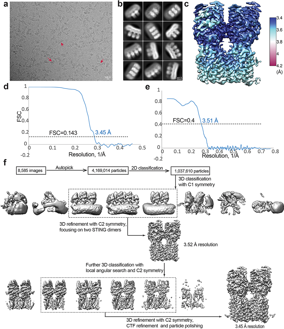
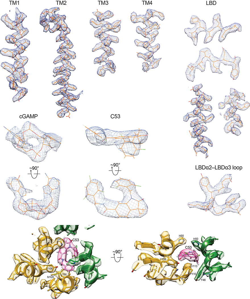
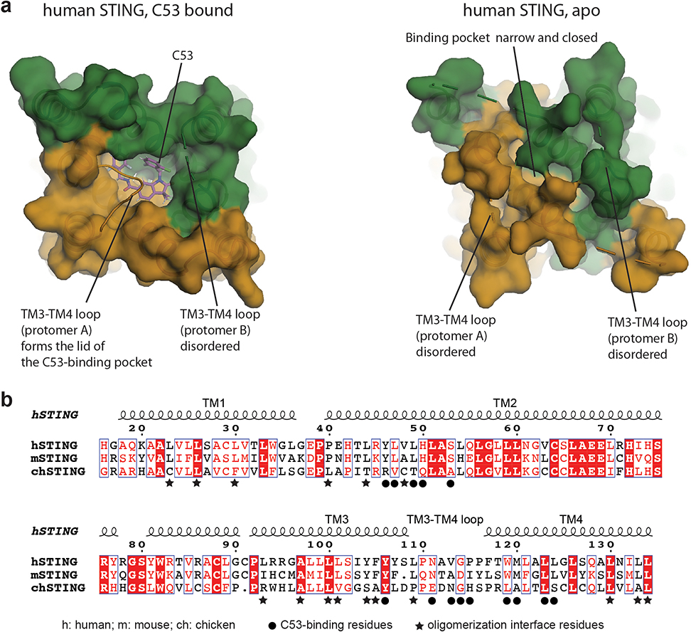
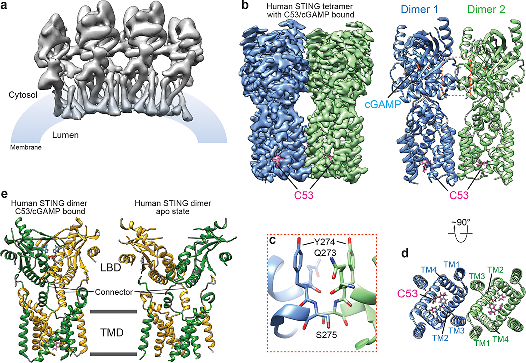
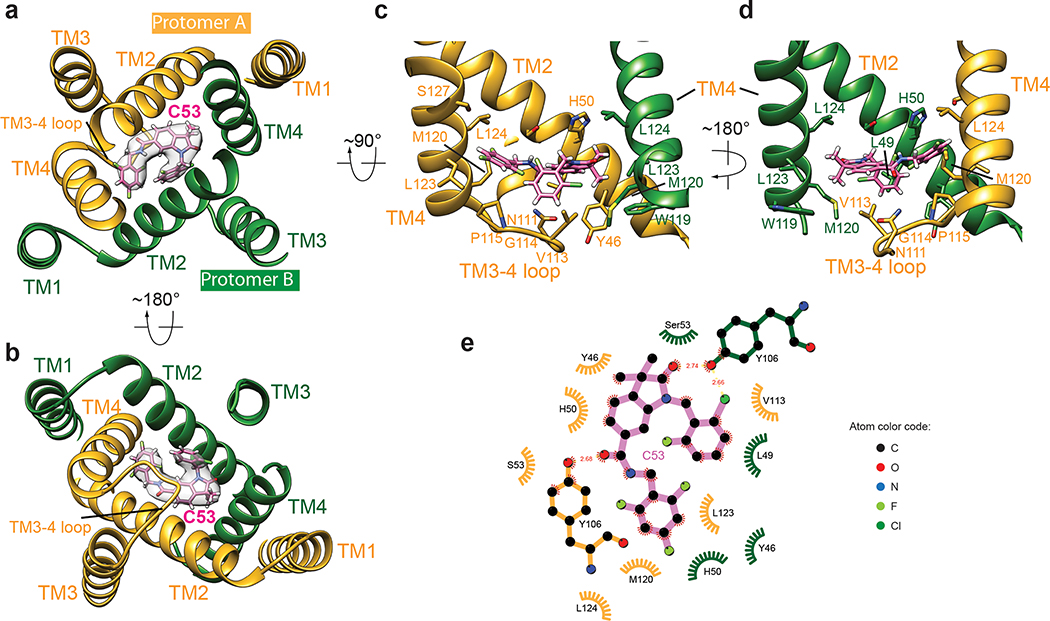
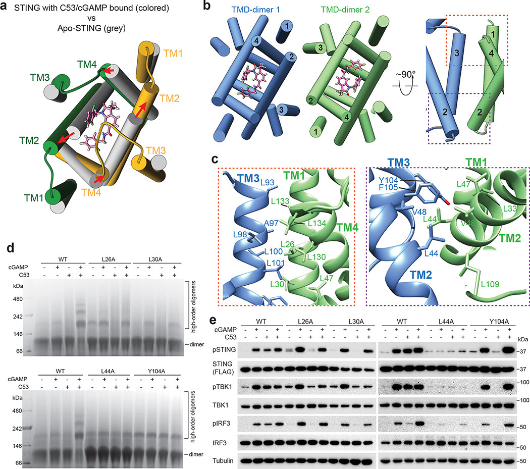
Stimulator of interferon genes (STING) is an adaptor protein in innate immunity against DNA viruses or bacteria1-5. STING-mediated immunity could be exploited in the development of vaccines or cancer immunotherapies. STING is a transmembrane dimeric protein that is located in the endoplasmic reticulum or in the Golgi apparatus. STING is activated by the binding of its cytoplasmic ligand-binding domain to cyclic dinucleotides that are produced by the DNA sensor cyclic GMP-AMP (cGAMP) synthase or by invading bacteria1,6,7. Cyclic dinucleotides induce a conformational change in the STING ligand-binding domain, which leads to a high-order oligomerization of STING that is essential for triggering the downstream signalling pathways8,9. However, the cGAMP-induced STING oligomers tend to dissociate in solution and have not been resolved to high resolution, which limits our understanding of the activation mechanism. Here we show that a small-molecule agonist, compound 53 (C53)10, promotes the oligomerization and activation of human STING through a mechanism orthogonal to that of cGAMP. We determined a cryo-electron microscopy structure of STING bound to both C53 and cGAMP, revealing a stable oligomer that is formed by side-by-side packing and has a curled overall shape. Notably, C53 binds to a cryptic pocket in the STING transmembrane domain, between the two subunits of the STING dimer. This binding triggers outward shifts of transmembrane helices in the dimer, and induces inter-dimer interactions between these helices to mediate the formation of the high-order oligomer. Our functional analyses show that cGAMP and C53 together induce stronger activation of STING than either ligand alone.


Conflict of interest statement

Competing interests. The authors declare no competing interests.

Figures

Extended Data Fig. 1.
Extended Data Fig. 1.. Purification of human STING and its interaction with C53 and cGAMP.
a, Gel filtration profile of human STING on a Superdex S200 10/300 column. b, SDS-PAGE analyses of fractions indicated by the bracket in a. c, Analyses of STING oligomerization by native gel. C53 and cGAMP together induced robust high-order oligomerization of purified STING solubilized in detergent solution, while either one alone failed to do so under the same condition. The results shown are representatives of three biological repeats.
Extended Data Fig. 2.
Extended Data Fig. 2.. Image processing procedure of human STING tetramer bound to both cGAMP and C53.
a, Motion corrected micrograph. Red arrows highlight high-order oligomers of STING. The curved overall shape of the oligomers is evident from these examples. b, 2D class averages of high-order oligomers of human STING. Large oligomers were segmented into particles containing four dimers at the maximum. c, Final 3D reconstruction of the tetramer colored based on local resolution. d, Gold-standard FSC curve of the final 3D reconstruction. e, FSC between the final map and the atomic model. f, Image processing procedure.
Extended Data Fig. 3.
Extended Data Fig. 3.. Sample density maps of various parts of the structure.
Various parts of the structure with the cryo-EM density overlayed are shown. Most of the protein sidechains are clearly visible. The high-quality of the densities for cGAMP and C53 allows unambiguous docking of the compounds.
Extended Data Fig. 4.
Extended Data Fig. 4.. C53-induced dilation of the binding pocket in the STING-TMD.
a, Comparison of the TMD of human STING in the C53-bound and the apo states. It is evident that the TMD pocket in apo-STING is much smaller and cannot accommodate C53, and binding of C53 induces dilation of this pocket. b, Sequence alignment of the TMD of STING from human (h), mouse (m) and chicken (ch). Black circles highlight residues in the C53-binding pocket. Stars highlight residues in the TMD-TMD interface that contribute to the oligomerization of STING.
Extended Data Fig. 5.
Extended Data Fig. 5.. Effects of mutations in the C53-binding site on STING oligomerization in cells.
a and c, Representative images of cells expressing STING wild type or the mutants in the binding pocket and the TM3-TM4 loop, respectively. Hela cells were transfected with GFP-tagged human STING wild type or mutants. Cells were stimulated with cGAMP, C53 or both. Localization of STING-GFP in cells was monitored by the fluorescence signal of GFP. Nuclei were stained with DAPI. The experiments were repeated three times. The images are representatives from these experiments. As expected, STING showed a diffuse pattern in cells in the absence of an agonist. Both C53 and cGAMP induced puncta formation of STING wild type. C53-induced puncta formation was reduced or abolished by the mutations. b and d, Quantification of STING puncta formation in cells expressing the wild type or mutants. The bar graph shows the individual data points, mean and s.e.m. of the percentage of cells with STING forming large puncta from the three biological repeats. Statistical significance p-values between the wild type and mutants were calculated by two-tailed Welch’s t-test. Scale bar, 10 μm. Source data of puncta counting results are provided in the source data file.
Extended Data Fig. 6.
Extended Data Fig. 6.. Further characterization of activation of STING in cells by C53 and cGAMP.
a and b, Activation of STING in cells by C53 and cGAMP at a series of concentrations. Notably, while cGAMP at 10 nM and C53 at 100 nM individually induced minimal phosphorylation of STING, the combination of the two led to robust activation of STING. c and d, Activation of STING by cGAMP or C53 with or without digitonin-mediated cell permeabilization. It is clear from these results that cGAMP-mediated activation of STING is dependent on digitonin-mediated permeabilization, whereas C53 can enter the cell and activate STING on its own. The experiments were carried out in a similar manner as in Fig. 3 and Fig. 4, and the results shown are representatives of three biological repeats. For gel source data, see Supplementary Figure 1.
Extended Data Fig. 7.
Extended Data Fig. 7.. Additional mutational analyses of STING activation by C53 and cGAMP.
a, C53 could induce phosphorylation of the R238A/Y240A mutant of STING in cells, which has its cGAMP-pocket disrupted and could not be activated by cGAMP. b, M120L, a mutation in the C53-binding pocket, reduced STING phosphorylation when cells were stimulated with cGAMP at a lower concentration (100 nM). c, L30A, a mutation in TMD-TMD oligomerization interface, reduced STING phosphorylation when cells were stimulated with cGAMP at a lower concentration (100 nM). These effects of M120L and L30A were suppressed when cells were treated with cGAMP at 1000 nM. These results suggest that both the TMD pocket and the TMD-TMD interface are important for cGAMP-mediated activation of STING, although high concentrations of cGAMP can overcome the detrimental effects of some mutations in these regions. The experiments were carried out in a similar manner as in Fig. 3 and Fig. 4, and the results shown are representatives of three biological repeats. For gel source data, see Supplementary Figure 1.
Extended Data Fig. 8.
Extended Data Fig. 8.. Effects of mutations in the TMD-TMD interface on STING oligomerization in cells.
a, Representative images of cells expressing STING wild type or the mutants in the TMD-TMD interface. Hela cells were transfected with GFP-tagged human STING wild type or mutants. Cells were stimulated with cGAMP, C53 or both. Localization of STING-GFP in cells was monitored by the fluorescence signal of GFP. Nuclei were stained with DAPI. The experiments were repeated three times. The images are representatives from these experiments. As expected, STING showed a diffuse pattern in cells in the absence of an agonist. Both C53 and cGAMP induced puncta formation of STING wild type, which was reduced or abolished by the mutants. b, Quantification of STING puncta formation in cells expressing the wild type or mutants. The bar graph shows the individual data points, mean and s.e.m. of the percentage of cells with STING forming large puncta from the three biological repeats. Statistical significance p-values between the wild type and mutants were calculated by two-tailed Welch’s t-test. Scale bar, 10 μm. Source data of puncta counting results are provided in the source data file.
Fig. 1.
Fig. 1.. Structure of the high-order oligomer of human STING bound to both cGAMP and C53.
a, Overall cryo-EM map of the oligomer containing four STING dimers. b, High-resolution cryo-EM map and atomic model of the STING tetramer. c, Expanded view of the inter-dimer interfaced mediated by the LBDα2-LBDα3 loop. The color scheme is the same as in b. d, C53-binding pocket in the TMD of STING viewed from the luminal side of ER/Golgi. The color scheme is the same as in b. e, Comparison of the human STING dimer bound to both cGAMP and C53 with the STING apo-state. The two subunits in the dimer are colored green and yellow respectively.
Fig. 2.
Fig. 2.. Interaction between STING and C53.
a, b, Overview of the C53-binding site in the TMD of one STING dimer, viewed from the cytosolic and luminal sides, respectively. Cryo-EM density for C53 is shown in semi-transparent white. c, d, Details of the binding site in two different views. Helices in front of C53 are omitted for clarity. e, Two-dimensional diagram of the interactions between C53 and STING. Hydrogen atoms are omitted.
Fig. 3.
Fig. 3.. Mutational analyses of the C53-binding site in human STING.
a, Effects of mutations in the C53-binding site on STING oligomerization induced by cGAMP and C53. Native gel results shown are representatives of three biological repeats. b, Effects of mutations in the C53-binding site on phosphorylation of STING, TBK1 and IRF3 induced by cGAMP and C53 in cells. HEK293T cells were transfected with human STING wild type or the mutants. Cells were stimulated with cGAMP, C53 or both and lysates were subjected to western blot analyses. The results shown are representatives of three biological repeats. For gel source data, see Supplementary Figure 1.
Fig. 4.
Fig. 4.. TMD-mediated interactions contribute to STING oligomerization.
a, C53 induces dilation of the TMD binding pocket in the STING dimer. A comparison with apo-STING shows that the TM helices in the C53-bound structure shift away from each other to enlarge the binding pocket. The structure is viewed from the ER/Golgi luminal side. b, Two orthogonal views of the TMD-TMD interface between the two STING dimers. TM3 from one dimer joins one set of TM1, TM2 and TM4 from the other dimer to form a four-helix bundle. c, Detailed views of the TMD-TMD interface. d, Effects of mutations in the TMD-TMD interface on STING oligomerization induced by cGAMP and C53. Native gel results shown are representatives of three biological repeats. e, Effects of mutations in the TMD-TMD interface on phosphorylation of STING, TBK1 and IRF3 induced by cGAMP and C53 in cells. HEK293T cells were transfected with human STING wild type or the mutants. Cells were stimulated with cGAMP, C53 or both and lysates were subjected to western blot analyses. The results shown are representatives of three biological repeats. For gel source data, see Supplementary Figure 1.

Similar articles

Cited by

References

    1. Zhang X, Bai XC & Chen ZJ Structures and Mechanisms in the cGAS-STING Innate Immunity Pathway. Immunity 53, 43–53, doi:10.1016/j.immuni.2020.05.013 (2020). - DOI - PubMed
    1. Ishikawa H & Barber GN STING is an endoplasmic reticulum adaptor that facilitates innate immune signalling. Nature 455, 674–678, doi:10.1038/nature07317 (2008). - DOI - PMC - PubMed
    1. Zhong B et al. The adaptor protein MITA links virus-sensing receptors to IRF3 transcription factor activation. Immunity 29, 538–550, doi:10.1016/j.immuni.2008.09.003 (2008). - DOI - PubMed
    1. Jin L et al. MPYS, a novel membrane tetraspanner, is associated with major histocompatibility complex class II and mediates transduction of apoptotic signals. Molecular and cellular biology 28, 5014–5026, doi:10.1128/MCB.00640-08 (2008). - DOI - PMC - PubMed
    1. Sun W et al. ERIS, an endoplasmic reticulum IFN stimulator, activates innate immune signaling through dimerization. Proc Natl Acad Sci U S A 106, 8653–8658, doi:10.1073/pnas.0900850106 (2009). - DOI - PMC - PubMed