. 2022 May;298(5):101847.
doi: 10.1016/j.jbc.2022.101847. Epub 2022 Mar 18.

Capsaicin inhibits intestinal Cl- secretion and promotes Na+ absorption by blocking TRPV4 channels in healthy and colitic mice

Affiliations

Capsaicin inhibits intestinal Cl- secretion and promotes Na+ absorption by blocking TRPV4 channels in healthy and colitic mice

Hanxing Wan et al. J Biol Chem. 2022 May.

Abstract

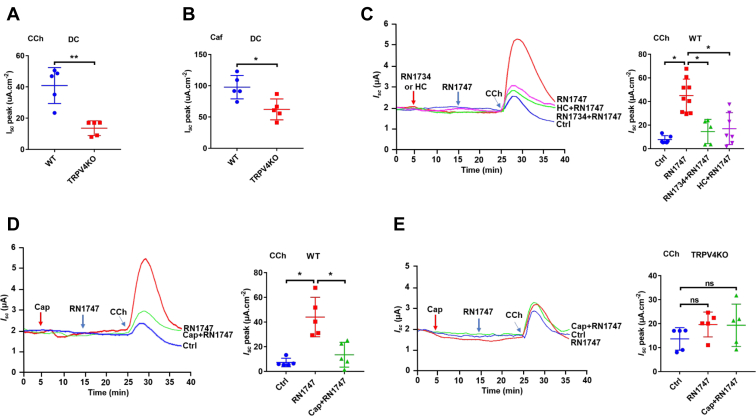
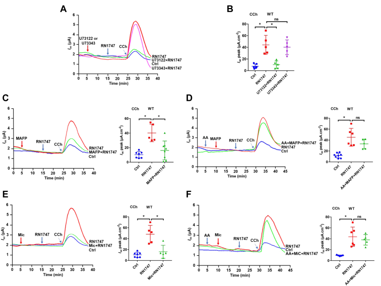
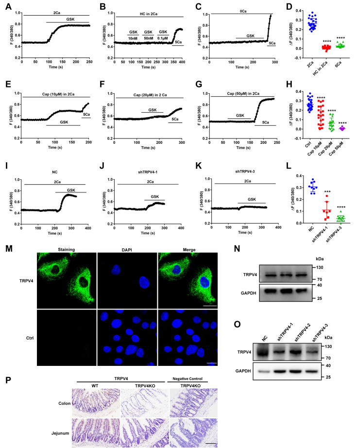
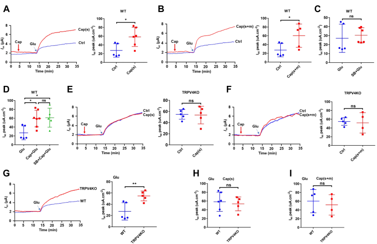
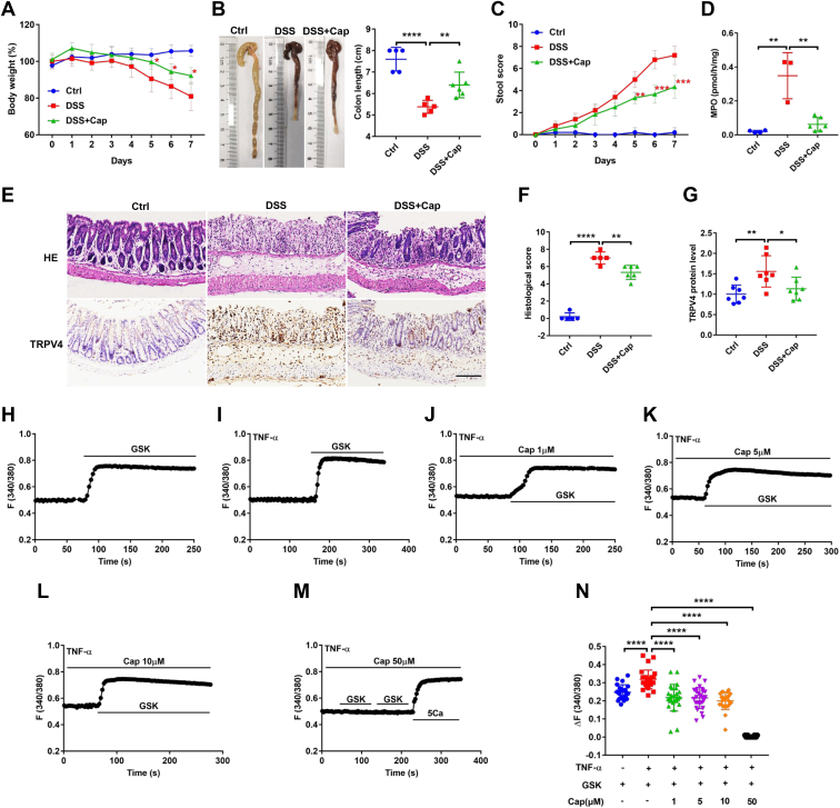
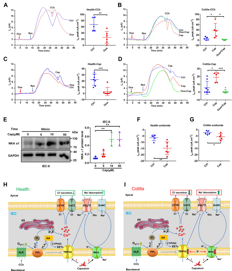
Although capsaicin has been studied extensively as an activator of the transient receptor potential vanilloid cation channel subtype 1 (TRPV1) channels in sensory neurons, little is known about its TRPV1-independent actions in gastrointestinal health and disease. Here, we aimed to investigate the pharmacological actions of capsaicin as a food additive and medication on intestinal ion transporters in mouse models of ulcerative colitis (UC). The short-circuit current (Isc) of the intestine from WT, TRPV1-, and TRPV4-KO mice were measured in Ussing chambers, and Ca2+ imaging was performed on small intestinal epithelial cells. We also performed Western blots, immunohistochemistry, and immunofluorescence on intestinal epithelial cells and on intestinal tissues following UC induction with dextran sodium sulfate. We found that capsaicin did not affect basal intestinal Isc but significantly inhibited carbachol- and caffeine-induced intestinal Isc in WT mice. Capsaicin similarly inhibited the intestinal Isc in TRPV1 KO mice, but this inhibition was absent in TRPV4 KO mice. We also determined that Ca2+ influx via TRPV4 was required for cholinergic signaling-mediated intestinal anion secretion, which was inhibited by capsaicin. Moreover, the glucose-induced jejunal Iscvia Na+/glucose cotransporter was suppressed by TRPV4 activation, which could be relieved by capsaicin. Capsaicin also stimulated ouabain- and amiloride-sensitive colonic Isc. Finally, we found that dietary capsaicin ameliorated the UC phenotype, suppressed hyperaction of TRPV4 channels, and rescued the reduced ouabain- and amiloride-sensitive Isc. We therefore conclude that capsaicin inhibits intestinal Cl- secretion and promotes Na+ absorption predominantly by blocking TRPV4 channels to exert its beneficial anti-colitic action.

Keywords: Na(+)/K(+)-ATPase; TRPV4 channels; epithelial Na(+) channels; short-circuit current; ulcerative colitis.


Conflict of interest statement

Conflicts of interests The authors declare that they have no conflicts of interest with the contents of this article.

Figures

Figure 1
Figure 1
Capsaicin inhibition of secretagogues-induced intestinal Iscvia a TRPV4-dependent but TRPV1-independent manner.AC, representative time courses and summary data showing the inhibitory effect of capsaicin (Cap) on carbachol (CCh, 100 μM, n = 6)- or caffeine (Caf, 10 mM, n = 6)-stimulated jejunal Isc after mucosal (m), serosal (s) addition, and mucosal plus serosal (m+s) addition of capsaicin (30 μM). DF, representative time courses and summary data showing the inhibitory effect of Cap on CCh (n = 6)- or Caf (n = 6)-stimulated distal colonic Isc after capsaicin addition as in AC. G and H, summary data showing the inhibitory effect of capsaicin (Cap, 30 μM) on carbachol (CCh, 100 μM, n = 5)- or caffeine (Caf, 10 mM, n = 5)-stimulated distal colonic Isc in the absence or the presence of SB705498 (SB, 5 μM) to both sides in WT mice. I and J, summary data showing the inhibitory effect of capsaicin on CCh (n = 7)- or Caf (n = 7)-stimulated jejunal Isc in TRPV1 KO mice. K and L, representative time courses and summary data showing the inhibitory effect of capsaicin on CCh (n = 5)- or Caf (n = 5)-stimulated distal colonic Isc in the absence or the presence of HC067047 (HC, 10 μM) to both sides in WT mice. MO, representative time courses and summary data showing the inhibitory effect of capsaicin on CCh (n = 5)- or Caf (n = 5)-stimulated distal colonic Isc in TRPV4 KO mice. P and Q, summary data showing the inhibitory effect of capsaicin on CCh (n = 5)- or Caf (n = 5)-stimulated jejunal Isc in TRPV4 KO mice. Ctrl represents as the control without capsaicin treatment, and m or s in parentheses represents mucosal or serosal addition of capsaicin, respectively. CCh and Caf both add to serosal side. The data are presented as mean ± SD. ∗p < 0.05, ∗∗p < 0.01, ∗∗∗p < 0.001, ∗∗∗∗p < 0.0001 were performed by Student’s t test. ns, no significant differences. Caf, caffeine; CCh, carbachol; Isc, short-circuit current; TRPV, transient receptor potential vanilloid.
Figure 2
Figure 2
Capsaicin inhibition of CCh-induced colonic Cl-secretion by selective blockade of TRPV4 channels.A and B, summary data comparing carbachol (CCh, 100 μM, n = 5)- or caffeine (Caf, 10 mM, n = 5)-stimulated distal colonic Isc between WT mice and TRPV4 KO mice. C, representative time courses and summary data showing the inhibitory effect of TRPV4 blockers RN1734 (50 μM, n = 5) and HC067047 (HC, 10 μM, n = 7) on CCh (50 μM, n = 6) -stimulated distal colonic Isc in the absence or the presence of TRPV4 activator RN1747 (40 μM) in WT mice. D, representative time courses and summary data showing the inhibitory effect of capsaicin (Cap, 30 uM, n = 5) on CCh (50 μM, n = 5) -stimulated distal colonic Isc in the absence or the presence of TRPV4 activator RN1747 (40 μM) in WT mice. E, representative time courses and summary data showing the effect of Cap (30 μM, n = 5) on CCh (100 μM, n = 5)-stimulated distal colonic Isc in the absence or the presence of TRPV4 activator RN1747 (40 μM, n = 5) in TRPV4 KO mice. Ctrl represents as the control with CCh treatment only. CCh adds to serosal side, RN1747, RN1734, and HC add to both sides. The data are presented as mean ± SD. ∗p < 0.05 and ∗∗p < 0.01 were performed by Student’s t test. ns, no significant differences. Caf, caffeine; CCh, carbachol; Isc, short-circuit current; TRPV, transient receptor potential vanilloid.
Figure 3
Figure 3
Contribution of TRPV4 channels to the PLC, PLA2, and CYP450 pathway-mediated colonic epithelial Cl-secretion.A and B, representative time courses and summary data showing the effect of TRPV4 activator RN1747 (40 μM) on CCh (50 μM)-stimulated distal colonic Isc (n = 5) in the absence or the presence of U73122 (30 μM) or U73343 (10 μM). C and D, representative time courses and summary data showing the effect of RN1747 on CCh-stimulated distal colonic Isc (n = 6) in the absence or the presence of MAFP (10 μM) or MAFP plus arachidonic acid (AA, 50 μM). E and F, representative time courses and summary data showing the effect of RN1747 on CCh-stimulated distal colonic Isc (n = 5) in the absence or the presence of miconazole (Mic, 10 μM) or Mic plus AA. Ctrl represents as the control with CCh treatment only. CCh adds to serosal side. RN1747, U73122, U73343, MAFP, AA, and Mic add to both sides. The data are presented as mean ± SD. ∗p < 0.05 was performed by Student’s t test. ns, no significant differences. CCh, carbachol; Isc, short-circuit current; MAFP, methyl arachidonyl fluorophosphonate; PLA, phospholipid A; PLC, phospholipid C; TRPV, transient receptor potential vanilloid.
Figure 4
Figure 4
Capsaicin blockade of Ca2+entry via TRPV4 channels and the protein expression of TRPV4 in IEC-6 cells and mouse intestinal tissues.AC, summary tracings of [Ca2+]cyt time course in response to GSK1016790A (GSK, 10–100 nM) and calcium (5 mM) in the absence (0Ca2+, n = 26) or the presence (2Ca2+, n = 23) of calcium and calcium plus HC067047 (HC, 5 μM, n = 19). D, summary data showing the peaks of GSK-increased [Ca2+]cyt signaling as in (AC). EG, summary tracings of [Ca2+]cyt time course in response to GSK (10 nM) in the absence or the presence of different dose of capsaicin (Cap, n = 26). H, summary data showing the peaks of GSK-increased [Ca2+]cyt signaling described as in (EG). IK, summary tracings of [Ca2+]cyt time course in response to GSK (10 nM) of NC and shTRPV4-1 or shTRPV4-3. L, summary data showing the peaks of GSK-increased [Ca2+]cyt signaling described as in (IK). The data are presented as mean ± SD. ∗∗∗p < 0.001 and ∗∗∗∗p < 0.0001 were performed by Student’s t test. M, immunostaining of TRPV4 proteins to confirm their expression in IEC-6 cells. The upper panels: the specific staining of TRPV4 proteins (in green) and merge with the nuclei of the cells stained with DAPI. The lower panel: the nuclei of the cells stained with DAPI (in blue) without primary antibodies against TRPV4 as a negative control. The scale bar represents 20 μm for each image. N and O, Western blotting analysis of TRPV4 proteins expression in IEC-6 cells (N) and shTRPV4 in IEC-6 cells (O). GAPDH was used as a loading control. Each one is the representative of all images taken from 3 independent experiments with similar results. P, immunohistological analysis on TRPV4 proteins in jejunal and colonic tissues from WT mice (the left panels) and TRPV4 KO mice (the middle panels). The right panels were without primary antibodies against TRPV4 in the intestinal tissues from TRPV4 KO mice as negative controls. The scale bar represents 100 μm for each image. [Ca2+]cyt, cytosolic Ca2+ concentrations; IECs, intestinal epithelial cells; TRPV, transient receptor potential vanilloid.
Figure 5
Figure 5
Capsaicin promotion of jejunal Na+absorption by rather blocking TRPV4 channels than activating TRPV1 channels.A and B, representative time courses and summary data showing the effect of capsaicin (Cap, 30 μM, n = 5) on glucose-induced jejunal Isc after its serosal (s) addition or serosal plus mucosal (s+m) addition in WT mice. C and D, summary data showing the effect of SB705498 (SB, 5 μM, n = 5) on glucose-induced jejunal Isc and the effect of Cap on the Isc (n = 5) in the absence or the presence of SB in WT mice. E and F, representative time courses and summary data showing the effect of Cap on glucose-induced jejunal Isc (n = 5) after its serosal (s) addition or serosal plus mucosal (s+m) addition in TRTPV4 KO mice. G, representative time courses and summary data comparing the glucose-induced jejunal Isc (n = 5) between WT mice and TRPV4 KO mice. H and I, summary data comparing the effect of Cap on glucose-induced jejunal Isc (n = 5) between WT mice and TRPV4 KO mice after its serosal (s) addition or serosal plus mucosal (s+m) addition. The data are presented as mean ± SD. ∗p < 0.05 and ∗∗p < 0.01 were performed by Student’s t test. ns, no significant differences. Isc, short-circuit current; TRPV, transient receptor potential vanilloid.
Figure 6
Figure 6
Anti-coliticeffect of capsaicin in mice and its inhibition on Ca2+signaling in IEC-6 cells pretreated with TNF-α.A and B, summary data showing the time course of body weight and mouse colon length after different treatments without (Ctrl) or with capsaicin (intragastrically (10 mg/kg) once per day for 7 days), DSS, or DSS + Cap (n = 5). C and D, summary data showing the time courses of stool score and MPO after different treatments as in A and B. E, histological examination and immunohistological analysis showing colonic micro-structure (the upper panels) and TRPV4 immunostaining (the lower panels) after different treatments as in A and B. The scale bar represents 100 μm for each image. F and G, summary data showing histological score and TRPV4 protein level after different treatments as in A and B. H and I, summary tracings of [Ca2+]cyt time course in response to GSK (10 nM) in IEC-6 cells without (n = 22) or with (n = 28) TNF-α (40 ng/ml) pretreatment. JM, summary tracings of [Ca2+]cyt time course showing the inhibitory effect of Cap at different doses on GSK-induced [Ca2+]cyt signaling in IEC-6 cells pretreated with TNF-α. N, summary data showing the inhibitory effect of Cap at different doses on peaks of GSK-increased [Ca2+]cyt signaling described as in HM. The data are presented as mean ± SD. ∗p < 0.05, ∗∗p < 0.01, ∗∗∗p < 0.001, and ∗∗∗∗p < 0.0001 were performed by Student’s t test. [Ca2+]cyt, cytosolic Ca2+ concentrations; DSS, dextran sodium sulfate; IECs, intestinal epithelial cells; MPO, myeloperoxidase; TRPV, transient receptor potential vanilloid.
Figure 7
Figure 7
Capsaicin stimulation of NKA and ENaC activities in the colonic epithelia of healthy and colitis mice and the proposed mechanisms of capsaicin action on intestinal epithelial ion transports.A, representative time courses and summary data showing the effect of CCh (100 μM, n = 5) on distal colonic Isc across the basolateral membrane in apically permeabilized epithelia in the absence or the presence of ouabain (Oua, 100 μM, n = 5) in healthy mice. B, representative time courses and summary data showing the effect of CCh on distal colonic Isc (n = 5) across the basolateral membrane in apically permeabilized epithelia in the absence or the presence of ouabain in colitic mice with or without capsaicin pretreated. C, representative time courses and summary data showing the effect of capsaicin (Cap, 30 μM, n = 7) on distal colonic Isc across the basolateral membrane in apically permeabilized epithelia in the absence or the presence of Oua (n = 7) in healthy mice. D, representative time courses and summary data showing the effect of Cap on distal colonic Isc across the basolateral membrane in apically permeabilized epithelia in the absence or the presence of Oua (n = 6) in colitic mice with or without capsaicin pretreated. E, Western blot showing that Cap induced NKAα1 in IEC-6 cells (n = 3 independent experiments). F, summary data showing the distal colonic Isc in response to amiloride (100 μM, n = 5) in the absence or the presence of capsaicin (30 μM, n = 5) in healthy mice. G, summary data showing the colonic Isc (n = 5) in response to amiloride in colitic mice with or without capsaicin pretreated. CCh and ouabain were added to serosal side. Amiloride was added to mucosal side, but capsaicin was added to both sides. The data are presented as mean ± SD. ∗p < 0.05, ∗∗p < 0.01, and ∗∗∗p < 0.001 were performed by Student’s t test. H, in health, after the PIP2 suppression on basolateral TRPV4 channels is relieved by the activation of PLC and PLA2 in cholinergic signaling pathway, Ca2+ enters via TRPV4 channels and ER/Ca2+ releases via IP3R. Cytosolic Ca2+ stimulates Ca2+-dependent Cl- secretion through apical CFTR and CaCC. TRPV4 channels could also be activated by their endogenous ligands CYP450 metabolites produced from AA/CYP450 pathway. Capsaicin attenuates Cl- secretion but stimulates Na+ absorption by blocking TRPV4 channels but stimulating NKA activity on the basolateral side of intestinal epithelial cells (IEC). I, in colitis, the expression and function of TRPV4 channels are enhanced but NKA activities are reduced by inflammatory factors, the Cl- secretion are increased, and Na+ absorption are decreased. Capsaicin ameliorates colitis and reduces diarrhea by: (1) suppressing hyperactivation of basolateral TRPV4 channels to reduce intestinal Cl- secretion and (2) blocking basolateral TRPV4 channels and stimulating NKA activity to increase intestinal Na+ absorption. AA, arachidonic acid; CaCC, Ca2+-activated Cl- channels; CFTR, cystic fibrosis transmembrane conductance regulator; CYP450, cytochrome P450; [Ca2+]cyt, cytosolic Ca2+ concentrations; ENaC, epithelial Na+ channels; ER, endoplasmic reticulum; IP3R, IP3 receptor; IF, inflammatory factors; NKA, Na+/K+-ATPase; SGLT1, Na+-glucose cotransporter 1; TRPV, transient receptor potential vanilloid.

Similar articles

Cited by

References

    1. Chang J.T. Pathophysiology of inflammatory bowel diseases. N. Engl. J. Med. 2020;383:2652–2664. - PubMed
    1. de Mattos B.R., Garcia M.P., Nogueira J.B., Paiatto L.N., Albuquerque C.G., Souza C.L., Fernandes L.G., Tamashiro W.M., Simioni P.U. Inflammatory bowel disease: An overview of immune mechanisms and biological treatments. Mediators Inflamm. 2015;2015:493012. - PMC - PubMed
    1. Keita Å V., Lindqvist C.M., Öst Å., Magana C.D.L., Schoultz I., Halfvarson J. Gut barrier dysfunction-a primary defect in twins with Crohn's disease predominantly caused by genetic predisposition. J. Crohn's colitis. 2018;12:1200–1209. - PMC - PubMed
    1. Michielan A., D'Incà R. Intestinal permeability in inflammatory bowel disease: Pathogenesis, clinical evaluation, and therapy of leaky gut. Mediators Inflamm. 2015;2015:628157. - PMC - PubMed
    1. Baumgart D.C., Le Berre C. Newer biologic and small-molecule therapies for inflammatory bowel disease. N. Engl. J. Med. 2021;385:1302–1315. - PubMed

Publication types