Review
. 2022 Jun 16;82(12):2315-2334.
doi: 10.1016/j.molcel.2022.02.021. Epub 2022 Mar 9.

The expanding universe of PARP1-mediated molecular and therapeutic mechanisms

Affiliations
Review

The expanding universe of PARP1-mediated molecular and therapeutic mechanisms

Dan Huang et al. Mol Cell. .

Abstract

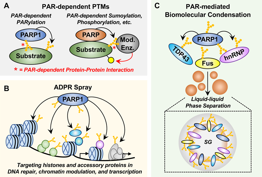
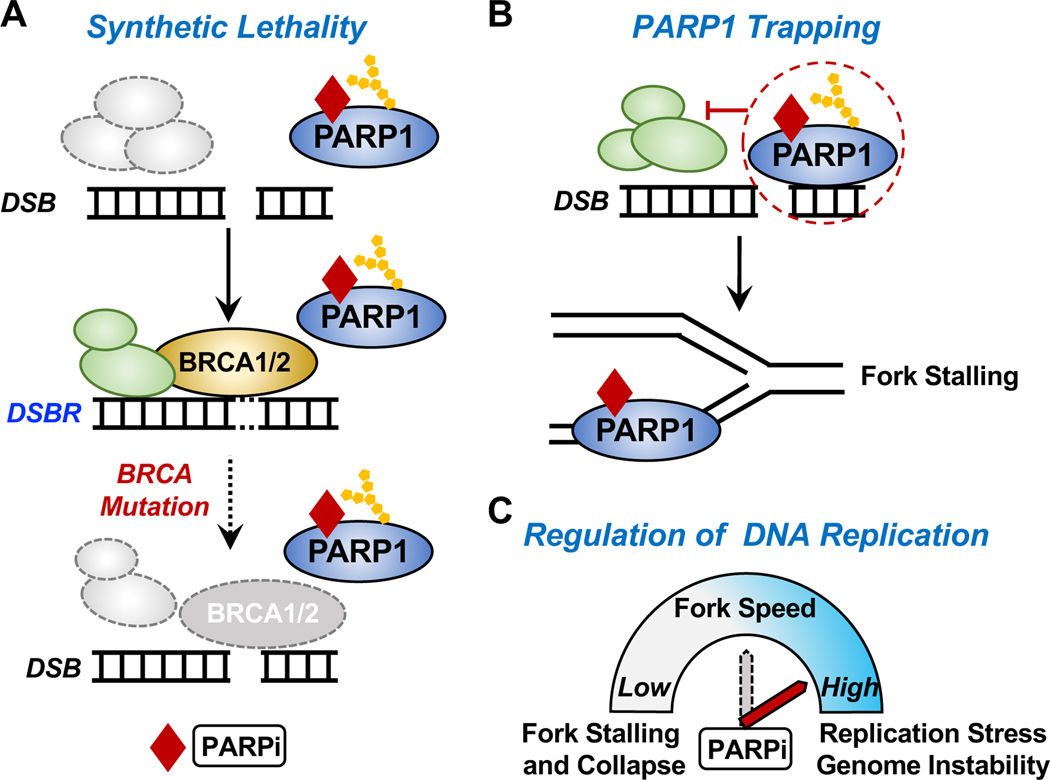
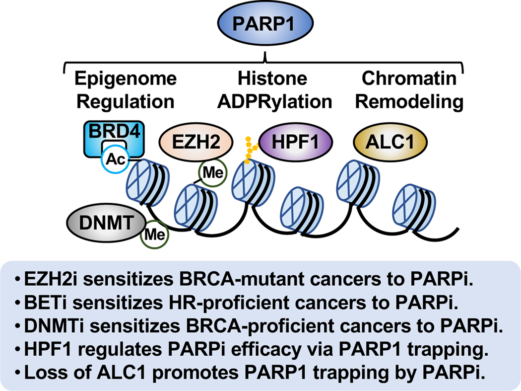
ADP-ribosylation (ADPRylation) is a post-translational modification of proteins catalyzed by ADP-ribosyl transferase (ART) enzymes, including nuclear PARPs (e.g., PARP1 and PARP2). Historically, studies of ADPRylation and PARPs have focused on DNA damage responses in cancers, but more recent studies elucidate diverse roles in a broader array of biological processes. Here, we summarize the expanding array of molecular mechanisms underlying the biological functions of nuclear PARPs with a focus on PARP1, the founding member of the family. This includes roles in DNA repair, chromatin regulation, gene expression, ribosome biogenesis, and RNA biology. We also present new concepts in PARP1-dependent regulation, including PAR-dependent post-translational modifications, "ADPR spray," and PAR-mediated biomolecular condensate formation. Moreover, we review advances in the therapeutic mechanisms of PARP inhibitors (PARPi) as well as the progress on the mechanisms of PARPi resistance. Collectively, the recent progress in the field has yielded new insights into the expanding universe of PARP1-mediated molecular and therapeutic mechanisms in a variety of biological processes.

Keywords: ADP-ribosylation; DNA damage response; DNA replication; PARP; PARP inhibitor; PARPi; PTM; RNA biology; biomolecular condensate; chromatin; gene regulation; histone; poly(ADP-ribose) polymerase; post-translational modification; ribosome biogenesis; therapeutic resistance; therapeutics.


Conflict of interest statement

Declaration of interests W.L.K. is a founder of Ribon Therapeutics and a founder, consultant, and SAB member of ARase Therapeutics. He is also a co-holder of U.S. Patent 9,599,606 covering a set of ADP-ribose detection reagents, which have been licensed to and is sold by EMD Millipore. D.H. declares no competing interests.

Figures

Figure 1.
Figure 1.. The expanding universe of PARP1 biology and tools used to investigate its functions.
(A) Structural and functional domains of PARP1, including (1) a DNA binding domain (DBD) containing three zinc finger (Zn) motifs and a nuclear localization signal (NLS), (2) an automodification domain containing a BRCA C-terminus (BRCT) motif, and (3) a catalytic domain containing a tryptophan (W)-glycine (G)-arginine (R) (WGR) motif and the PARP catalytic site signature motif. (B) Schematic illustrating how the understanding of PARP1 biology, as well as the methods and tools used to study it, has evolved over time from the original focus on protein ADPRylation and DNA repair.
Figure 2.
Figure 2.. Dynamics of PARP-mediated ADPRylation.
(A) Chemical structures of mono(ADP-ribose) (MAR) and poly(ADP-ribose) (PAR). The positions on the ribose moieties where the PAR polymer is elongated and branched are indicated. (B) Dynamics of PARP-mediated ADPRylation. PARP family members serve as “writers” of ADPRylation by catalytically transferring ADPR moieties from NAD+ to substrate proteins. The ADPR units can be hydrolyzed and removed by “erasers,” such as PARG, TARG1, ARH1, ARH3, MacroD1, and MacroD2. The ADPR units on target proteins can be recognized by “readers” containing macrodomains, WWE domains, PAR-binding zinc fingers (PBZs), or PAR-binding motifs. The NAD+ required for ADPRylation is supplied through NAD+ synthesis pathways catalyzed by “feeders” - the nicotinamide mononucleotide adenylyl transferases (NMNATs).
Figure 3.
Figure 3.. Roles of PARP1 in DNA damage repair.
PARP1 binds to sites of DNA damage, is rapidly activated, and catalyzes autoADPRylation, as well as transmodification of local substrate proteins, playing important roles in various DNA repair pathways. (A) Activated PARP1 at sites of single-strand breaks (SSBs) recruits DNA repair effectors, such as X-ray repair cross-complementing protein 1 (XRCC1), to mediate DNA repair. AutoADPRylation causes the dissociation of PARP1 from the DNA damage site, which is ultimately required for the efficient DNA repair. (B) In response to double-strand breaks (DSBs), activated PARP1 recruits the MRE11-RAD50NBS1 (MRN) complex and other repair factors to the sites of damage for homologous recombination (HR)-mediated repair. PARP1 also differentially regulates classical and alternative non-homologous end-joining (NHEJ) repair pathways. PARP1 binds to DSBs in direct competition with Ku70/80 proteins, thus inhibiting the classical pathway of NHEJ that utilizes Ku, DNA-PKcs, DNA ligase IV, and XRCC4. Conversely, PARP1 mediates the recruitment of MRN and CtIP to DSBs to promotes alternative NHEJ (Alt-EJ). (C) PARP1 links DNA damage to DNA replication by regulating DNA replication fork progression during replication stress. PARP1 is activated at the stalled replication forks in response to replication stress. Activated PARP1 may promote replication fork reversal or stabilization by multiple distinct mechanisms, such as (1) inhibiting the ATP-dependent DNA helicase Q1 (RECQ1), which is responsible for restart of the stalled replication fork or (2) inhibiting fork degradation by MRN and EXO1 through the recruitment of BRCA1. Conflicting studies have suggested an active role of PARP1 in replication fork restart through MRE11 based on the prevention or delay of replication restart upon PARP1 depletion, but the evidence directly linking PARP1 and MRE11 during fork restart is limited.
Figure 4.
Figure 4.. Biological functions of PARP1 in the nucleus.
PARP1 regulates a broad array of processes in the nucleus, such as DNA repair, chromatin regulation, gene expression, ribosome biogenesis, and RNA processing. (A) Roles of PARP1 in DNA damage repair networks, including single-strand break (SSB) repair, base excision repair (BER), nucleotide excision repair (NER), and DSB repair through both homologous recombination (HR) and alternative end-joining (Alt-EJ) pathways. (B) Roles of PARP1 in chromatin regulation. PARP1 modulates the structure and function of chromatin through the (1) regulation of chromatin composition, including histone variants and linker histone; (2) regulation of histone ADPRylation and other histone PTMs, including phosphorylation, acetylation and methylation; and (3) regulation of chromatin remodelers. (C) Roles of PARP1 in ribosome biogenesis. SnoRNA-mediated activation of PARP1 promotes PARylation of DDX21, a DEAD-box RNA helicase in the nucleolus, resulting in the enhanced rDNA transcription and subsequent protein synthesis. (D) Roles of PARP1 in RNA biology. PARP1 regulates multiple steps of RNA processing by interacting with and modifying RNA binding proteins and RNA processing factors.
Figure 5.
Figure 5.. Emerging concepts and mechanisms of PARP1-dependent regulation.
(A) PAR-dependent PTMs. PAR-dependent protein-protein interactions can drive substrates to their cognate modifying enzymes. Examples include (1) “PAR-dependent PARylation,” where a substrate protein interacts with automodified PARP1 through PAR, leading to subsequent PARylation of the substrate protein by PARP1 and (2) other “PAR-dependent post-translational modifications,” where a PARP (e.g., PARP1, PARP5) interacts with and PARylates a substrate protein, which promotes PAR-dependent interactions with a modifying enzyme, leading to posttranslational modification (e.g., ubiquitylation, phosphorylation) of the PARP substrate protein by that modifying enzyme. (B) ADPR spray. ADPRylation events mediated by nuclear PARP1 may drive a high density “ADPR spray” across key histone and accessory proteins in processes including DNA repair, chromatin modulation, and transcription. (C) PAR-mediated biomolecular condensate formation and function. Free or protein-linked PAR may regulate the formation or dissociation of biomolecular condensates. In this example, PAR facilitates the phase separation of FUS, TDP43, and hnRNPA1 to form stress granules (SG) in the pathogenesis of amyotrophic lateral sclerosis (ALS) and frontotemporal dementia (FTD).
Figure 6.
Figure 6.. Mechanisms of action of PARP inhibitors.
(A) Synthetic lethality. The simultaneous inhibition of PARP1 activity in DNA repair by PARP inhibitors (PARPi) and loss of BRCA1/2 functionality (e.g., through genetic lesions) leads to the failure of DNA damage repair and subsequent cell death. (B) PARPi-induced PARP1 “trapping.” Some PARPi “trap” PARP1 on DNA, thus preventing autoPARylation of PARP1 and release from the site of damaged DNA. This interferes with the catalytic cycle of PARP1 and efficient DNA damage repair. PARP1 trapping acts as a replication barrier, which impairs the progression of DNA replication forks and leads to fork stalling. (C) Regulation of DNA replication progression by PARPi. Activated PARP1 promotes replication fork reversal and reduces replication fork speed upon replication stress (see Figure 3C). In this regard, PARPi increases replication fork speed, leading to replication stress and genome instability.
Figure 7.
Figure 7.. PARPi sensitivity and chromatin regulation.
The multifaceted roles of PARP1 in chromatin regulation impact the sensitivity and efficacy of PARPi. This may involve effects of PARP1 on epigenome regulation, histone ADPRylation, and chromatin remodeling.

Similar articles

Cited by

References

    1. Ahel D, Horejsi Z, Wiechens N, Polo SE, Garcia-Wilson E, Ahel I, Flynn H, Skehel M, West SC, Jackson SP, et al. (2009). Poly(ADP-ribose)-dependent regulation of DNA repair by the chromatin remodeling enzyme ALC1. Science 325, 1240–1243.10.1126/science.1177321. - DOI - PMC - PubMed
    1. Alemasova EE, and Lavrik OI (2019). Poly(ADP-ribosyl)ation by PARP1: reaction mechanism and regulatory proteins. Nucleic Acids Res 47, 3811–3827. 10.1093/nar/gkz120. - DOI - PMC - PubMed
    1. Altmeyer M., Neelsen KJ., Teloni F., Pozdnyakova I., Pellegrino S., Grofte M., Rask MD., Streicher W., Jungmichel S., Nielsen ML., and Luka J. (2015). Liquid demixing of intrinsically disordered proteins is seeded by poly(ADP-ribose). Nat Commun 6, 8088. 10.1038/ncomms9088. - DOI - PMC - PubMed
    1. Ame JC, Rolli V, Schreiber V, Niedergang C, Apiou F, Decker P, Muller S, Hoger T, Menissier-de Murcia J, and de Murcia G. (1999). PARP-2, A novel mammalian DNA damage-dependent poly(ADP-ribose) polymerase. J Biol Chem 274, 17860–17868. 10.1074/jbc.274.25.17860. - DOI - PubMed
    1. Ame JC, Spenlehauer C, and de Murcia G. (2004). The PARP superfamily. Bioessays 26, 882893. 10.1002/bies.20085. - DOI - PubMed

Publication types

LinkOut - more resources