

Extracellular vesicle PD-L1 in reshaping tumor immune microenvironment: biological function and potential therapy strategies
- PMID: 35090497
- PMCID: PMC8796536
- DOI: 10.1186/s12964-021-00816-w
Extracellular vesicle PD-L1 in reshaping tumor immune microenvironment: biological function and potential therapy strategies
Abstract
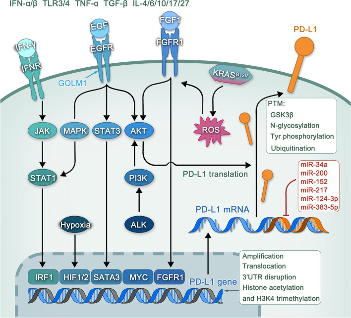
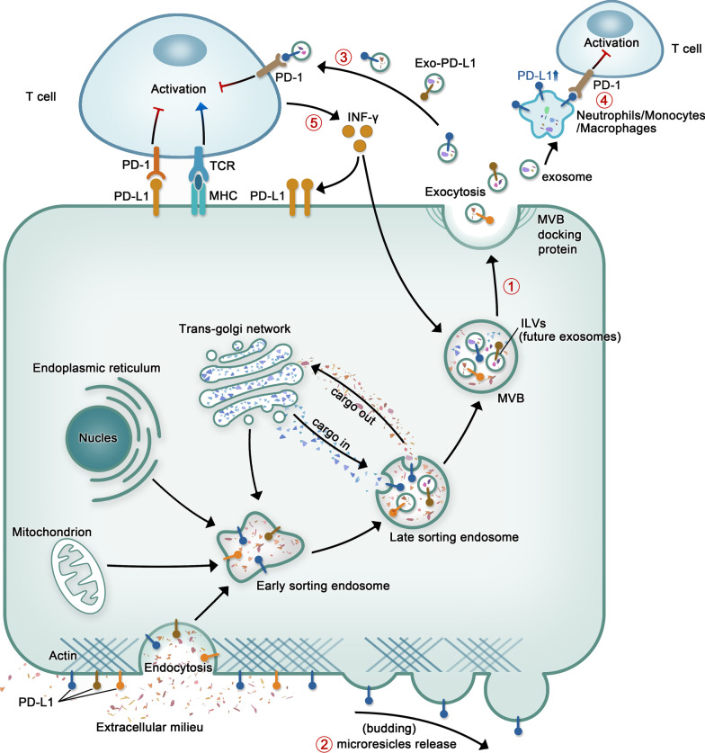
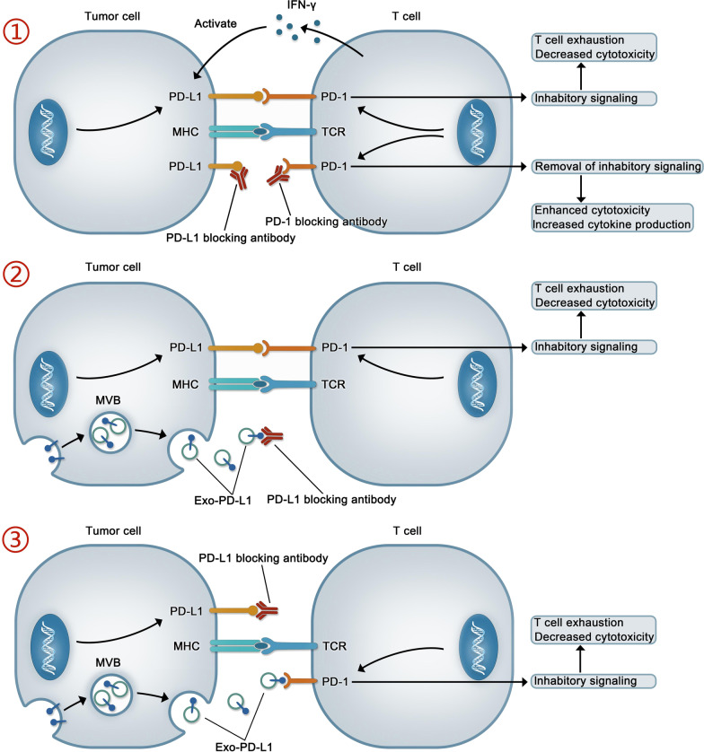
Programmed cell death 1 ligand 1 (PD-L1) is the ligand for programmed death protein-1 (PD-1), is associated with immunosuppression. Signaling via PD-1/PD-L1 will transmits negative regulatory signals to T cells, inducing T-cell inhibition, reducing CD8+ T-cell proliferation, or promoting T-cell apoptosis, which effectively reduces the immune response and leads to large-scale tumor growth. Accordingly, many antibody preparations targeting PD-1 or PD-L1 have been designed to block the binding of these two proteins and restore T-cell proliferation and cytotoxicity of T cells. However, these drugs are ineffective in clinical practice. Recently, numerous of studies have shown that, in addition to the surface of tumor cells, PD-L1 is also found on the surface of extracellular vesicles secreted by these cells. Extracellular vesicle PD-L1 can also interact with PD-1 on the surface of T cells, leading to immunosuppression, and has been proposed as a potential mechanism underlying PD-1/PD-L1-targeted drug resistance. Therefore, it is important to explore the production, regulation and tumor immunosuppression of PD-L1 on the surface of tumor cells and extracellular vesicles, as well as the potential clinical application of extracellular vesicle PD-L1 as tumor biomarkers and therapeutic targets. Video Abstract.
Keywords: Biomarker; Extracellular vesicles; Immune escape; Immunotherapy; PD-L1.
© 2022. The Author(s).
Conflict of interest statement
The authors declare that they have no competing interests.
Figures
Similar articles
-
Extracellular vesicles containing PD-L1 contribute to CD8+ T-cell immune suppression and predict poor outcomes in small cell lung cancer.Clin Exp Immunol. 2022 May 12;207(3):307-317. doi: 10.1093/cei/uxac006. Clin Exp Immunol. 2022. PMID: 35553630 Free PMC article.
-
Macitentan improves antitumor immune responses by inhibiting the secretion of tumor-derived extracellular vesicle PD-L1.Theranostics. 2022 Jan 31;12(5):1971-1987. doi: 10.7150/thno.68864. eCollection 2022. Theranostics. 2022. PMID: 35265193 Free PMC article.
-
EV PD-L1 is Correlated With Clinical Features and Contributes to T Cell Suppression in Pediatric Thyroid Cancer.J Clin Endocrinol Metab. 2020 Aug 1;105(8):dgaa309. doi: 10.1210/clinem/dgaa309. J Clin Endocrinol Metab. 2020. PMID: 32459310
-
Tumor-derived exosomes in the PD-1/PD-L1 axis: Significant regulators as well as promising clinical targets.J Cell Physiol. 2021 Jun;236(6):4138-4151. doi: 10.1002/jcp.30197. Epub 2020 Dec 4. J Cell Physiol. 2021. PMID: 33275291 Review.
-
The role of exosomal PD-L1 in tumor progression and immunotherapy.Mol Cancer. 2019 Oct 23;18(1):146. doi: 10.1186/s12943-019-1074-3. Mol Cancer. 2019. PMID: 31647023 Free PMC article. Review.
Cited by
-
Exosomes Derived from Immune Cells: The New Role of Tumor Immune Microenvironment and Tumor Therapy.Int J Nanomedicine. 2022 Dec 21;17:6527-6550. doi: 10.2147/IJN.S388604. eCollection 2022. Int J Nanomedicine. 2022. PMID: 36575698 Free PMC article. Review.
-
Challenges and the Evolving Landscape of Assessing Blood-Based PD-L1 Expression as a Biomarker for Anti-PD-(L)1 Immunotherapy.Biomedicines. 2022 May 20;10(5):1181. doi: 10.3390/biomedicines10051181. Biomedicines. 2022. PMID: 35625917 Free PMC article. Review.
-
Influencing immunity: role of extracellular vesicles in tumor immune checkpoint dynamics.Exp Mol Med. 2024 Nov;56(11):2365-2381. doi: 10.1038/s12276-024-01340-w. Epub 2024 Nov 11. Exp Mol Med. 2024. PMID: 39528800 Free PMC article. Review.
-
Regulation of Extracellular Vesicle-Mediated Immune Responses against Antigen-Specific Presentation.Vaccines (Basel). 2022 Oct 10;10(10):1691. doi: 10.3390/vaccines10101691. Vaccines (Basel). 2022. PMID: 36298556 Free PMC article. Review.
-
Does Elevated Pre-Treatment Plasma PD-L1 Level Indicate an Increased Tumor Burden and Worse Prognosis in Metastatic Colorectal Cancer?J Clin Med. 2022 Aug 17;11(16):4815. doi: 10.3390/jcm11164815. J Clin Med. 2022. PMID: 36013050 Free PMC article.
References
-
- Xu R, Rai A, Chen MS, Suwakulsiri W, Greening DW, Simpson RJ. Extracellular vesicles in cancer—implications for future improvements in cancer care. Nat Rev Clin Oncol. 2018;15:617–638. - PubMed
Publication types
MeSH terms
Substances
Grants and funding
- 81472302/National Natural Science Foundation of China
- 82003040/Innovative Research Group Project of the National Natural Science Foundation of China
- 2020-BS-103/Natural Science Foundation of Liaoning Province
- 2020M681016/China Postdoctoral Science Foundation
- XLYC1902050/Xingliaoyingcaijihua Project of Liaoning Province
LinkOut - more resources
Full Text Sources
Research Materials
