

Injury-Induced Cellular Plasticity Drives Intestinal Regeneration
- PMID: 34915204
- PMCID: PMC8803615
- DOI: 10.1016/j.jcmgh.2021.12.005
Injury-Induced Cellular Plasticity Drives Intestinal Regeneration
Abstract
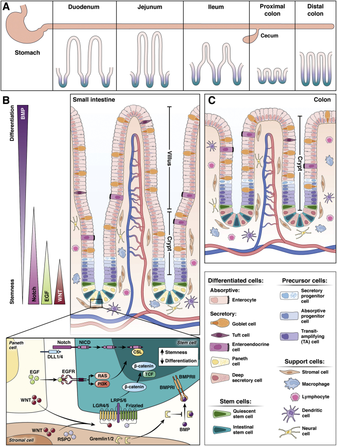
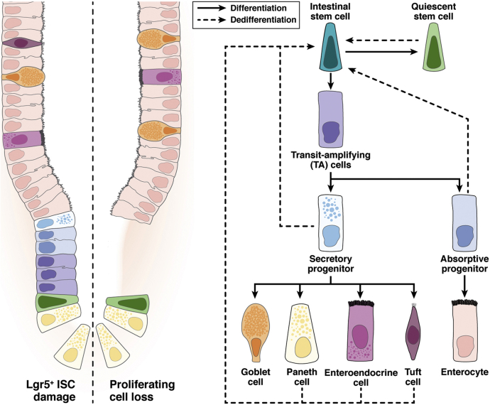
The epithelial lining of the intestine, particularly the stem cell compartment, is affected by harsh conditions in the luminal environment and also is susceptible to genotoxic agents such as radiation and chemotherapy. Therefore, the ability for intestinal epithelial cells to revert to a stem cell state is an important physiological damage response to regenerate the intestinal epithelium at sites of mucosal injury. Many signaling networks involved in maintaining the stem cell niche are activated as part of the damage response to promote cellular plasticity and regeneration. The relative contribution of each cell type and signaling pathway is a critical area of ongoing research, likely dependent on the nature of injury as well as the regional specification within the intestine. Here, we review the current understanding of the multicellular cooperation to restore the intestinal epithelium after damage.
Keywords: Cellular Plasticity; Intestinal Homeostasis; Intestinal Regeneration; Stem Cell Niche.
Copyright © 2022 The Authors. Published by Elsevier Inc. All rights reserved.
Figures
Similar articles
-
ANGPTL2 expression in the intestinal stem cell niche controls epithelial regeneration and homeostasis.EMBO J. 2017 Feb 15;36(4):409-424. doi: 10.15252/embj.201695690. Epub 2017 Jan 2. EMBO J. 2017. PMID: 28043948 Free PMC article.
-
The Intestinal Stem Cell Niche: Homeostasis and Adaptations.Trends Cell Biol. 2018 Dec;28(12):1062-1078. doi: 10.1016/j.tcb.2018.08.001. Epub 2018 Sep 5. Trends Cell Biol. 2018. PMID: 30195922 Free PMC article. Review.
-
Plasticity of Intestinal Epithelium: Stem Cell Niches and Regulatory Signals.Int J Mol Sci. 2020 Dec 31;22(1):357. doi: 10.3390/ijms22010357. Int J Mol Sci. 2020. PMID: 33396437 Free PMC article. Review.
-
TNFAIP8 controls murine intestinal stem cell homeostasis and regeneration by regulating microbiome-induced Akt signaling.Nat Commun. 2020 May 22;11(1):2591. doi: 10.1038/s41467-020-16379-2. Nat Commun. 2020. PMID: 32444641 Free PMC article.
-
Cellular Plasticity of Defa4Cre-Expressing Paneth Cells in Response to Notch Activation and Intestinal Injury.Cell Mol Gastroenterol Hepatol. 2019;7(3):533-554. doi: 10.1016/j.jcmgh.2018.11.004. Epub 2018 Nov 27. Cell Mol Gastroenterol Hepatol. 2019. PMID: 30827941 Free PMC article.
Cited by
-
JAK/STAT signaling promotes the emergence of unique cell states in ulcerative colitis.Stem Cell Reports. 2024 Aug 13;19(8):1172-1188. doi: 10.1016/j.stemcr.2024.06.006. Epub 2024 Jul 18. Stem Cell Reports. 2024. PMID: 39029458 Free PMC article.
-
Neural cell state shifts and fate loss in ageing and age-related diseases.Nat Rev Neurol. 2023 Jul;19(7):434-443. doi: 10.1038/s41582-023-00815-0. Epub 2023 Jun 2. Nat Rev Neurol. 2023. PMID: 37268723 Free PMC article. Review.
-
Lgr5+ intestinal stem cells are required for organoid survival after genotoxic injury.bioRxiv [Preprint]. 2024 Apr 25:2024.04.08.588400. doi: 10.1101/2024.04.08.588400. bioRxiv. 2024. Update in: Development. 2024 Dec 1;151(23):dev202941. doi: 10.1242/dev.202941. PMID: 38645040 Free PMC article. Updated. Preprint.
-
Lactobacillus salivarius Promotion of Intestinal Stem Cell Activity in Hens Is Associated with Succinate-Induced Mitochondrial Energy Metabolism.mSystems. 2022 Dec 20;7(6):e0090322. doi: 10.1128/msystems.00903-22. Epub 2022 Nov 22. mSystems. 2022. PMID: 36413033 Free PMC article.
-
p53 suppresses MHC class II presentation by intestinal epithelium to protect against radiation-induced gastrointestinal syndrome.Nat Commun. 2024 Jan 2;15(1):137. doi: 10.1038/s41467-023-44390-w. Nat Commun. 2024. PMID: 38167344 Free PMC article.
References
-
- Barker N. Adult intestinal stem cells: critical drivers of epithelial homeostasis and regeneration. Nat Rev Mol Cell Biol. 2014;15:19–33. - PubMed
-
- Barker N., van Es J.H., Kuipers J., Kujala P., van den Born M., Cozijnsen M., Haegebarth A., Korving J., Begthel H., Peters P.J., Clevers H. Identification of stem cells in small intestine and colon by marker gene Lgr5. Nature. 2007;449:1003–1007. - PubMed
-
- Bottcher A., Buttner M., Tritschler S., Sterr M., Aliluev A., Oppenlander L., Burtscher I., Sass S., Irmler M., Beckers J., Ziegenhain C., Enard W., Schamberger A.C., Verhamme F.M., Eickelberg O., Theis F.J., Lickert H. Non-canonical Wnt/PCP signalling regulates intestinal stem cell lineage priming towards enteroendocrine and Paneth cell fates. Nat Cell Biol. 2021;23:23–31. - PubMed
-
- Herring C.A., Banerjee A., McKinley E.T., Simmons A.J., Ping J., Roland J.T., Franklin J.L., Liu Q., Gerdes M.J., Coffey R.J., Lau K.S. Unsupervised trajectory analysis of single-cell RNA-seq and imaging data reveals alternative tuft cell origins in the gut. Cell Syst. 2018;6:37–51 e9. - PMC - PubMed
Publication types
MeSH terms
Grants and funding
LinkOut - more resources
Full Text Sources
