. 2022 Feb 1;132(3):e144339.
doi: 10.1172/JCI144339.

An alternatively spliced STING isoform localizes in the cytoplasmic membrane and directly senses extracellular cGAMP

Affiliations

An alternatively spliced STING isoform localizes in the cytoplasmic membrane and directly senses extracellular cGAMP

Xiaobo Li et al. J Clin Invest. .

Abstract

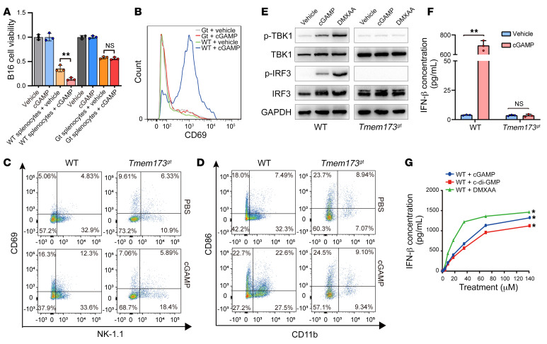
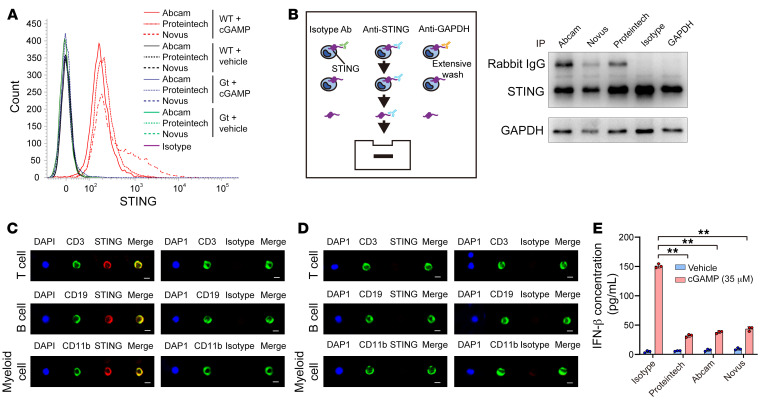
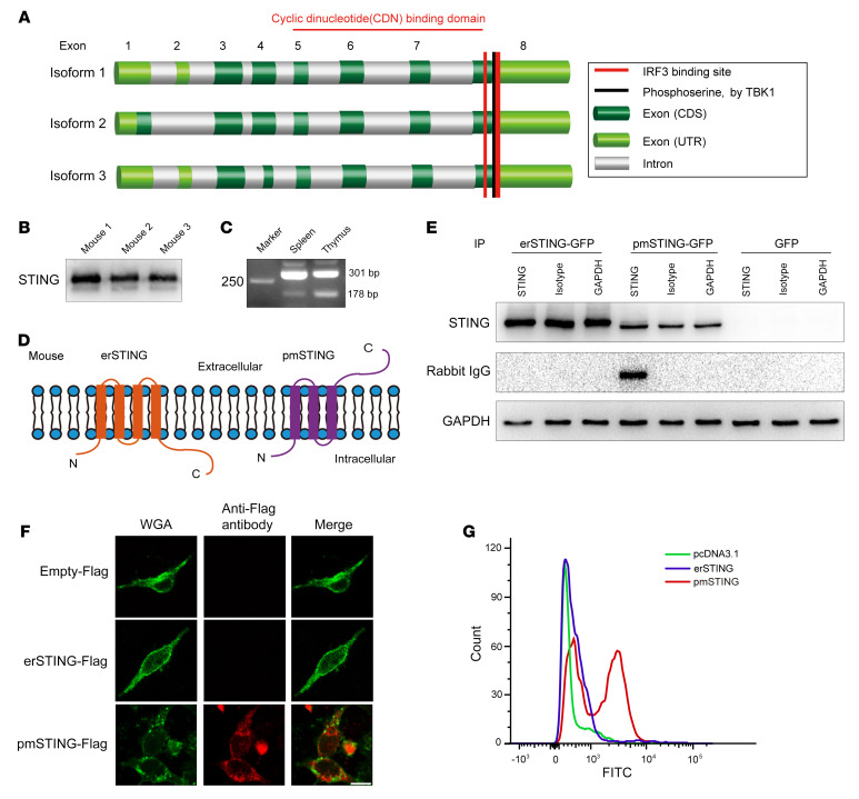
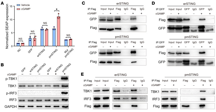
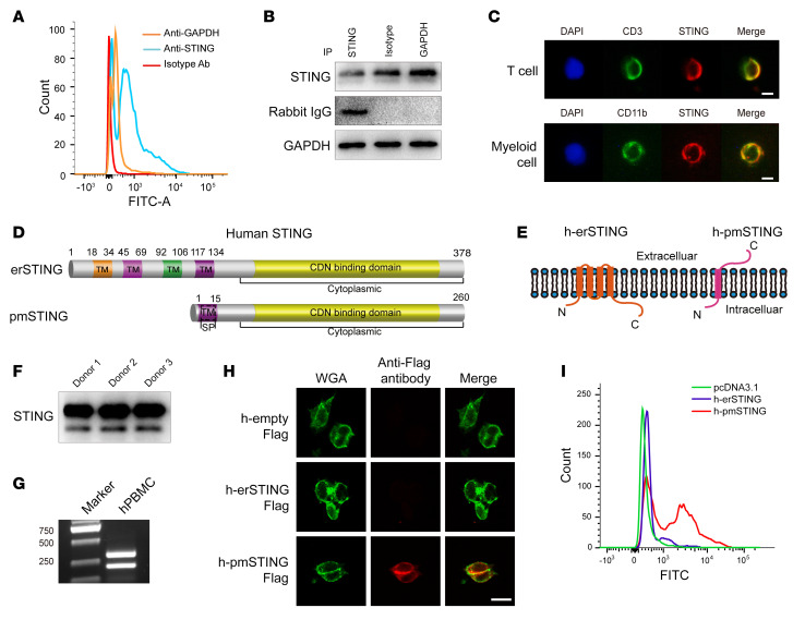
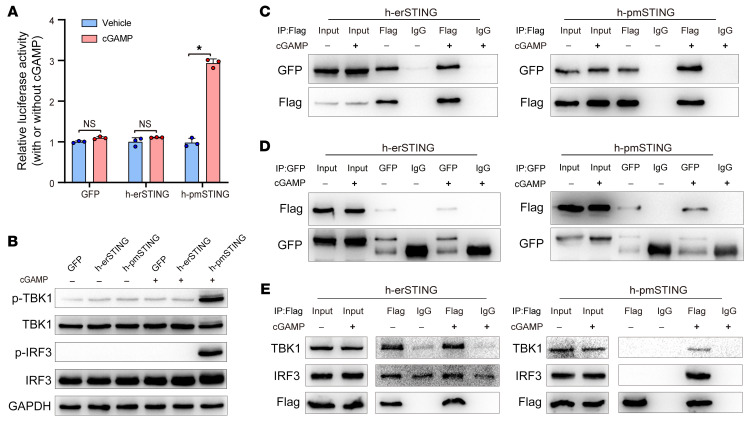
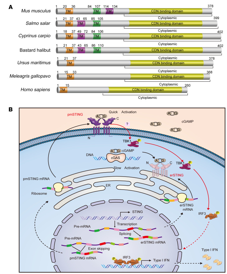
It has been revealed that 2'3'-cyclic-GMP-AMP (cGAMP), a second messenger that activates the antiviral stimulator of IFN genes (STING), elicits an antitumoral immune response. Since cGAMP cannot cross the cell membrane, it is not clear how intracellular STING has been activated by extracellular cGAMP until SLC19A1 was identified as an importer to transport extracellular cGAMP into the cytosol. However, SLC19A1-deficient cells also sense extracellular cGAMP, suggesting the presence of mechanisms other than the facilitating transporters for STING sensing extracellular cGAMP. Here, using immunoprecipitation, immunofluorescence, and flow cytometry, we identified an alternatively spliced STING isoform, plasmatic membrane STING (pmSTING), that localized in the plasma membrane with its C-terminus outside the cell, due to a lack of 1 transmembrane domain in its N-terminus compared with canonical STING. Further studies showed that extracellular cGAMP not only promoted the dimerization of pmSTING and interaction of pmSTING with TANK-binding kinase 1 (TBK1) and IFN regulatory factor 3 (IRF3), but also enhanced the phosphorylation of TBK1 and IRF3 and the production of IFN in pmSTING-transfected cells. Additionally, we also identified similar pmSTING isoforms in other species including human. This study suggests a conserved role for pmSTING in sensing extracellular cGAMP and provides insight into the role of cGAMP as an immunotransmitter.

Keywords: Cancer immunotherapy; Cellular immune response; Immunology; Innate immunity; Oncology.


Figures

Figure 1
Figure 1. Extracellular cGAMP activates immune responses in a STING-dependent manner.
(A) The effect of cGAMP on the viability of B16 cells cocultured with or without splenocytes from WT or STING-deficient mice (Tmem173gt) was assessed by MTT assay (n = 3). **P < 0.01, by 2-tailed, paired Student’s t test. (B) Expression of CD69 in WT or Tmem173gt splenocytes treated with vehicle or cGAMP was detected by flow cytometry. (C) Expression of CD69 in WT or Tmem173gt NK cells treated with vehicle or cGAMP was detected by flow cytometry. (D) Expression of CD86 in WT or Tmem173gt myeloid cells treated with vehicle or cGAMP was detected by flow cytometry. (E) Expression of p-TBK1 and p-IRF3 was detected by Western blotting in WT or Tmem173gt splenocytes treated with vehicle, cGAMP, or DMXAA, respectively. (F) Production of IFN-β in WT or Tmem173gt splenocytes treated with vehicle or cGAMP was detected by ELISA (n = 3). **P < 0.01, by 2-tailed, paired Student’s t test. (G) Production of IFN-β in WT splenocytes treated with different concentrations of cGAMP, c-di-GMP, or DMXAA was detected by ELISA (n = 3). *P < 0.05, by 2-way ANOVA. Data are presented as the mean ± SD.
Figure 2
Figure 2. Identification of a cell surface STING projecting its C-terminus outside of splenocytes.
(A) A cell surface STING with its C-terminus outside of mouse splenocytes was detected with 3 antibodies against the STING C-terminal epitope using flow cytometry. (B) WT splenocytes were incubated with the indicated antibodies and then washed and lysed. Immunoblotting was performed to detect the existence of IgG in the cell lysate using a second antibody against rabbit IgG. (C) Colocalization of cell surface STING with surface proteins of T cells (CD3), B cells (CD19), and myeloid cells (CD11b) from C57BL/6 mice was detected using confocal microscopy. Scale bars: 5 μm. (D) Expression of cell surface STING and surface proteins of T cells (CD3), B cells (CD19), and myeloid cells (CD11b) from STING-deficient mice (Tmem173gt) using confocal microscopy. Scale bars: 5 μm. (E) Extracellular cGAMP–induced production of IFN-β was detected by ELISA in WT splenocytes preincubated with the indicated antibodies (n = 3). **P < 0.01, by 1-way ANOVA followed by Dunnett’s test for comparison with the isotype antibody and the cGAMP treatment group.
Figure 3
Figure 3. An alternatively spliced STING isoform with 3 TM domains localizes in the plasma membrane of mouse splenocytes.
(A) Predicted exon structure and schematic of the functional domains in the C-terminus of mouse Tmem173 transcript variants based on NCBI’s GENE database. (B) Different STING isoforms were detected by immunoblotting in splenocytes from 3 individual C57BL/6 mice. (C) Two STING isoforms with a different N-terminus were detected by reverse transcription PCR (RT-PCR) in mouse spleen and thymus. (D) Predicted plasma membrane topology of detected mouse STING isoforms. (E) B16Tmem173–/– cells transfected with erSTING-EGFP, pmSTING-EGFP, or EGFP were incubated with the indicated antibodies and then washed and lysed. Immunoblotting was performed to detect IgG in the cell lysate using a secondary antibody against rabbit IgG. (F and G) B16Tmem173–/– cells were transfected with erSTING-Flag, pmSTING-Flag, or a vector plasmid, respectively. An antibody against Flag was used to detect Flag projecting outside cells using immunofluorescence (F) and flow cytometry (G), respectively. Scale bars: 20 μm.
Figure 4
Figure 4. Mouse pmSTING isoform directly senses extracellular cGAMP and activates TBK1/IRF3/IFN signaling.
(A) B16Tmem173–/– cells stably expressing SEAP to monitor IFN activity were transfected with erSTING-Flag, pmSTING-Flag, or mutated pmSTING-Flag (Mut pmSTING-Flag) at the TBK1 phosphorylation site (Ser316R). SEAP was detected in these cells after treatment with vehicle or cGAMP (n = 3). *P < 0.05, by 2-tailed, paired Student’s t test. (B) Western blotting was performed to detect p-TBK1 and p-IRF3 in B16Tmem173–/– cells transfected with erSTING-Flag or pmSTING-Flag upon treatment with vehicle or cGAMP. (C and D) B16Tmem173–/– cells were transfected with both erSTING-Flag and erSTING-EGFP or both pmSTING-Flag and pmSTING-EGFP, and then treated with vehicle or cGAMP. Immunoprecipitation using anti-Flag antibody (C) or anti-GFP antibody (D) was performed to detect pmSTING or erSTING dimerization in response to extracellular cGAMP. (E) B16Tmem173–/– cells were transfected with erSTING-Flag or pmSTING-Flag and then treated with vehicle or cGAMP. Immunoprecipitation using anti-Flag antibody was performed to detect the interaction between pmSTING (or erSTING) and TBK1 or IRF3, respectively.
Figure 5
Figure 5. An alternatively spliced isoform of human STING localizes in the plasma membrane.
(A) Flow cytometry was performed to detect cell surface STING with its C-terminus outside human PBMCs. (B) Human PBMCs were incubated with the indicated antibodies and then washed and lysed. Immunoblotting was performed to detect IgG in the cell lysate using a secondary antibody against rabbit IgG. (C) Colocalization of cell surface STING with surface protein of T cells (CD3) and myeloid cells (CD11b) from human PBMCs was detected by confocal microscopy. Scale bars: 10 μm. (D) Exon structure of the predicted human TMEM173 transcript variants based on the NCBI’s GENE database. (E) Predicted plasma membrane topology of STING isoforms in homo sapiens. (F) Two STING isoforms were detected by an antibody against the STING C-terminal epitope using immunoblotting of human PBMCs from 3 healthy donors. (G) Two STING isoforms with a different N-terminus were detected in human PBMCs (hPBMC) by RT-PCR. (H and I) 293T cells were transfected with h-erSTING-Flag, h-pmSTING-Flag, or a vector plasmid, respectively. An antibody against Flag was used to detect Flag projecting outside of cells by immunofluorescence (H) and flow cytometry (I), respectively. Scale bar: 20 μm.
Figure 6
Figure 6. Human pmSTING isoform also directly senses extracellular cGAMP and activates TBK1/IRF3/IFN signaling.
(A) 293T cells were cotransfected with h-erSTING-Flag or h-pmSTING-Flag and a luciferase reporter to detect IFN production. Luciferase activity was detected in these cells after treatment with vehicle or cGAMP (n = 3). *P < 0.05, by 2-tailed, paired Student’s t test. (B) Western blotting was performed to detect p-TBK1 and p-IRF3 levels in 293T cells transfected with h-erSTING-Flag or h-pmSTING-Flag upon treatment with vehicle or cGAMP. (C and D) 293T cells were transfected with both h-erSTING-Flag and h-erSTING-EGFP or both h-pmSTING-Flag and h-pmSTING-EGFP, and then treated with vehicle or cGAMP. Immunoprecipitation using anti-Flag antibody (C) or anti-GFP antibody (D) was performed to detect the dimerization of h-pmSTING or h-erSTING in response to extracellular cGAMP. (E) 293T cells were transfected with h-erST1 ING-Flag or h-pmSTING-Flag and then treated with vehicle or cGAMP. Immunoprecipitation using anti-Flag antibody was performed to detect the interaction between h-pmSTING (or h-erSTING) and TBK1 or IRF3, respectively.
Figure 7
Figure 7. Schematic of the predicted pmSTING in various species and model of pmSTING sensing extracellular cGAMP and prompting IFN production.
(A) Multiple protein sequence alignment of STING proteins orthologous to the mouse pmSTING isoform. The STING proteins in different species shown here meet the following criteria: (a) possess an odd number of predicted TM domains; and (b) possess a C-terminal domain identical to that of the respective species’ canonical STING protein. (B) Model of the generation of 2 alternatively spliced STING isoforms and how they sense extracellular and intracellular cGAMP, respectively. Two STING transcripts generated by alternative splicing are translated in the cytoplasm and transported to the pmSTING and the endoplasmic reticulum (erSTING), respectively. Upon binding the intracellular cGAMP synthesized by cytosolic DNA–activated cGAS, erSTING undergoes homodimerization and translocates from endoplasmic reticulum to the perinuclear area, where it recruits and activates TBK1, which phosphorylates the transcription factor IRF3 and results in the translocation of IRF3 from the cytoplasm to the nucleus to induce the transcription of IFN and other immune cytokines. By contrast, the extracellular cGAMP released by dead cells directly binds pmSTING and causes homodimerization and translocation of pmSTING from the plasmatic membrane to the perinuclear area, where it activates TBK1/IRF3/IFN signaling.

Similar articles

Cited by

References

    1. Zhu Y, et al. STING: a master regulator in the cancer-immunity cycle. Mol Cancer. 2019;18(1):152. doi: 10.1186/s12943-019-1087-y. - DOI - PMC - PubMed
    1. Jin L, et al. MPYS, a novel membrane tetraspanner, is associated with major histocompatibility complex class II and mediates transduction of apoptotic signals. Mol Cell Biol. 2008;28(16):5014–5026. doi: 10.1128/MCB.00640-08. - DOI - PMC - PubMed
    1. Wu J, et al. Cyclic GMP-AMP is an endogenous second messenger in innate immune signaling by cytosolic DNA. Science. 2013;339(6121):826–830. doi: 10.1126/science.1229963. - DOI - PMC - PubMed
    1. Du M, Chen ZJ. DNA-induced liquid phase condensation of cGAS activates innate immune signaling. Science. 2018;361(6403):704–709. doi: 10.1126/science.aat1022. - DOI - PMC - PubMed
    1. Civril F, et al. Structural mechanism of cytosolic DNA sensing by cGAS. Nature. 2013;498(7454):332–337. doi: 10.1038/nature12305. - DOI - PMC - PubMed

Publication types

Substances

Grants and funding

Tongsen Zheng