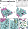
Viral proteases: Structure, mechanism and inhibition
- PMID: 34861941
- PMCID: PMC8595904
- DOI: 10.1016/bs.enz.2021.09.004
Viral proteases: Structure, mechanism and inhibition
Abstract
Viral proteases are diverse in structure, oligomeric state, catalytic mechanism, and substrate specificity. This chapter focuses on proteases from viruses that are relevant to human health: human immunodeficiency virus subtype 1 (HIV-1), hepatitis C (HCV), human T-cell leukemia virus type 1 (HTLV-1), flaviviruses, enteroviruses, and coronaviruses. The proteases of HIV-1 and HCV have been successfully targeted for therapeutics, with picomolar FDA-approved drugs currently used in the clinic. The proteases of HTLV-1 and the other virus families remain emerging therapeutic targets at different stages of the drug development process. This chapter provides an overview of the current knowledge on viral protease structure, mechanism, substrate recognition, and inhibition. Particular focus is placed on recent advances in understanding the molecular basis of diverse substrate recognition and resistance, which is essential toward designing novel protease inhibitors as antivirals.
Keywords: Coronavirus; Drug resistance; Enterovirus; Flavivirus; HCV; HIV-1; HTLV-1; Protease inhibitors; Substrate envelope; Viral protease.
Copyright © 2021 Elsevier Inc. All rights reserved.
Figures


Similar articles
-
Avoiding Drug Resistance by Substrate Envelope-Guided Design: Toward Potent and Robust HCV NS3/4A Protease Inhibitors.mBio. 2020 Mar 31;11(2):e00172-20. doi: 10.1128/mBio.00172-20. mBio. 2020. PMID: 32234812 Free PMC article.
-
Viral protease inhibitors.Handb Exp Pharmacol. 2009;189(189):85-110. doi: 10.1007/978-3-540-79086-0_4. Handb Exp Pharmacol. 2009. PMID: 19048198 Free PMC article. Review.
-
A low-background, fluorescent assay to evaluate inhibitors of diverse viral proteases.J Virol. 2023 Aug 31;97(8):e0059723. doi: 10.1128/jvi.00597-23. Epub 2023 Aug 14. J Virol. 2023. PMID: 37578235 Free PMC article.
-
Peptidomimetic therapeutic agents targeting the protease enzyme of the human immunodeficiency virus and hepatitis C virus.Acc Chem Res. 2008 Oct;41(10):1252-63. doi: 10.1021/ar8000519. Epub 2008 Aug 6. Acc Chem Res. 2008. PMID: 18681464
-
Viral proteases as therapeutic targets.Mol Aspects Med. 2022 Dec;88:101159. doi: 10.1016/j.mam.2022.101159. Epub 2022 Nov 29. Mol Aspects Med. 2022. PMID: 36459838 Free PMC article. Review.
Cited by
-
Repositioning of anti-infective compounds against monkeypox virus core cysteine proteinase: a molecular dynamics study.Mol Divers. 2024 Dec;28(6):4113-4135. doi: 10.1007/s11030-023-10802-8. Epub 2024 Apr 23. Mol Divers. 2024. PMID: 38652365
-
Exploration of Microbially Derived Natural Compounds against Monkeypox Virus as Viral Core Cysteine Proteinase Inhibitors.Viruses. 2023 Jan 16;15(1):251. doi: 10.3390/v15010251. Viruses. 2023. PMID: 36680291 Free PMC article.
-
Potential Resistance of SARS-CoV-2 Main Protease (Mpro) against Protease Inhibitors: Lessons Learned from HIV-1 Protease.Int J Mol Sci. 2022 Mar 23;23(7):3507. doi: 10.3390/ijms23073507. Int J Mol Sci. 2022. PMID: 35408866 Free PMC article. Review.
-
Breaking the Chain: Protease Inhibitors as Game Changers in Respiratory Viruses Management.Int J Mol Sci. 2024 Jul 25;25(15):8105. doi: 10.3390/ijms25158105. Int J Mol Sci. 2024. PMID: 39125676 Free PMC article. Review.
-
Repurposing Anti-Dengue Compounds against Monkeypox Virus Targeting Core Cysteine Protease.Biomedicines. 2023 Jul 18;11(7):2025. doi: 10.3390/biomedicines11072025. Biomedicines. 2023. PMID: 37509664 Free PMC article.
References
-
- Brik A., Wong C.H. HIV-1 protease: mechanism and drug discovery. Org. Biomol. Chem. 2003;1(1):5–14. - PubMed
-
- Prabu-Jeyabalan M., Nalivaika E., Schiffer C.A. Substrate shape determines specificity of recognition for HIV-1 protease: analysis of crystal structures of six substrate complexes. Structure. 2002;10(3):369–381. - PubMed
-
- Chellappan S., Kairys V., Fernandes M.X., Schiffer C., Gilson M.K. Evaluation of the substrate envelope hypothesis for inhibitors of HIV-1 protease. Proteins. 2007;68(2):561–567. - PubMed
MeSH terms
Substances
Grants and funding
LinkOut - more resources
Full Text Sources
Medical
