Recent advances in nanotherapeutics for the treatment of burn wounds
- PMID: 34778468
- PMCID: PMC8579746
- DOI: 10.1093/burnst/tkab026
Recent advances in nanotherapeutics for the treatment of burn wounds
Abstract
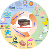
Moderate or severe burns are potentially devastating injuries that can even cause death, and many of them occur every year. Infection prevention, anti-inflammation, pain management and administration of growth factors play key roles in the treatment of burn wounds. Novel therapeutic strategies under development, such as nanotherapeutics, are promising prospects for burn wound treatment. Nanotherapeutics, including metallic and polymeric nanoformulations, have been extensively developed to manage various types of burns. Both human and animal studies have demonstrated that nanotherapeutics are biocompatible and effective in this application. Herein, we provide comprehensive knowledge of and an update on the progress of various nanoformulations for the treatment of burn wounds.
Keywords: Burn wounds; Metal and metal oxide nanotherapeutics; Polymeric nanotherapeutics; Therapeutic mechanism; Wound healing.
© The Author(s) 2021. Published by Oxford University Press.
Figures


Similar articles
-
Polymer-based Nanotherapeutics for Burn Wounds.Curr Pharm Biotechnol. 2022;23(12):1460-1482. doi: 10.2174/1389201022666210927103755. Curr Pharm Biotechnol. 2022. PMID: 34579630 Review.
-
Topical treatment for facial burns.Cochrane Database Syst Rev. 2020 Jul 29;7(7):CD008058. doi: 10.1002/14651858.CD008058.pub3. Cochrane Database Syst Rev. 2020. PMID: 32725896 Free PMC article.
-
[Clinical study of cell sheets containing allogeneic keratinocytes and fibroblasts for the treatment of partial-thickness burn wounds].Zhonghua Shao Shang Za Zhi. 2020 Mar 20;36(3):171-178. doi: 10.3760/cma.j.cn501120-20191113-00426. Zhonghua Shao Shang Za Zhi. 2020. PMID: 32241042 Chinese.
-
[Research advances on the prevention and treatment strategies of burn wound progressive deepening].Zhonghua Shao Shang Za Zhi. 2021 Dec 20;37(12):1199-1204. doi: 10.3760/cma.j.cn501120-20200828-00396. Zhonghua Shao Shang Za Zhi. 2021. PMID: 34937157 Review. Chinese.
-
[Advances in the research of early deepening mechanism and prevention measures of burn wounds].Zhonghua Shao Shang Za Zhi. 2019 Mar 20;35(3):229-232. doi: 10.3760/cma.j.issn.1009-2587.2019.03.014. Zhonghua Shao Shang Za Zhi. 2019. PMID: 30897873 Chinese.
Cited by
-
Chitosan-insulin nano-formulations as critical modulators of inflammatory cytokines and Nrf-2 pathway to accelerate burn wound healing.Discov Nano. 2023 Dec 12;18(1):154. doi: 10.1186/s11671-023-03941-2. Discov Nano. 2023. PMID: 38087141 Free PMC article.
-
Polymeric biomaterials-based tissue engineering for wound healing: a systemic review.Burns Trauma. 2023 Feb 7;11:tkac058. doi: 10.1093/burnst/tkac058. eCollection 2023. Burns Trauma. 2023. PMID: 36761088 Free PMC article.
-
Nanocomposites used in the treatment of skin lesions: a scoping review.Rev Esc Enferm USP. 2024 May 13;58:e20230338. doi: 10.1590/1980-220X-REEUSP-2023-0338en. eCollection 2024. Rev Esc Enferm USP. 2024. PMID: 38743957 Free PMC article. Review.
-
An Overview of Recent Developments in the Management of Burn Injuries.Int J Mol Sci. 2023 Nov 15;24(22):16357. doi: 10.3390/ijms242216357. Int J Mol Sci. 2023. PMID: 38003548 Free PMC article. Review.
-
Polymer-Based Functional Materials Loaded with Metal-Based Nanoparticles as Potential Scaffolds for the Management of Infected Wounds.Pharmaceutics. 2024 Jan 23;16(2):155. doi: 10.3390/pharmaceutics16020155. Pharmaceutics. 2024. PMID: 38399218 Free PMC article. Review.
References
-
- Guttman-Yassky E, Zhou L, Krueger JG. The skin as an immune organ: tolerance versus effector responses and applications to food allergy and hypersensitivity reactions. J Allergy Clin Immunol. 2019;144:362–74. - PubMed
-
- Peck MD. Epidemiology of burns throughout the world. Part I: distribution and risk factors. Burns. 2011;37:1087–100. - PubMed
-
- Wang Y, Beekman J, Hew J, Jackson S, Issler-Fisher AC, Parungao R, et al. . Burn injury: challenges and advances in burn wound healing, infection, pain and scarring. Adv Drug Deliv Rev. 2018;123:3–17. - PubMed
-
- Davies A, Spickett-Jones F, Jenkins ATA, Young AE. A systematic review of intervention studies demonstrates the need to develop a minimum set of indicators to report the presence of burn wound infection. Burns. 2020;46:1487–97. - PubMed
-
- Ahmadi AR, Chicco M, Huang J, Qi L, Burdick J, Williams GM, et al. . Stem cells in burn wound healing: a systematic review of the literature. Burns. 2019;45:1014–23. - PubMed
Publication types
LinkOut - more resources
Full Text Sources

