Morphological Aspects and Viremia Analysis of BALB/c Murine Model Experimentally Infected with Dengue Virus Serotype 4
- PMID: 34696384
- PMCID: PMC8538460
- DOI: 10.3390/v13101954
Morphological Aspects and Viremia Analysis of BALB/c Murine Model Experimentally Infected with Dengue Virus Serotype 4
Abstract
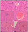
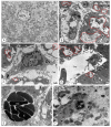
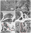
Ever since its brief introduction in the Brazilian territory in 1981, dengue virus serotype 4 (DENV-4) remained absent from the national epidemiological scenario for almost 25 years. The emergence of DENV-4 in 2010 resulted in epidemics in most Brazilian states. DENV-4, however, remains one of the least studied among the four DENV serotypes. Despite being known as a mild serotype, DENV-4 is associated with severe cases and deaths and deserves to be investigated; however, the lack of suitable experimental animal models is a limiting factor for pathogenesis studies. Here, we aimed to investigate the susceptibility and potential tropism of DENV-4 for liver, lung and heart of an immunocompetent mice model, and to evaluate and investigate the resulting morphological and ultrastructural alterations upon viral infection. BALB/c mice were inoculated intravenously with non-neuroadapted doses of DENV-4 isolated from a human case. The histopathological analysis of liver revealed typical alterations of DENV, such as microsteatosis, edema and vascular congestion, while in lung, widespread areas of hemorrhage and interstitial pneumonia were observed. While milder alterations were present in heart, characterized by limited hemorrhage and discrete presence of inflammatory infiltrate, the disorganization of the structure of the intercalated disc is of particular interest. DENV-4 RNA was detected in liver, lung, heart and serum of BALB/c mice through qRT-PCR, while the NS3 viral protein was observed in all of the aforementioned organs through immunohistochemistry. These findings indicate the susceptibility of the model to the serotype and further reinforce the usefulness of BALB/c mice in studying the many alterations caused by DENV.
Keywords: BALB/c mice; DENV-4; heart; histopathology; liver; lung; ultrastructure.
Conflict of interest statement
The authors declare no conflict of interest exists.
Figures


Similar articles
-
Impact of dengue virus (serotype DENV-2) infection on liver of BALB/c mice: A histopathological analysis.Tissue Cell. 2017 Feb;49(1):86-94. doi: 10.1016/j.tice.2016.11.005. Epub 2016 Nov 23. Tissue Cell. 2017. PMID: 28034555
-
Morphological studies in a model for dengue-2 virus infection in mice.Mem Inst Oswaldo Cruz. 2006 Dec;101(8):905-15. doi: 10.1590/s0074-02762006000800014. Mem Inst Oswaldo Cruz. 2006. PMID: 17293987
-
Phenotypic characterization of patient dengue virus isolates in BALB/c mice differentiates dengue fever and dengue hemorrhagic fever from dengue shock syndrome.Virol J. 2011 Aug 11;8:398. doi: 10.1186/1743-422X-8-398. Virol J. 2011. PMID: 21835036 Free PMC article.
-
First detection of dengue virus in the saliva of immunocompetent murine model.Mem Inst Oswaldo Cruz. 2018 Feb 5;113(4):e170208. doi: 10.1590/0074-02760170208. Mem Inst Oswaldo Cruz. 2018. PMID: 29412340 Free PMC article.
-
Viral kinetics of primary dengue virus infection in non-human primates: a systematic review and individual pooled analysis.Virology. 2014 Mar;452-453:237-46. doi: 10.1016/j.virol.2014.01.015. Epub 2014 Feb 14. Virology. 2014. PMID: 24606701 Free PMC article. Review.
Cited by
-
Primary infection of BALB/c mice with a dengue virus type 4 strain leads to kidney injury.Mem Inst Oswaldo Cruz. 2023 May 8;118:e220255. doi: 10.1590/0074-02760220255. eCollection 2023. Mem Inst Oswaldo Cruz. 2023. PMID: 37162062 Free PMC article.
-
Suppression of TGF-β/Smad2 signaling by GW788388 enhances DENV-2 clearance in macrophages.J Med Virol. 2022 Sep;94(9):4359-4368. doi: 10.1002/jmv.27879. Epub 2022 Jun 2. J Med Virol. 2022. PMID: 35596058 Free PMC article.
References
-
- Stanaway J.D., Shepard D.S., Undurraga E.A., Halasa Y.A., Coffeng L.E., Brady O.J., Hay S.I., Bedi N., Bensenor I.M., Castañeda-Orjuela C.A., et al. The global burden of dengue: An analysis from the Global Burden of Disease Study 2013. Lancet Infect. Dis. 2016;16:712–723. doi: 10.1016/S1473-3099(16)00026-8. - DOI - PMC - PubMed
-
- Secretaria de Vigilância em Saúde/Ministério da Saúde Boletim Epidemiológico-Volume 43-nº 1–2012. [(accessed on 17 June 2021)]; Dengue: Situação Epidemiológica (de Janeiro a Abril de 2012) Available online: https://antigo.saude.gov.br/images/pdf/2014/julho/23/BE-2012-43--1--pag-....
Publication types
MeSH terms
Substances
LinkOut - more resources
Full Text Sources

