. 2021 Sep 1;207(5):1310-1321.
doi: 10.4049/jimmunol.2100297. Epub 2021 Aug 11.

FcRn-Targeted Mucosal Vaccination against Influenza Virus Infection

Affiliations

FcRn-Targeted Mucosal Vaccination against Influenza Virus Infection

Susan Park Ochsner et al. J Immunol. .

Abstract

The respiratory tract is constantly exposed to various airborne pathogens. Most vaccines against respiratory infections are designed for the parenteral routes of administration; consequently, they provide relatively minimal protection in the respiratory tract. A vaccination strategy that aims to induce the protective mucosal immune responses in the airway is urgently needed. The FcRn mediates IgG Ab transport across the epithelial cells lining the respiratory tract. By mimicking this natural IgG transfer, we tested whether FcRn delivers vaccine Ags to induce a protective immunity to respiratory infections. In this study, we designed a monomeric IgG Fc fused to influenza virus hemagglutinin (HA) Ag with a trimerization domain. The soluble trimeric HA-Fc were characterized by their binding with conformation-dependent HA Abs or FcRn. In wild-type, but not FcRn knockout, mice, intranasal immunization with HA-Fc plus CpG adjuvant conferred significant protection against lethal intranasal challenge with influenza A/PR/8/34 virus. Further, mice immunized with a mutant HA-Fc lacking FcRn binding sites or HA alone succumbed to lethal infection. Protection was attributed to high levels of neutralizing Abs, robust and long-lasting B and T cell responses, the presence of lung-resident memory T cells and bone marrow plasma cells, and a remarkable reduction of virus-induced lung inflammation. Our results demonstrate for the first time, to our knowledge, that FcRn can effectively deliver a trimeric viral vaccine Ag in the respiratory tract and elicit potent protection against respiratory infection. This study further supports a view that FcRn-mediated mucosal immunization is a platform for vaccine delivery against common respiratory pathogens.


Figures

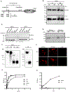
Figure 1.
Figure 1.. Expression and characterization of the trimeric HA-Fc fusion proteins.
(A). Schematic illustration of the genetic fusion of influenza HA, the T4 fibritin foldon domain (Fd), and murine Fcγ2a cDNA to create a trimeric HA-Fc fusion gene. Mutations were made in the Fcγ2a fragment using site-directed mutagenesis by replacing Cys224, Cys227, and Cys229 respectively with a Ser residue to abolish Fc dimerization, and replacing Glu318, Lys320, and Lys322 with an Ala residue to deplete complement C1q binding site. His310/Gln311 (HQ) and His433/Asn434 (HN) residues were replaced with Ala310/Asp311 (AD) and Ala433/Gln434 (AQ) to eliminate FcRn binding sites, this plasmid was designated as HA-Fc/mut. (B). The HA-Fc fusion protein secreted by a stable CHO cell line. The HA-Fc were subjected to SDS-PAGE and Western blot analyses and detected by either goat anti-mouse IgG-Fc (top panel) or an anti-HA mAb (bottom panel). The fusion protein was shown as a monomer under both non-reducing (NR) and reducing (R) conditions. (C). FcRn binding of the HA-Fc. CHO cells expressing mouse FcRn and β2m were incubated with 3 μg HA-Fc/wt, HA-Fc/mut, or HA protein for 1 hr at 4°C under pH 6.0 or pH 7.4 condition. After washing, the cells were lysed with 0.5% CHAPS in cold PBS (pH 6.0 or 7.4). Samples were subjected to Western blot analyses. The HA-Fc or HA (top) or mouse FcRn (bottom) was detected with anti-HA or anti-mouse FcRn primary Ab and HRP-conjugated secondary Ab. (D). The HA-Fc/wt and HA-Fc/mut were purified by affinity chromatography and visualized with Coomassie blue staining. (E). Western blot analysis of the purified HA-Fc that was cross-linked with BS3. The BS3 -treated (lane 1) or -untreated (lane 2) samples were separated by SDS-PAGE under reducing conditions followed by Western blotting using anti-HA Ab (RA5-22, mouse IgG1). (F). Stable CHO cell lines expressing HA-Fc/wt and HA-Fc/mut were probed with conformation-dependent anit-HA mAbs. CHO cells were transfected with HA-Fc plasmids and fixed with 4% paraformaldehyde. Cells were then incubated with HA-specific mAb 6F12 (top panel) or KB2 (bottom panel) and visualized using immunofluorescence staining. (G). Interactions of the purified HA-Fc with a panel of HA stalk-specific and conformation-dependent Abs CR6261, FI6v3, 6F12, or CR8020. The specific binding was detected by the ELISA method. HIV gp120 specific IgG mAb B12 was used as a negative control. Representative images of three experiment
Figure 2.
Figure 2.. FcRn-mediated respiratory immunization induces HA-specific Ab and T cell immune responses.
Five μg of HA-Fc/wt, HA-Fc/mut, HA, or PBS in combination with 10 μg of CpG was i.n. administered into wild-type (WT) or FcRn knockout (KO) mice. One-way ANOVA with Dunnett’s multiple comparison tests was used. Values marked with asterisks in the figures: *, P < 0.05; **, P < 0.01, ***, P<0.001. Immunization conditions are displayed on the bottom. (A). Measurement of anti-influenza HA-specific IgG Ab titers in serum after the booster immunization. Influenza HA-specific Ab titers were measured by coating with HA protein in ELISA 14 days after boosting. The IgG titers were measured in 10 representative mouse sera. The data represent mean ± S.E.M. (B). Test of neutralizing Ab activity in the immunized sera. Two weeks after boost, sera sampled from 13 to 20 mice per group were heat-inactivated, diluted twofold in PBS with antibiotics/antimycotics. Influenza PR8 (100 TCID50) was added and incubated at 37°C for 1 hr. The mixture was added to MDCK cells and incubated at 37°C and subsequently removed after 1 hr. The serum-free Opti-MEM containing 1 μg/ml TPCK-trypsin was added to cells. After incubation at 37°C for 72 hr, an HA assay was performed on the supernatant. The neutralization Ab titers were expressed as the reciprocal of the twofold serial dilution preventing the appearance of the agglutination of the erythrocytes of chicken. Each assay was performed in triplicate. (C, D, E, & F). The percentage of IFN-γ and TNF-α producing T cells in the lung 7 days after the boost. The lung lymphocytes from the immunized mice were stimulated for 10 hr with purified HA or medium control. Lymphocytes were gated by forward and side scatters and T cells labeled with anti-CD3 and identified by their respective surface markers CD4 and CD8 and intracellular IFN-γ or TNF-α staining. Numbers represent the percentage of IFN-γ+ CD4+ (C), TNF-α+ CD4+ (D), IFN-γ+ CD8+ (E), or TNF-α+ CD8+ (F) T cells. Isotype controls included FITC-mouse-IgG1 with baseline response. Flow cytometry plots are representative of two independent experiments with 4 immunized mice pooled in each group. Graphical data is the average percentage of the two experiments.
Figure 3.
Figure 3.. FcRn-mediated intranasal vaccination significantly induced HA-specific local immune responses in the respiratory tract.
(A & B). Measurement of anti-influenza HA-specific Ab titers in nasal washings (A, IgA), and BAL (B, IgG) after the boost. Influenza HA-specific Abs were measured by ELISA 14 days after boost. The Ab titer was measured in 10 representative mouse samples. The data represent mean values for each group (±S.E.M.). One-way ANOVA with Dunnett's multiple comparison tests was used. (C). Accumulation of activated B cells in germinal centers (GCs) in the mediastinal lymph nodes (MedLNs) and spleens. Representative flow cytometric analyses of GC B cells among CD19+B220+ B cells in the MedLNs and spleens 10 days after the boost. B220+PNAhigh cells are B cells that exhibit the phenotypic attributes of GC B cells. The GC staining in the spleen was used as a positive control. GC B cells are pooled from individual mice because of the limited cell numbers isolated from each MedLN. Numbers are the percentage of activated GC B cells (PNA+FAS+) among gated B cells and are representative of three independent experiments.
Figure 4.
Figure 4.. FcRn-targeted respiratory immunization engenders protective immunity to intranasal (i.n.) challenge with virulent influenza virus.
(A). Body-weight changes following the influenza challenge. Two weeks after the boost, groups of 13-20 mice (HA-Fc/wt=19, HA-Fc/mut=15, HA=15, HA-Fc/wt/KO=13, PBS=20) were i.n. challenged with PR8 virus (5 MLD50) and weighed daily for 14 days. Mice were deceased or humanely euthanized if more than 25% of initial body weight was lost. The data is representative of three similar experiments with the data combined. (B) Mean survival following influenza challenge. Two weeks after the boost, groups of 13-20 mice were i.n. challenged with PR8 virus (5 MLD50) and weighed daily for 14 days. Mice were humanely euthanized if more than 25% of initial body weight was lost. The percentage of mice from protection after the challenge was shown by the Kaplan-Meier survival curve. The data is representative of at least three similar experiments. Statistical differences were determined using multiple Mantel-Cox tests. (C) Mean of viral titers in the lungs following influenza virus challenge. The virus titers in the lungs of the immunized and control mice (n=4-5) were determined 4 days after lethal challenge. Supernatants of the lung homogenates were added onto MDCK cells and incubated for three days. The viral titers were measured by 50% endpoint dilution assay along with an HA assay.
Figure 5.
Figure 5.. Gross- and histopathology of the lungs from the challenged mice.
(A). Lungs were collected from 6- to 14-day period post-challenge based on a 25% body-weight loss endpoint. The lungs from uninfected mice were included as normal control (n=3). The lung sections were stained with Hematoxylin-Eosin (H & E) to determine the level of inflammation in the lungs (10x). The representative slides were shown on the right. (B). The inflammatory responses for each lung section were scored in a blinded manner. Statistical differences were determined by one-way ANOVA with Tukey’s multiple comparison tests. All scale bars represent 50 μm, 10 X magnification.
Figure 6.
Figure 6.. Increased memory immune responses in FcRn-mediated respiratory immunization.
(A) The duration of influenza-specific serum IgG response. Influenza HA-specific IgG was quantified by ELISA in serum by endpoint titer from 8-10 mice at 8 weeks after the boost. Influenza HA-specific IgG Ab was not detectable (ND) in PBS-immunized mice. Statistical differences were determined using one-way ANOVA with Dunnett's multiple comparison tests. (B & C). Measurement of anti-influenza HA-specific Ab titers in nasal washings (B, IgA), and BAL (C, IgG) after the boost immunization. Influenza HA-specific Abs were measured 8 weeks after boosting by ELISA. The Ab titer was measured in 5 representative mouse samples. The data represent mean values for each group (±S.E.M.). (D) Long-lived influenza HA-specific Ab-secreting cells in the bone marrow. Bone marrow cells (BMCs) removed 8 weeks after the boost was placed on HA-coated plates and quantified by ELISPOT analysis of IgG-secreting plasma cells. Data were pooled from two separate experiments with 5 immunized mice pooled in each group. The graphs were plotted based on the average ELISPOT for four replicate wells for each experiment. The asterisk denotes statistics significant differences (P<0.05). (E+F). Induction of tissue-resident memory (TRM) T cells in mouse lungs. An additional group of mice that were i.m. immunized HA-Fc/wt was included as a parenteral route control. The CD3+CD4+CD69+CD11a+ (E) or CD3+CD8+CD69+CD103+ (F) TRM T cells in the lungs were assessed 8 weeks after the boost by FACS. Flow cytometry plots are representative of two independent experiments with 4 immunized mice pooled in each group. Numbers in the quadrants represent the percentage of TRM T lymphocytes.
Figure 7.
Figure 7.. FcRn-targeted respiratory immunization induces protective memory immune responses to resist challenge by influenza virus.
(A). Body-weight changes following the influenza virus challenge. Eight weeks after the boost, groups of mice (HA-Fc/wt=17, HA-Fc/mut=16, HA=13, HA-Fc/wt/KO=11, PBS=17) were i.n. challenged with PR8 virus (5 MLD50) and weighed daily for 14 days. Mice were deceased or humanely euthanized if more than 25% of initial body weight was lost. (B). Mean survival following influenza virus challenge in mice 8 weeks following the boost. The immunized mice were i.n. challenged with 5 MLD50 of PR8 virus and weighed daily for 14 days. Mice were deceased or humanely euthanized if more than 25% of initial body weight was lost. The percentage of mice protected on the indicated days is calculated as the number of mice survived divided by the number of mice in each group (n=5), as shown by the Kaplan-Meier survival curve. Statistical differences were determined using multiple Mantel-Cox tests. The data represent combined data of two independent animal experiments.

Similar articles

Cited by

References

    1. Allie SR, and Randall TD. 2017. Pulmonary immunity to viruses. Clin. Sci 131: 1737–1762. - PubMed
    1. Iwasaki A, Foxman EF, and Molony RD. 2017. Early local immune defences in the respiratory tract. Nat. Rev. Immunol 17: 7–20. - PMC - PubMed
    1. Barría MI, Garrido JL, Stein C, Scher E, Ge Y, Engel SM, Kraus TA, Banach D, and Moran TM. 2013. Localized Mucosal Response to Intranasal Live Attenuated Influenza Vaccine in Adults. J. Infect. Dis 207: 115–124. - PMC - PubMed
    1. Chiu C, and Openshaw PJ. 2015. Antiviral B cell and T cell immunity in the lungs. Nat. Immunol 16: 18–26. - PMC - PubMed
    1. Mueller SN, Gebhardt T, Carbone FR, and Heath WR. 2013. Memory T Cell Subsets, Migration Patterns, and Tissue Residence. Annu. Rev. Immunol 31: 137–161. - PubMed

Publication types

MeSH terms

Substances