. 2021 Aug 10;118(32):e2101675118.
doi: 10.1073/pnas.2101675118.

The human cytomegalovirus protein pUL13 targets mitochondrial cristae architecture to increase cellular respiration during infection

Affiliations

The human cytomegalovirus protein pUL13 targets mitochondrial cristae architecture to increase cellular respiration during infection

Cora N Betsinger et al. Proc Natl Acad Sci U S A. .

Abstract

Viruses modulate mitochondrial processes during infection to increase biosynthetic precursors and energy output, fueling virus replication. In a surprising fashion, although it triggers mitochondrial fragmentation, the prevalent pathogen human cytomegalovirus (HCMV) increases mitochondrial metabolism through a yet-unknown mechanism. Here, we integrate molecular virology, metabolic assays, quantitative proteomics, and superresolution confocal microscopy to define this mechanism. We establish that the previously uncharacterized viral protein pUL13 is required for productive HCMV replication, targets the mitochondria, and functions to increase oxidative phosphorylation during infection. We demonstrate that pUL13 forms temporally tuned interactions with the mitochondrial contact site and cristae organizing system (MICOS) complex, a critical regulator of cristae architecture and electron transport chain (ETC) function. Stimulated emission depletion superresolution microscopy shows that expression of pUL13 alters cristae architecture. Indeed, using live-cell Seahorse assays, we establish that pUL13 alone is sufficient to increase cellular respiration, not requiring the presence of other viral proteins. Our findings address the outstanding question of how HCMV targets mitochondria to increase bioenergetic output and expands the knowledge of the intricate connection between mitochondrial architecture and ETC function.

Keywords: HCMV; metabolism; mitochondria; pUL13; proteomics.


Conflict of interest statement

The authors declare no competing interest.

Figures
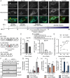
Fig. 1.
Fig. 1.
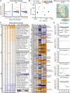
pUL13 localizes to the mitochondria and is required for productive virus replication. (A) Confocal analysis of pUL13 localization in fibroblasts labeled with mito-BFP (mitochondria) and infected with UL13-YFP HCMV. (Scale bars, 5 μm.) Temporal stages of HCMV replication are shown. (B) Schematics of the UL13 genetic locus of WT, UL13M, and ∆UL13 HCMV. (C) Extracellular HCMV titers at 144 hpi during WT or UL13M infection. (D) pUL13 protein levels during WT, UL13M, and ∆UL13 infection at 96 hpi, measured by targeted MS. Peptide sequences and locations within the pUL13 sequence are shown. n = 4 peptides. (E) Extracellular HCMV titers during WT or ∆UL13 infection. (F) Representative Western blot showing IE (IE1), DE (pUL26), and L (pUL99) virus protein levels during WT or ∆UL13 infection. (G) Cell-associated virus genomes produced during ∆UL13 and WT infection, quantified using qPCR. (H) Cell-associated virus titers collected during WT or ∆UL13 infection. (I) Particle:PFU ratio of virus produced during WT or ∆UL13 infection. n = 3 biological replicates for C, E, G, H, and I. Significance determined by Student’s t test. *P ≤ 0.05, **P ≤ 0.01, ***P ≤ 0.001, and ****P ≤ 0.0001; ns, P > 0.05. Error bars indicate the SD.
Fig. 2.
Fig. 2.
∆UL13 infection results in altered temporal viral protein levels and decreased abundance of cellular metabolic proteins. (A) The relative abundances of viral proteins detected by TMT-MS (n = 95 proteins), shown as fold change during ∆UL13 relative to WT infection. Proteins passing 1.25-fold change at any time point are highlighted in red (increased) and blue (decreased). (B) Principal component analysis of virus protein abundances at each analyzed time point during WT or ∆UL13 infection. (C) Relative abundances of host proteins during ∆UL13 relative to WT infection. The average k-means cluster profile and enriched gene ontology terms are shown. Proteins passing the 1.25-fold change and P value < 0.05 cutoffs at any time point are shown in the second heatmap (Center), organized by biological process. (D) Schematic of the ETC and ATP synthase within the mitochondrial inner membrane. IMS, intermembrane space. (E) The relative temporal abundances of ETC and ATP synthase protein subunits, shown organized by complex. Proteins passing the 1.25-fold change and P value < 0.05 cutoffs at any time point are labeled.
Fig. 3.
Fig. 3.
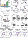
pUL13 increases oxidative phosphorylation during HCMV infection. (A) Diagram illustrating the method of converting OCR into the six parameters of ETC function. Reproduced from ref. with permission, © Agilent Technologies, Inc. (B) HCMV rewires the metabolism of infected cells, increasing metabolic flux through glycolysis and glutaminolysis (6, 7). Dashed lines indicate multistep pathways; solid lines indicate single-step pathways. (C) OCR of mock, WT, and ∆UL13-infected cells 72 hpi using either pyruvate or glutamine as the respiratory substrate. The carbon source was injected at 0 min; vertical lines indicate the injection time of oligomycin, FCCP, and Antimycin A + Rotenone. (D) Radar charts showing parameters of ETC function during mock, WT, and ∆UL13 infection at 48 and 72 hpi using either pyruvate or glutamine as the respiratory substrate. Solid lines indicate the mean, and shaded regions indicate the SD. (E) ECAR at 48 or 72 hpi during mock, WT, or ∆UL13 infection following the provision of pyruvate or glutamine as the respiratory substrate. The carbon source and ETC modulators were injected as in C. (F) LC-MS quantification of intracellular metabolites related to ATP metabolism. Metabolites were extracted from mock, WT, or ∆UL13 infected cells at 48 and 72 hpi. The energy charge was calculated from the abundance of ATP, ADP, and AMP as described in ref. . n = 3 biological replicates. For CF: Significance was determined by one-way ANOVA using Tukey’s test for post hoc analysis. *P ≤ 0.05, **P ≤ 0.01, ***P ≤ 0.001, and ****P ≤ 0.0001. Error bars indicate the SD. n ≥ 4 biological replicates, unless otherwise indicated.
Fig. 4.
Fig. 4.
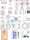
pUL13 interacts with components of the ETC and associated cristae-shaping proteins during infection. (A) IP-MS workflow for studying pUL13 interactions during infection. (B) InterViSTA (27) spatiotemporal analysis of pUL13 interactions. (C) Functional network of pUL13 interactions during infection, depicting only proteins that passed specificity criteria. Bars indicate the scaled protein abundance, normalized to bait (pUL13), at each time point. (D) Schematic of cristae-remodeling proteins and complexes identified in the pUL13 interactome. (E) Heatmap of pUL13 interactions, validated and quantified using PRM following IP-MS of pUL13 during UL13-YFP infection, scaled from minimum to maximum association with pUL13. White spaces indicate lack of detection. n = 2 to 3 peptides per protein. (F) PRM analysis of pUL13 abundance in endogenous anti-IMMT IP samples during WT HCMV infection. Protein abundances were quantified using two unique peptides per protein and normalized to IMMT abundance. Error bars denote the SD. n = 2 biological replicates.
Fig. 5.
Fig. 5.
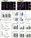
pUL13 alters cristae architecture and is sufficient for increased OXPHOS. (A) Confocal and STED analysis of pUL13 localization and cristae architecture in pUL13-3xFLAG and YFP-3xFLAG stably expressing fibroblasts. Cristae were visualized using an antibody against COXIV, a component of ETC complex IV. Yellow boxes indicate the regions sampled for STED analysis. Mitochondrial sections (boxed in red) have been enlarged to highlight cristae ultrastructure. (Scale bars, 5 μm.) (B) Extracellular HCMV titers during WT or ∆UL13 infection of pUL13-3xFLAG and YFP-3xFLAG stably expressing fibroblasts. n = 3 biological replicates. Significance was determined by one-way ANOVA. (C) Quantification of cristae morphology in STED images acquired from pUL13-3xFLAG and YFP-3xFLAG stably expressing fibroblasts. Error bars indicate a 95% CI. (D) OCR of pUL13-3xFLAG and YFP-3xFLAG stably expressing cells using either pyruvate or glutamine as the respiratory substrate. The carbon source and ETC modulators were injected as in Fig 3C. (E) Parameters of ETC function in pUL13-3xFLAG and YFP-3xFLAG stably expressing cells. n = 5 biological replicates for D and E. Significance was determined by Student’s t test for CE. *P ≤ 0.05, **P ≤ 0.01, ***P ≤ 0.001, and ****P ≤ 0.0001; n.s., P > 0.05. Error bars indicate the SD unless otherwise stated.
Fig. 6.
Fig. 6.
Proposed model for the pUL13-induced increase in mitochondrial bioenergetics during HCMV infection. OMM, outer mitochondrial membrane; IMS, intermembrane space; IMM, inner mitochondrial membrane.

Similar articles

Cited by

References

    1. Sanchez E. L., Lagunoff M., Viral activation of cellular metabolism. Virology 479–480, 609–618 (2015). - PMC - PubMed
    1. Eisenreich W., Rudel T., Heesemann J., Goebel W., How viral and intracellular bacterial pathogens reprogram the metabolism of host cells to allow their intracellular replication. Front. Cell. Infect. Microbiol. 9, 42 (2019). - PMC - PubMed
    1. Khan M., Syed G. H., Kim S. J., Siddiqui A., Mitochondrial dynamics and viral infections: A close nexus. Biochim. Biophys. Acta 1853 (10 Pt B), 2822–2833 (2015). - PMC - PubMed
    1. McCormick A. L., Smith V. L., Chow D., Mocarski E. S., Disruption of mitochondrial networks by the human cytomegalovirus UL37 gene product viral mitochondrion-localized inhibitor of apoptosis. J. Virol. 77, 631–641 (2003). - PMC - PubMed
    1. Munger J., Bajad S. U., Coller H. A., Shenk T., Rabinowitz J. D., Dynamics of the cellular metabolome during human cytomegalovirus infection. PLoS Pathog. 2, e132 (2006). - PMC - PubMed

Publication types

MeSH terms