. 2022 Jan 20;29(1):84-97.e8.
doi: 10.1016/j.chembiol.2021.07.007. Epub 2021 Jul 30.

A photo-cross-linking GlcNAc analog enables covalent capture of N-linked glycoprotein-binding partners on the cell surface

Affiliations

A photo-cross-linking GlcNAc analog enables covalent capture of N-linked glycoprotein-binding partners on the cell surface

Han Wu et al. Cell Chem Biol. .

Abstract

N-glycans are displayed on cell-surface proteins and can engage in direct binding interactions with membrane-bound and secreted glycan-binding proteins (GBPs). Biochemical identification and characterization of glycan-mediated interactions is often made difficult by low binding affinities. Here we describe the metabolic introduction of a diazirine photo-cross-linker onto N-acetylglucosamine (GlcNAc) residues of N-linked glycoproteins on cell surfaces. We characterize sites at which diazirine-modified GlcNAc is incorporated, as well as modest perturbations to glycan structure. We show that diazirine-modified GlcNAc can be used to covalently cross-link two extracellular GBPs, galectin-1 and cholera toxin subunit B, to cell-surface N-linked glycoproteins. The extent of cross-linking correlates with display of the preferred glycan ligands for the GBPs. In addition, covalently cross-linked complexes could be isolated, and protein components of cross-linked N-linked glycoproteins were identified by proteomics analysis. This method may be useful in the discovery and characterization of binding interactions that depend on N-glycans.

Keywords: N-glycan; cholera; cross-linking; diazirine; galectin; glycosylation; glycosyltransferase.


Conflict of interest statement

Declaration of interests K.W.M. acknowledges ownership interest and roles as president and CEO of Glyco Expression Technologies, Inc., a biotechnology spinout commercializing recombinant glycosyltransferases, and may conceivably profit from the results described herein. J.J.K. is a member of the advisory board for Cell Chemical Biology. Other authors declare no competing interests.

Figures

Figure 1.
Figure 1.. Strategy for metabolic production of photocrosslinking N-glycans.
N-glycans are synthesized in the ER and Golgi through the sequential actions of enzymes, including multiple GlcNAc-transferases. N-linked glycoproteins are displayed on the cell surface, where glycans are recognized by extracellular GBPs. Individual binding interactions are typically low-affinity, although high avidity binding can be achieved through multivalency. To introduce diazirine, cells expressing UAP1(F383G) are cultured with Ac3GlcNDAz-1P(AcSATE)2. Ac3GlcNDAz-1P(AcSATE)2 is deprotected, potentially by intracellular esterases, and transformed to UDP-GlcNDAz. In this work, we asked whether UDP-GlcNDAz enters the Golgi and is a substrate for Golgi GlcNAc-transferases. Cell surface N-linked glycoproteins displaying the diazirine modification can be activated by UV light, resulting in covalent crosslinking to extracellular GBPs. See also Fig. S1.
Figure 2.
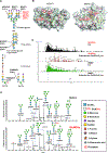
Figure 2.. GlcNDAz is incorporated into N-glycans.
(A) Biosynthetic roles of GlcNAc-transferases were examined using recombinant Golgi GlcNAc-transferases (Fig. S2). Percentages represent relative activity of GlcNAc transferases in using UDP-GlcNDAz as a substrate compared to UDP-GlcNAc. Percentages are averaged from two trials. (B) Modeled structures of human MGAT1 and MGAT2 (PDB 5VCM) (Kadirvelraj et al., 2018) in complex with UDP-GlcNAc. The human MGAT1 structure was modeled based on the structure of the rabbit MGAT1:UDP-GlcNAc:Mn2+ (PDB 1FOA). (Unligil et al., 2000) A magenta sphere marks the methyl group to which the diazirine is attached in GlcNDAz. (C) Glycopeptides from T84 UAP1(F383G) cells treated with Ac3GlcNDAz-1P(AcSATE)2 or DMSO were analyzed by mass spectrometry, and ion chromatograms were extracted for the GlcNDAz – N2 ion. Chromatograms are from single trial. (D) Comparison of glycoforms detected on peptide 38VVRPDSELGERPPEDNQSFQYDHEAFLGK66 from human reticulocalbin-1 from T84 UAP1(F383G) cells treated with Ac3GlcNDAz-1P(AcSATE)2 or vehicle. Red asterisks label peaks (m/z 4305.80, 4467.85, 4952.17, 5115.22) assigned to other co-eluting glycopeptides. Spectrum is from single trial. See also Fig. S2, Fig. S3, Fig. S4, and Data S1.
Figure 3.
Figure 3.. GlcNDAz-containing glycopeptides.
Glycopeptides extracted from T84 UAP1(F383G) cells treated with Ac3GlcNDAz-1P(AcSATE)2 were identified by mass spectrometry. Possible glycan structures are proposed based on glycan composition, MS2 analysis, and biosynthesis pathways. We make the assumption that GlcNDAz is most likely to be found attached to α1–6Man in the N-glycan core, but data do not exclude other possible antennary arrangements. Data are from single trial.
Figure 4.
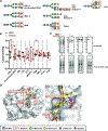
Figure 4.. Effects of GlcNDAz incorporation on N-glycan structure
(A) Specificity of lectins used in (B) and (C). (B) Lectin binding to T84 UAP1(F383G) cells treated with Ac3GlcNDAz-1P(AcSATE)2 at non-saturating concentrations (Fig. S3) and normalized to vehicle-treated cells. Lectin concentration used: GSL II (1 μg/ml), succinylated WGA (0.1 μg/ml), DSL (1 μg/ml), LEL (1 μg/ml), ECA (0.1 μg/ml), galectin-1 (1 μg/ml), SNA (3 μg/ml), MAL II (3 μg/ml), PHA-L (3 μg/ml), CTB (3 μg/ml). Data represent mean ±SD from four biological replicates, each of which is averaged from three technical replicates. (C) Lectin blot of total lysates from T84 UAP1(F383G) cells treated with Ac3GlcNDAz-1P(AcSATE)2 or vehicle. Blots are representative of biological triplicate. (D) Structure of human B4GALT1 (overlay of PDB 2AEC (Ramasamy et al., 2005) and PDB 1TVY (Ramakrishnan et al., 2006)) in complex with UDP-Gal and trisaccharide acceptor containing terminal GlcNAc is shown with zoom-in for the area indicated by the dotted box. A magenta sphere marks the methyl group to which the diazirine is attached in GlcNDAz. See also Fig. S5.
Figure 5.
Figure 5.. GlcNDAz can be used to covalently capture glycan-mediated binding interactions.
(A) Scheme of crosslinking experiment. (B) Scheme of detecting crosslinking complexes by immunoblot analysis of total cell lysates. Crosslinking of human galectin-1 (~15 kDa) and CTB (~14 kDa) to larger cell surface glycoproteins results in a shift of galectin-1 and CTB to apparent higher molecular weight. (C) Crosslinking of galectin-1 to glycoproteins from T84 UAP1(F383G) cells. (D) Crosslinking of cholera toxin to glycoproteins from T84 UAP1(F383G) cells. (E) Crosslinking of galectin-1 with pre-treatment with 40 μg/ml ECA lectin. (F) Crosslinking of galectin-1 in presence of 100 mM of mono- or disaccharides, or with pretreatment of cells with 1 μg/ml of kifunensine for 72 h (G) Crosslinking of galectin-1 to glycoproteins with pre-treatment by StcE. (H) Crosslinking of CTB with cells cultured with 2F-fucose for 72 h, pretreatment with 40 μg/ml of lectin AAL, or in presence of 100 mM of l-fucose, but not d-fucose. All blots are representative of biological triplicate.
Figure 6.
Figure 6.. GlcNDAz-enabled proteomics facilitates identification of candidate galectin-1 binding partners.
(A) Scatter-plots of proteins from T84 UAP1(F383G) cells identified in the three major crosslinked bands in two biological replicates. Abundance and fold-enrichment represent the average of two biological replicates. Proteins detected in crosslinked but not control samples are shown on the right of each plot. Proteins detected in crosslinked samples in two replicates but detected in control samples in only one replicate are represented with blue symbols. For these proteins, fold-enrichment from the trial in which proteins were identified in both crosslinked and control samples was used in the plot. (B) Venn diagram of proteins identified from each region in two replicates. (C) – (E) CEA (C), CDCP1 (D) and LY75 (E) were enriched by immunoprecipitation with antiCEA, anti-CDCP1 and anti-LY75 antibodies and crosslinked complexes were detected with anti-galectin-1 antibody by immunoblot. Confirmation of successful immunoprecipitation is presented in Fig. S6. See also Table S1. Figures (C)–(E) are representative of biological duplicate using 100 μg/ml galectin-1 for crosslinking. In a third trial conducted with 30 μg/ml galectin-1, similar results albeit with fainter crosslinked bands were obtained.
Figure 7.
Figure 7.. GlcNDAz-mediated crosslinking of LAMP1 to galectin-1 occurs in cell lysates but not intact cells.
(A) T84 UAP1(F383G) cells were cultured with 100 μM of Ac3GlcNDAz-1P(AcSATE)2 or vehicle. Galectin-1 was added to a final concentration of 100 μg/mL, then samples were UV irradiated. Cells were lysed, and immunopurified for LAMP1. Immunoblot analysis was performed with a galectin-1 antibody. Data are representative of biological duplicate using 100 μg/ml galectin-1 for crosslinking. In a third trial conducted with 30 μg/ml galectin-1, similar results were obtained. (B) T84 UAP1(F383G) cells were cultured with 100 μM of Ac3GlcNDAz-1P(AcSATE)2 or vehicle. Cells were lysed, then 30 μg/mL of galectin-1 was added and UV irradiation applied. Immunopurification was performed with a LAMP1 antibody. Immunoblot analysis was performed with a galectin-1 antibody. Data are representative of biological triplicate. See also Fig. S7.

Similar articles

Cited by

References

    1. Aits S, Kricker J, Liu B, Ellegaard A-M, Hämälistö S, Tvingsholm S, Corcelle-Termeau E, Høgh S, Farkas T, Holm Jonassen A, et al. (2015). Sensitive detection of lysosomal membrane permeabilization by lysosomal galectin puncta assay. Autophagy 11, 1408–1424. - PMC - PubMed
    1. Boyce M, Carrico IS, Ganguli AS, Yu S-H, Hangauer MJ, Hubbard SC, Kohler JJ, and Bertozzi CR (2011). Metabolic cross-talk allows labeling of O-linked beta-N-acetylglucosamine-modified proteins via the N-acetylgalactosamine salvage pathway. Proc. Natl. Acad. Sci. U.S.A 108, 3141–3146. - PMC - PubMed
    1. Camby I, Le Mercier M, Lefranc F, and Kiss R (2006). Galectin-1: a small protein with major functions. Glycobiology 16, 137R–157R. - PubMed
    1. Cervin J, Wands AM, Casselbrant A, Wu H, Krishnamurthy S, Cvjetkovic A, Estelius J, Dedic B, Sethi A, Wallom K-L, et al. (2018). GM1 ganglioside-independent intoxication by Cholera toxin. PLoS Pathog. 14, e1006862. - PMC - PubMed
    1. Cummings RD, and Kornfeld S (1984). The distribution of repeating [Gal beta 1,4GlcNAc beta 1,3] sequences in asparagine-linked oligosaccharides of the mouse lymphoma cell lines BW5147 and PHAR 2.1. J Biol Chem 259, 6253–6260. - PubMed

Publication types